Trisomy 18 occurs in around 1 in 5,000 live births.[3] Many of those affected die before birth.[3] Some studies suggest that more babies that survive to birth are female.[2] Survival beyond a year of life is around 5–10%.[3] It is named after the English geneticist John Hilton Edwards, who first described the syndrome in 1960.[5]
Signs and symptoms
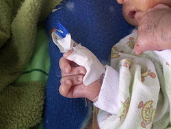
Clenched hand and overlapping fingers: index finger overlaps third finger and fifth finger overlaps fourth finger, characteristically seen in trisomy 18. This is caused by congenital joint contracture.
In utero, the most common characteristic is cardiac anomalies, followed by central nervous system anomalies such as head shape abnormalities. The most common intracranial anomaly is the presence of choroid plexus cysts, which are pockets of fluid on the brain. These are not problematic in themselves, but their presence may be a marker for trisomy 18.[9][10] Sometimes, excess amniotic fluid or polyhydramnios is exhibited.[7] Although uncommon in the syndrome, trisomy 18 causes a large portion of prenatally diagnosed cases of Dandy–Walker malformation.[11][12]
Trisomy 18 is a chromosomal abnormality characterized by the presence of an extra copy of genetic material on the 18th chromosome, either in whole (trisomy 18) or in part (such as due to translocations). The additional chromosome usually occurs before conception. The effects of the extra copy vary greatly, depending on the extent of the extra copy, genetic history, and chance. Trisomy 18 occurs in all human populations but is more prevalent in female offspring.[13]
A typical egg or sperm cell contains individual chromosomes, each of which contributes to the 23 pairs of chromosomes needed to form a normal cell with a typical human karyotype of 46 chromosomes. Numerical errors can arise at either of the two meiotic divisions and cause the failure of a chromosome to segregate into the daughter cells (nondisjunction). This results in an extra chromosome, making the haploid number 24 rather than 23. Fertilization of eggs or insemination by sperm that contain an extra chromosome results in trisomy, or three copies of a chromosome rather than two.[14]
Trisomy 18 (47,XX,+18) is caused by a meiotic nondisjunction event. In nondisjunction, a pair of chromosomes fails to separate during cell division; thus, a gamete (i.e., a sperm or egg cell) is produced with an extra copy of a chromosome (for a total of 24 chromosomes). When combined with a normal gamete from the other parent, the resulting embryo has 47 chromosomes, with three copies of the problematic chromosome (in this case, chromosome 18). (Although an embryo could inherit a trisomy from both parents, it is, as a rule, extremely rare, and worse in terms of clinical perspective and prognosis.)
A small percentage of cases occur when only some of the body's cells have an extra copy of chromosome 18, resulting in a mixed population of cells with a differing number of chromosomes. Such cases are sometimes called mosaic trisomy 18. Very rarely, a piece of chromosome 18 becomes attached to another chromosome (translocated) before or after conception. Affected individuals have two copies of chromosome 18 plus extra material from chromosome 18 attached to another chromosome. With a translocation, a person has a partial trisomy for chromosome 18, and the abnormalities are often less severe than for the typical trisomy 18.[15]
Levels of PAPP-A, AFP, and uE3 are generally decreased during pregnancy, and free beta HCG is elevated.[16]
Prognosis
About 60% of pregnancies that are affected do not result in a live birth.[13] Major causes of death include hypoxia and heart abnormalities. It is impossible to predict an exact prognosis during pregnancy or the neonatal period.[13] Half of the live infants do not survive beyond the first week of life without interventions.[17] The median lifespan is five to 15 days without interventions.[18][19] About 8–12% of infants survive longer than 1 year without interventions.[20][21][bettersourceneeded] One percent of children live to age 10.[13] However, a retrospective Canadian study of 254 children with trisomy 18 demonstrated ten-year survival of 9.8%, and another found that 68.6% of children with surgical intervention survived infancy.[21] Though rare, some persons with Trisomy 18 survive into their twenties and thirties with the current eldest being well over 50 years. Current ongoing research at the University of Michigan shows survival rate with full interventions is about 90% until the first birthday, and 80% until 5 years. [22]
Epidemiology
Trisomy 18 occurs in about 1 in 5,000 live births, but more pregnancies are affected by the syndrome as the majority of those diagnosed with the condition prenatally will not survive to birth.[3] Although women in their teens, 20s, and early 30s may conceive babies with trisomy 18, the risk increases with age. The average maternal age for conceiving a child with this disorder is 32.5.[23]
History
Trisomy 18 was first identified by John Hilton Edwards in 1960, although he originally believed it to be caused by a trisomy of chromosome 18.[24]Klaus Patau and Eeva Therman reported another two cases shortly thereafter.[25] They identified the extra chromosome as being part of what Patau's lab called "group E", containing chromosomes 16, 17, and 18, but were unable to determine which chromosome was responsible at the time. Analyzing 5 more cases, they were able to determine that the extra chromosome was chromosome 18.[26]
↑Jorde, Lynn B.; Carey, John C.; Bamshad, Michael J. (2009). Medical Genetics (4ed.). Elsevier Health Sciences. p.109. ISBN978-0323075763. Archived from the original on 2016-10-02.
12"Trisomy 18". Medline. Archived from the original on 2008-10-01. Retrieved 2008-07-24.
↑Hurt K, Sottner O, Záhumenský J, etal. (2007). "[Choroid plexus cysts and risk of trisomy 18. Modifications regarding maternal age and markers]". Ceska Gynekol (in Czech). 72 (1): 49–52. PMID17357350.
↑Papp C, Ban Z, Szigeti Z, Csaba A, Beke A, Papp Z (2007). "Role of second trimester sonography in detecting trisomy 18: a review of 70 cases". J Clin Ultrasound. 35 (2): 68–72. doi:10.1002/jcu.20290. PMID17206726. S2CID23836946.
↑Smith, David W.; Patau, Klaus; Therman, Eeva; Inhorn, Stanley L. (September 1, 1960). "A new autosomal trisomy syndrome: multiple congenital anomalies caused by an extra chromosome". The Journal of Pediatrics. 57 (3): 338–345. doi:10.1016/S0022-3476(60)80241-7. PMID13831938.
↑Patau, Klaus; Therman, Eeva; Smith, David W.; DeMars, Robert I. (January 19, 1961). "Trisomy for chromosome no. 18 in man". Chromosoma. 12 (1). Berlin: 280–5. doi:10.1007/BF00328924. PMID13733243. S2CID2105207.
This page is based on this Wikipedia article Text is available under the CC BY-SA 4.0 license; additional terms may apply. Images, videos and audio are available under their respective licenses.