ATP citrate synthase

Last updated
ATP citrate synthase
Identifiers
EC no. 2.3.3.8
CAS no. 9027-95-6
Databases
IntEnz IntEnz view
BRENDA BRENDA entry
ExPASy NiceZyme view
KEGG KEGG entry
MetaCyc metabolic pathway
PRIAM profile
PDB structures RCSB PDB PDBe PDBsum
Gene Ontology AmiGO / QuickGO
Search
PMC articles
PubMed articles
NCBI proteins
Human ATP citrate lyase
6hxh.png
Crystal structure of human ATP citrate lyase in complex with citrate, coenzyme A and Mg.ADP. [1]
Identifiers
SymbolACLY
Alt. symbolsACL
NCBI gene 47
HGNC 115
OMIM 108728
PDB 3MWE, 3PFF, 5TDE, 5TDF, 5TDM, 5TDZ, 5TE1, 5TEQ, 5TES, 5TET, 6HXH, 6HXK, 6HXL, 6HXM, 6O0H, 6QFB 3MWD, 3MWE, 3PFF, 5TDE, 5TDF, 5TDM, 5TDZ, 5TE1, 5TEQ, 5TES, 5TET, 6HXH, 6HXK, 6HXL, 6HXM, 6O0H, 6QFB
RefSeq NM_001096
UniProt P53396
Other data
EC number 2.3.3.8
Locus Chr. 17 q21.2
Search for
Structures Swiss-model
Domains InterPro

ATP citrate synthase (also ATP citrate lyase (ACLY)) is an enzyme that in animals represents an important step in fatty acid biosynthesis. [2] By converting citrate to acetyl-CoA, the enzyme links carbohydrate metabolism, which yields citrate as an intermediate, with fatty acid biosynthesis, which consumes acetyl-CoA. [3] In plants, ATP citrate lyase generates cytosolic acetyl-CoA precursors of thousands of specialized metabolites, including waxes, sterols, and polyketides. [4]

Contents

Function

ATP citrate lyase is the primary enzyme responsible for the synthesis of cytosolic acetyl-CoA in many tissues. The enzyme is a tetramer of apparently identical subunits. In animals, the product, acetyl-CoA, is used in several important biosynthetic pathways, including lipogenesis and cholesterogenesis. [5] It is activated by insulin. [6]

In plants, ATP citrate lyase generates acetyl-CoA for cytosolically-synthesized metabolites; Acetyl-CoA is not transported across subcellular membranes of plants. Such metabolites include: elongated fatty acids (used in seed oils, membrane phospholipids, the ceramide moieties of sphingolipids, cuticle, cutin, and suberin); flavonoids; malonic acid; acetylated phenolics, alkaloids, isoprenoids, anthocyanins, and sugars; and, mevalonate-derived isoprenoids (e.g., sesquiterpenes, sterols, brassinosteroids); malonyl and acyl-derivatives (d-amino acids, malonylated flavonoids, acylated, prenylated and malonated proteins). [4] De novo fatty acid biosynthesis in plants occurs in plastids; thus, ATP citrate lyase is not relevant to this pathway.

Reaction

ATP citrate lyase is responsible for catalyzing the conversion of citrate and Coenzyme A (CoA) to acetyl-CoA and oxaloacetate, driven by hydrolysis of ATP. [3] In the presence of ATP and CoA, citrate lyase catalyzes the cleavage of citrate to yield acetyl CoA, oxaloacetate, adenosine diphosphate (ADP), and orthophosphate (Pi):

citrate + ATP + CoA → oxaloacetate + Acetyl-CoA + ADP + Pi

This enzyme was formerly given the EC number 4.1.3.8. [7]

Location

The enzyme is cytosolic in plants [4] and animals. [8] [9]

Structure

The enzyme is composed of two subunits in green plants (including Chlorophyceae, Marchantimorpha, Bryopsida, Pinaceae, monocotyledons, and eudicots), species of fungi, glaucophytes, Chlamydomonas , and prokaryotes.

Animal ACL enzymes are homomeric; a fusion of the ACLA and ACLB genes probably occurred early in the evolutionary history of this kingdom. [4]

The mammalian ATP citrate lyase has a N-terminal citrate-binding domain that adopts a Rossmann fold, followed by a CoA binding domain and CoA-ligase domain and finally a C-terminal citrate synthase domain. The cleft between the CoA binding and citrate synthase domains forms the active site of the enzyme, where both citrate and acetyl-coenzyme A bind.

In 2010, a structure of truncated human ATP citrate lyase was determined using X-ray diffraction to a resolution of 2.10 Å. [3] In 2019, a full length structure of human ACLY in complex with the substrates coenzyme A, citrate and Mg.ADP was determined by X-ray crystallography to a resolution of 3.2 Å. [1] Moreover, in 2019 a full length structure of ACLY in complex with an inhibitor was determined by cryo-EM methods to a resolution of 3.7 Å. [10] Additional structures of heteromeric ACLY-A/B from the green sulfur bacteria Chlorobium limicola and the archaeon Methanosaeta concilii show that the architecture of ACLY is evolutionarily conserved. [1] Full length ACLY structures showed that the tetrameric protein oligomerizes via its C-terminal domain. The C-terminal domain had not been observed in the previously determined truncated crystal structures. The C-terminal region of ACLY assembles in a tetrameric module that is structurally similar to citryl-CoA lyase (CCL) found in deep branching bacteria. [1] [11] This CCL module catalyses the cleavage of the citryl-CoA intermediate into the products acetyl-CoA and oxaloacetate. In 2019, cryo-EM structures of human ACLY, alone or bound to substrates or products were reported as well. [12] [13] ACLY forms a homotetramer with a rigid citrate synthase homology (CSH) module, flanked by four flexible acetyl-CoA synthetase homology (ASH) domains; CoA is bound at the CSH–ASH interface in mutually exclusive productive or unproductive conformations. The structure of a catalytic mutant of ACLY in the presence of ATP, citrate and CoA substrates reveals a CoA and phosphor-citrate intermediate in the N-terminal domain. Cryo-EM structures of products bound ACLY and substrates bound ACLY were also determined at 3.0 Å and 3.1 Å. An EM structure of mutant E599Q in complex with CoA and phospho-citrate intermediate was determined at resolution of 2.9 Å. Comparison between these structures of apo-ACLY and ligands bound ACLY demonstrated conformational changes on ASH domain (N-terminal domain) when different ligands bind.

Pharmacology

The enzyme's action can be inhibited by the coenzyme A-conjugate of bempedoic acid, a compound which lowers LDL cholesterol in humans. [14] The drug was approved by the Food and Drug Administration in February 2020 for use in the United States.

Related Research Articles

<span class="mw-page-title-main">Citric acid cycle</span> Interconnected biochemical reactions releasing energy

The citric acid cycle—also known as the Krebs cycle, Szent-Györgyi-Krebs cycle or the TCA cycle (tricarboxylic acid cycle)—is a series of biochemical reactions to release the energy stored in nutrients through the oxidation of acetyl-CoA derived from carbohydrates, fats, and proteins. The chemical energy released is available under the form of ATP. The Krebs cycle is used by organisms that respire (as opposed to organisms that ferment) to generate energy, either by anaerobic respiration or aerobic respiration. In addition, the cycle provides precursors of certain amino acids, as well as the reducing agent NADH, that are used in numerous other reactions. Its central importance to many biochemical pathways suggests that it was one of the earliest components of metabolism. Even though it is branded as a 'cycle', it is not necessary for metabolites to follow only one specific route; at least three alternative segments of the citric acid cycle have been recognized.

<span class="mw-page-title-main">Glycolysis</span> Series of interconnected biochemical reactions

Glycolysis is the metabolic pathway that converts glucose into pyruvate and, in most organisms, occurs in the liquid part of cells. The free energy released in this process is used to form the high-energy molecules adenosine triphosphate (ATP) and reduced nicotinamide adenine dinucleotide (NADH). Glycolysis is a sequence of ten reactions catalyzed by enzymes.

<span class="mw-page-title-main">Acetyl-CoA</span> Chemical compound

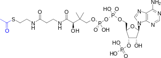
Acetyl-CoA is a molecule that participates in many biochemical reactions in protein, carbohydrate and lipid metabolism. Its main function is to deliver the acetyl group to the citric acid cycle to be oxidized for energy production.

<span class="mw-page-title-main">Biological carbon fixation</span> Series of interconnected biochemical reactions

Biological carbon fixation or сarbon assimilation is the process by which inorganic carbon is converted to organic compounds by living organisms. The compounds are then used to store energy and as structure for other biomolecules. Carbon is primarily fixed through photosynthesis, but some organisms use a process called chemosynthesis in the absence of sunlight.

<span class="mw-page-title-main">Oxaloacetic acid</span> Organic compound

Oxaloacetic acid (also known as oxalacetic acid or OAA) is a crystalline organic compound with the chemical formula HO2CC(O)CH2CO2H. Oxaloacetic acid, in the form of its conjugate base oxaloacetate, is a metabolic intermediate in many processes that occur in animals. It takes part in gluconeogenesis, the urea cycle, the glyoxylate cycle, amino acid synthesis, fatty acid synthesis and the citric acid cycle.

<span class="mw-page-title-main">Mitochondrial matrix</span> Space within the inner membrane of the mitochondrion

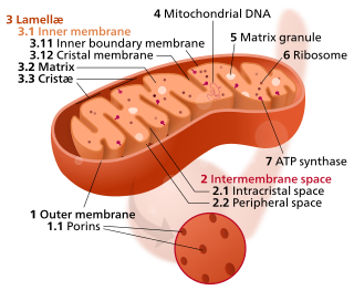
In the mitochondrion, the matrix is the space within the inner membrane. The word "matrix" stems from the fact that this space is viscous, compared to the relatively aqueous cytoplasm. The mitochondrial matrix contains the mitochondrial DNA, ribosomes, soluble enzymes, small organic molecules, nucleotide cofactors, and inorganic ions.[1] The enzymes in the matrix facilitate reactions responsible for the production of ATP, such as the citric acid cycle, oxidative phosphorylation, oxidation of pyruvate, and the beta oxidation of fatty acids.

Fatty acid metabolism consists of various metabolic processes involving or closely related to fatty acids, a family of molecules classified within the lipid macronutrient category. These processes can mainly be divided into (1) catabolic processes that generate energy and (2) anabolic processes where they serve as building blocks for other compounds.

<span class="mw-page-title-main">Glyoxylate cycle</span> Series of interconnected biochemical reactions

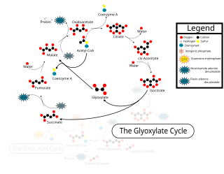
The glyoxylate cycle, a variation of the tricarboxylic acid cycle, is an anabolic pathway occurring in plants, bacteria, protists, and fungi. The glyoxylate cycle centers on the conversion of acetyl-CoA to succinate for the synthesis of carbohydrates. In microorganisms, the glyoxylate cycle allows cells to use two carbons, such as acetate, to satisfy cellular carbon requirements when simple sugars such as glucose or fructose are not available. The cycle is generally assumed to be absent in animals, with the exception of nematodes at the early stages of embryogenesis. In recent years, however, the detection of malate synthase (MS) and isocitrate lyase (ICL), key enzymes involved in the glyoxylate cycle, in some animal tissue has raised questions regarding the evolutionary relationship of enzymes in bacteria and animals and suggests that animals encode alternative enzymes of the cycle that differ in function from known MS and ICL in non-metazoan species.
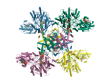
<span class="mw-page-title-main">Citrate synthase</span> Enzyme found in humans

The enzyme citrate synthase E.C. 2.3.3.1 ] exists in nearly all living cells and stands as a pace-making enzyme in the first step of the citric acid cycle. Citrate synthase is localized within eukaryotic cells in the mitochondrial matrix, but is encoded by nuclear DNA rather than mitochondrial. It is synthesized using cytoplasmic ribosomes, then transported into the mitochondrial matrix.

In biochemistry, fatty acid synthesis is the creation of fatty acids from acetyl-CoA and NADPH through the action of enzymes called fatty acid synthases. This process takes place in the cytoplasm of the cell. Most of the acetyl-CoA which is converted into fatty acids is derived from carbohydrates via the glycolytic pathway. The glycolytic pathway also provides the glycerol with which three fatty acids can combine to form triglycerides, the final product of the lipogenic process. When only two fatty acids combine with glycerol and the third alcohol group is phosphorylated with a group such as phosphatidylcholine, a phospholipid is formed. Phospholipids form the bulk of the lipid bilayers that make up cell membranes and surrounds the organelles within the cells. In addition to cytosolic fatty acid synthesis, there is also mitochondrial fatty acid synthesis (mtFASII), in which malonyl-CoA is formed from malonic acid with the help of malonyl-CoA synthetase (ACSF3), which then becomes the final product octanoyl-ACP (C8) via further intermediate steps.

<span class="mw-page-title-main">Long-chain-fatty-acid—CoA ligase</span> Class of enzymes

The long chain fatty acyl-CoA ligase is an enzyme of the ligase family that activates the oxidation of complex fatty acids. Long chain fatty acyl-CoA synthetase catalyzes the formation of fatty acyl-CoA by a two-step process proceeding through an adenylated intermediate. The enzyme catalyzes the following reaction,

<span class="mw-page-title-main">Thiolase</span> Enzymes

Thiolases, also known as acetyl-coenzyme A acetyltransferases (ACAT), are enzymes which convert two units of acetyl-CoA to acetoacetyl CoA in the mevalonate pathway.

<span class="mw-page-title-main">Isocitrate lyase</span>

Isocitrate lyase, or ICL, is an enzyme in the glyoxylate cycle that catalyzes the cleavage of isocitrate to succinate and glyoxylate. Together with malate synthase, it bypasses the two decarboxylation steps of the tricarboxylic acid cycle and is used by bacteria, fungi, and plants.

<span class="mw-page-title-main">Methylisocitrate lyase</span>

The enzyme methylisocitrate lyase catalyzes the chemical reaction

In enzymology, a citrate (Re)-synthase (EC 2.3.3.3) is an enzyme that catalyzes the chemical reaction

<span class="mw-page-title-main">Hydroxymethylglutaryl-CoA synthase</span> Class of enzymes

In molecular biology, hydroxymethylglutaryl-CoA synthase or HMG-CoA synthase EC 2.3.3.10 is an enzyme which catalyzes the reaction in which acetyl-CoA condenses with acetoacetyl-CoA to form 3-hydroxy-3-methylglutaryl-CoA (HMG-CoA). This reaction comprises the second step in the mevalonate-dependent isoprenoid biosynthesis pathway. HMG-CoA is an intermediate in both cholesterol synthesis and ketogenesis. This reaction is overactivated in patients with diabetes mellitus type 1 if left untreated, due to prolonged insulin deficiency and the exhaustion of substrates for gluconeogenesis and the TCA cycle, notably oxaloacetate. This results in shunting of excess acetyl-CoA into the ketone synthesis pathway via HMG-CoA, leading to the development of diabetic ketoacidosis.

<span class="mw-page-title-main">Malate synthase</span> Class of enzymes

In enzymology, a malate synthase (EC 2.3.3.9) is an enzyme that catalyzes the chemical reaction

Glutaminolysis (glutamine + -lysis) is a series of biochemical reactions by which the amino acid glutamine is lysed to glutamate, aspartate, CO2, pyruvate, lactate, alanine and citrate.

<span class="mw-page-title-main">Citrate synthase family</span>

In molecular biology, the citrate synthase family of proteins includes the enzymes citrate synthase EC 2.3.3.1, and the related enzymes 2-methylcitrate synthase EC 2.3.3.5 and ATP citrate lyase EC 2.3.3.8.

<span class="mw-page-title-main">Citrate–malate shuttle</span> Series of chemical reactions

The citrate-malate shuttle is a series of chemical reactions, commonly referred to as a biochemical cycle or system, that transports acetyl-CoA in the mitochondrial matrix across the inner and outer mitochondrial membranes for fatty acid synthesis. Mitochondria are enclosed in a double membrane. As the inner mitochondrial membrane is impermeable to acetyl-CoA, the shuttle system is essential to fatty acid synthesis in the cytosol. It plays an important role in the generation of lipids in the liver.

References

  1. 1 2 3 4 Verschueren KH, Blanchet C, Felix J, Dansercoer A, De Vos D, Bloch Y, et al. (April 2019). "Structure of ATP citrate lyase and the origin of citrate synthase in the Krebs cycle" (PDF). Nature. 568 (7753): 571–575. Bibcode:2019Natur.568..571V. doi:10.1038/s41586-019-1095-5. PMID   30944476. S2CID   92999924.
  2. Elshourbagy NA, Near JC, Kmetz PJ, Wells TN, Groot PH, Saxty BA, et al. (March 1992). "Cloning and expression of a human ATP-citrate lyase cDNA". European Journal of Biochemistry. 204 (2): 491–9. doi: 10.1111/j.1432-1033.1992.tb16659.x . PMID   1371749.
  3. 1 2 3 Sun T, Hayakawa K, Bateman KS, Fraser ME (August 2010). "Identification of the citrate-binding site of human ATP-citrate lyase using X-ray crystallography". The Journal of Biological Chemistry. 285 (35): 27418–28. doi: 10.1074/jbc.M109.078667 . PMC   2930740 . PMID   20558738.
  4. 1 2 3 4 Fatland BL, Ke J, Anderson MD, Mentzen WI, Cui LW, Allred CC, et al. (October 2002). "Molecular characterization of a heteromeric ATP-citrate lyase that generates cytosolic acetyl-coenzyme A in Arabidopsis". Plant Physiology. 130 (2): 740–56. doi:10.1104/pp.008110. PMC   166603 . PMID   12376641.
  5. "Entrez Gene: ATP citrate lyase".
  6. Guay C, Madiraju SR, Aumais A, Joly E, Prentki M (December 2007). "A role for ATP-citrate lyase, malic enzyme, and pyruvate/citrate cycling in glucose-induced insulin secretion". The Journal of Biological Chemistry. 282 (49): 35657–65. doi: 10.1074/jbc.M707294200 . PMID   17928289.
  7. ATP+Citrate+Lyase at the U.S. National Library of Medicine Medical Subject Headings (MeSH)
  8. Zaidi N, Swinnen JV, Smans K (2012). "ATP-Citrate Lyase: A Key Player in Cancer Metabolism". Cancer Research. 72 (15): 3709–3714. doi: 10.1158/0008-5472.CAN-11-4112 . PMID   22787121.
  9. Pietrocola F, Galluzzi L, Bravo-San Pedro JM, Madeo F, Kroemer G (2015). "Acetyl Coenzyme A: A Central Metabolite and Second Messenger". Cell Metabolism. 21 (6): 805–821. doi: 10.1016/j.cmet.2015.05.014 . PMID   26039447.
  10. Wei J, Leit S, Kuai J, Therrien E, Rafi S, Harwood HJ, et al. (April 2019). "An allosteric mechanism for potent inhibition of human ATP-citrate lyase". Nature. 568 (7753): 566–570. Bibcode:2019Natur.568..566W. doi:10.1038/s41586-019-1094-6. PMID   30944472. S2CID   93000843.
  11. Aoshima M, Ishii M, Igarashi Y (May 2004). "A novel enzyme, citryl-CoA lyase, catalysing the second step of the citrate cleavage reaction in Hydrogenobacter thermophilus TK-6". Molecular Microbiology. 52 (3): 763–70. doi:10.1111/j.1365-2958.2004.04010.x. PMID   15101982. S2CID   32105039.
  12. Wei X, Schultz K, Bazilevsky GA, Vogt A, Marmorstein R (January 2020). "Molecular basis for acetyl-CoA production by ATP-citrate lyase". Nature Structural & Molecular Biology. 27 (1): 33–41. doi:10.1038/s41594-019-0351-6. PMC   8436250 . PMID   31873304.
  13. Wei X, Schultz K, Bazilevsky GA, Vogt A, Marmorstein R (May 2020). "Author Correction: Molecular basis for acetyl-CoA production by ATP-citrate lyase". Nature Structural & Molecular Biology. 27 (5): 511–513. doi: 10.1038/s41594-020-0421-9 . PMC   8439269 . PMID   32242119.
  14. Ray KK, Bays HE, Catapano AL, Lalwani ND, Bloedon LT, Sterling LR, et al. (CLEAR Harmony Trial) (March 2019). "Safety and Efficacy of Bempedoic Acid to Reduce LDL Cholesterol". The New England Journal of Medicine. 380 (11): 1022–1032. doi: 10.1056/NEJMoa1803917 . hdl: 10044/1/68213 . PMID   30865796.

Further reading

This article incorporates text from the United States National Library of Medicine, which is in the public domain.