Ancylostoma caninum

Last updated

Ancylostoma caninum
Anterior end of A. caninum.jpg
Scientific classification OOjs UI icon edit-ltr.svg
Domain: Eukaryota
Kingdom: Animalia
Phylum: Nematoda
Class: Chromadorea
Order: Rhabditida
Family: Ancylostomatidae
Genus: Ancylostoma
Species:
A. caninum
Binomial name
Ancylostoma caninum
(Ercolani, 1859)

Ancylostoma caninum is a species of nematode known as a hookworm, which principally infects the small intestine of dogs. [1] [2] [3] The result of A. caninum infection ranges from asymptomatic cases to death of the dog; better nourishment, increasing age, prior A. caninum exposure, or vaccination are all linked to improved survival. [2] [4] [5] [6] Other hosts include carnivores such as wolves, foxes, and cats, with a small number of cases having been reported in humans. [1] [2]

Contents

Warm and moist conditions are important to allow survival of A. caninum during the free-living stages of its lifecycle, so it is largely restricted to temperate, tropical, and subtropical regions. [3] [7] In parts of the world where these climatic requirements are met such as Sri Lanka, Southeast Asia, and Malaysia, A. caninum is the main cause of hookworm disease in canines. [5] [7]

Morphology

Mouth and teeth of A. caninum Anterior end of A. caninum.jpg
Mouth and teeth of A. caninum
A. caninum egg Canine hookworm egg 1.JPG
A. caninum egg

A. caninum females are typically 14–16 mm (0.55–0.63 in) long and 0.5 mm (0.02 in) wide, while the males are smaller at 10–12 mm (0.39–0.47 in) in length and 0.36 mm (0.01 in) in width. [2] [3] Males have a copulatory bursa , which consists of spine-like spicules positioned on three muscular rays that grasp the female during mating. [1] [2] [3] As with other nematodes, the sperm lack flagella. [1] The copulatory bursa is a unique feature of Strongylida members, thus making it a useful means for identifying members of this suborder; it is also used to distinguish members within the suborder due to differences in bursa appearance between species. [2] The vulva of A. caninum females is located at the boundary of the second and final thirds of the body. [1]

The teeth of A. caninum are found in the buccal capsule and divided into three sets. [1] [2] Two ventral sets form a lower-jaw equivalent, while a further set projects from the dorsal side and loosely equates to an upper jaw. [2] Each ventral set has three points, with those furthest to the sides being the largest. [1] [8] While the ventral sets are prominent, the dorsal set is hidden deeper in the buccal capsule. [1]

A. caninum bends its head end upward (dorsally), which has been noted to be a potential source of confusion when determining how the hookworm is oriented. [2] If it has recently ingested blood, A. caninum is red in colour; if not, it appears grey. [1] A. caninum has an alimentary canal made up of an esophagus, intestine, and rectum – the esophagus is highly muscular, reflecting its role in pulling intestinal mucosa into the body when it feeds. [2] [3] Esophageal and anal rings of A. caninum are the source of nerve fibres that extend throughout the body to innervate sensory organs, including amphids and phasmids. [1] [3]

Eggs are laid by the females, typically when at the eight-cell stage. [3] Eggs are 38–43 μm in width, with thin walls. [3] [5]

Distribution

Freezing, temperatures exceeding 37 °C (99 °F), drying, or exposing A. caninum to sunlight all give reduced survival of the free-living stage, with rates of infection rising with temperature, provided 37 °C is not exceeded. [1] [2] A. caninum is, therefore, largely restricted to warm, moist climates, though infections are seen in the United States and southern Canada where the temperature is suboptimal. [2] Specific niches are also able to satisfy the environmental requirements of A. caninum, despite not necessarily being in the tropics, such as mines. [2]

Lifecycle

Transmission via the environment

Eggs are excreted from the host in the feces and typically hatch within a day on moist, warm soil into larvae with a non-living cuticle layer. [1] [2] [5] By 4–5 days, the larvae have moulted twice and are now able to infect a host. [2] Migration occurs from the feces into the surrounding soil. [2] Two routes of infection from the environment exist. The first route involves penetration of skin at hair follicles or sweat glands, especially between the footpads where contact with soil is frequent and the skin is thinner than otherwise. [2] Secretion of a protease by A. caninum is thought to aid this process. [2] The larvae then migrate through the dermis of the skin, enter the circulatory system and are carried to the lungs. [2] A. caninum larvae exit the blood at the lungs, move from the alveoli up through the trachea and are swallowed to end up in the intestine. [2]

The second and more common route to the small intestine is by direct ingestion of A. caninum by the host, but the subsequent process is identical in either case. [2] [3] During this third stage of the larva, male or female reproductive organs become established. [3] Larvae of this stage have been shown to secrete a molecule (Ac-asp-2) related to venom allergens in response to host-specific signals; this is thought to have a possible role in helping with the infection process. [9] A third and final moulting occurs, resulting in the mature form of A. caninum, which then feeds on mucosa and blood of the small intestinal wall. [2] The trigger of feeding is understood to be a receptor-mediated response; however, the detail of this process has yet to be established. [10] Sexual reproduction also occurs in the intestine to produce a further round of eggs to complete the cycle. [2] Females are thought to produce a pheromone which attracts males and are able to lay about 10,000 eggs per day. [1] [11]

Direct transmission

Direct transmission between hosts is also possible. Larvae having accessed through the skin may avoid exit via the lungs and remain in circulation for transport around the body. [3] At the uterine artery of a pregnant female, the larvae are able to cross the placenta to cause prenatal infection of foetuses. [3] Larvae of an infected foetus move to the liver until birth, when migration continues with movement to the intestine via the circulation and lungs as previously described. [3] Alternatively, A. caninum larvae evading exit from the circulation at the lungs may instead be carried to the mammary glands and transmitted from the mother in her colostrum or milk to her pups; infection then proceeds in an identical manner as infection by ingestion from the environment. [3] [5] Infected mothers have been found to only rarely give prenatal transmission to pups, while the likelihood of causing transmission via the lactational route during nursing is much higher. [12]

Pathogenesis

Damage during migration to intestine

Pair of A. caninum hookworms. Hookworms.JPG
Pair of A. caninum hookworms.

A. caninum larvae cause damage to the host at the point of entry through the skin, leaving a wound vulnerable to secondary infections. [2] As the larvae migrate through the skin, an inflammatory response, dermatitis, is often stimulated, which can be exacerbated in hosts which have hypersensitive responses. [2] [5] Further damage is caused when the larvae leave the circulation and enter the lung, with the amount of damage dependent on the extent of the infection; pneumonia and coughing are common consequences. [2]

Damage once in intestine

Once in the gut, A. caninum attaches to and ingests the mucosal lining along with some consumption of blood; up to 0.1 ml in 24hrs. [2] [5] In a 24hr period A. caninum typically feeds from six sites. [2] This damage to the mucosa compromises the body's defences and can result in secondary infections by microbes. [7] A group of anticoagulant proteins called A. caninum anticoagulant proteins (AcAPs), which inhibit a range of blood coagulation factors such as Xa, are used by A. caninum to help in the feeding process by preventing clotting and increasing blood loss. [13] [14] These AcAPs are among the most powerful natural anticoagulants that exist and are a key reason for anemia being caused and blood being observed in the faeces of infected hosts. [5] [14] Blood losses peak just prior to egg production by the females because this is when their requirements for food are greatest; the amount that they are eating is also peaking, so maximal damage to the intestine is being caused. [3]

Diagnosis

Analysis of faeces [15] is the definitive method by which a suspected A. caninum infection is confirmed. [2] The faeces are sampled and examined microscopically for the characteristic ovular, thin-shelled eggs of A. caninum. [5] Absence of eggs in faeces does not rule out infection; a significant delay of at least 5 weeks exists between initial infection and excretion of eggs in the faeces (larvae must fully mature and reproduce before eggs can be laid). [3] [5] In fact, pups frequently die before passing of eggs in the faeces begins. [5] Using the number of eggs in stool samples as an indicator of the extent of infestation requires care to be taken because females have been shown to produce fewer eggs when the overall number of worms increases. [16]

Signs and symptoms expected to be observed are lethargy, weight loss, weakness, roughness of the hair coat, and pale mucous membranes indicative of anemia. [2] [5] Well-fed, older dogs with smaller infestations may present few or even none of these symptoms. [2] [5] Diarrhoea is rare, but stools are typically black due to the blood-derived haemoglobin present in them. [2]

The disease resulting from such A. caninum infection is referred to by the general term "hookworm disease" or the more specific terms "ancylostomiasis" and "ancylostomosis," [17] which recognise the genus of the causative nematode. [2]

Prevention and control

Dog kennel: keeping kennels clean reduces risk of A. caninum infection. Dog kennel Mason.jpg
Dog kennel: keeping kennels clean reduces risk of A. caninum infection.

A clean environment minimises the risk of A. caninum infection; this can include regularly washed concrete or gravel in kennels instead of soil. [2] [5] Females are typically checked prior to using them for breeding purposes for nematodes such as A. caninum and birth and suckling can be restricted to sanitised areas to lower the risk of health complications to the pups. [5] When infection of a pregnant dog is known or suspected fenbendazole or ivermectin can be administered to the female to help avoid transmission to the pups. [5]

Canines have been seen to develop significant resistance to A. caninum naturally with age; this protection develops faster in and fully mature females show substantially greater resistance than fully mature dogs. [4] Specifically the age-related resistance means A. caninum takes longer to reach sexual maturity in older animals and fewer larvae fully develop. [11]

Vaccination

Numerous vaccines have been developed with varying success against A. caninum. Use of an enzyme important in the worm's feeding process is popular, with one example being AcCP2, a protease, which, when used to vaccinate dogs, gives a strong antibody response, a lowering of numbers of eggs found in stools and a decrease in intestinal worm size. [18] These effects are attributed to reduced AcCP2 activity upon antibody binding. [18] A similar approach has been taken using another A. caninum digestive enzyme, AcGST1, but it failed to give statistically significant results in dogs. [19]

An alternative approach has been to disrupt the migratory ability of A. caninum; this was done successfully using the AcASP1 protein of A. caninum, which gives an increase in antibody levels of all subclasses, as well as a reduced worm burden. [6] Other studies using the same vaccine have shown 79% reduction of worm burden [20] resulting from this approach. [21]

Animals with prior exposure to A. caninum show enhanced resistance, but careful removal of all worms from the previous infection results in loss of this resistance. [11] Studies in mice show resistance due to past exposure can protect against otherwise lethal worm doses and that this is a general form of resistance – defense is offered against subsequent infections via either mouth or skin. [22]

Medication

Drugs used in treatment of A. caninum infections of dogs include: dichlorvos, fenbendazole, flubendazole, mebendazole, nitroscanate, piperazine, pyrantel, milbemycin, moxidectin, diethylcarbamazine, oxibendazole, and ivermectin. [5]

In humans

In inappropriate hosts such as humans, A. caninum is able to enter the skin, but cannot proceed into the circulation and on to the intestine; instead, the disease dermal larva migrans results, caused by movement of the nematode within the skin and which can persist for several months without intervention. [1]

While access to the intestine is not possible via this route, it can occur via ingestion; in a report of 93 enteritis cases in northern Queensland, Australia, which were possibly caused by A. caninum infection, all those interviewed described behaviour consistent with A. caninum exposure and a colonoscopy of one patient gave positive identification of an adult A. caninum worm. [23] [24] Since then, work has shown A. caninum can easily go unnoticed or fail to be preserved in specimens, making the true incidence of infection in humans likely to be higher than is officially recorded. [25]

Economic burden

The animals affected by A. caninum infection are not used for food or labour purposes, thus the economic burden from animal illness is low. [1] Human A. caninum infections are likely underestimated and misdiagnosed; the economic impact caused by missing work due to infection [26] may be underestimated and significant. [25]

Related Research Articles

<i>Strongyloides stercoralis</i> Species of worm

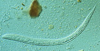
Strongyloides stercoralis is a human pathogenic parasitic roundworm causing the disease strongyloidiasis. Its common name in the US is threadworm. In the UK and Australia, however, the term threadworm can also refer to nematodes of the genus Enterobius, otherwise known as pinworms.

<span class="mw-page-title-main">Hookworm infection</span> Disease caused by intestinal parasites

Hookworm infection is an infection by a type of intestinal parasite known as a hookworm. Initially, itching and a rash may occur at the site of infection. Those only affected by a few worms may show no symptoms. Those infected by many worms may experience abdominal pain, diarrhea, weight loss, and tiredness. The mental and physical development of children may be affected. Anemia may result.

<i>Haemonchus contortus</i> Species of roundworm

Haemonchus contortus, also known as the barber's pole worm, is a very common parasite and one of the most pathogenic nematodes of ruminants. Adult worms attach to abomasal mucosa and feed on the blood. This parasite is responsible for anemia, oedema, and death of infected sheep and goats, mainly during summer in warm, humid climates.

<i>Necator americanus</i> Species of hookworm

Necator americanus is a species of hookworm commonly known as the New World hookworm. Like other hookworms, it is a member of the phylum Nematoda. It is an obligatory parasitic nematode that lives in the small intestine of human hosts. Necatoriasis—a type of helminthiasis—is the term for the condition of being host to an infestation of a species of Necator. Since N. americanus and Ancylostoma duodenale are the two species of hookworms that most commonly infest humans, they are usually dealt with under the collective heading of "hookworm infection". They differ most obviously in geographical distribution, structure of mouthparts, and relative size.

<i>Ascaris suum</i> Species of roundworm

Ascaris suum, also known as the large roundworm of pig, is a parasitic nematode that causes ascariasis in pigs. While roundworms in pigs and humans are today considered as two species with different hosts, cross-infection between humans and pigs is possible; some researchers have thus argued they are the same species. Ascariasis is associated with contact to pigs and pig manure in Denmark.

<span class="mw-page-title-main">Parasitic worm</span> Large type of parasitic organism

Parasitic worms, also known as helminths, are large macroparasites; adults can generally be seen with the naked eye. Many are intestinal worms that are soil-transmitted and infect the gastrointestinal tract. Other parasitic worms such as schistosomes reside in blood vessels.

<i>Ancylostoma duodenale</i> Species of the roundworm genus Ancylostoma

Ancylostoma duodenale is a species of the roundworm genus Ancylostoma. It is a parasitic nematode worm and commonly known as the Old World hookworm. It lives in the small intestine of hosts such as humans, cats and dogs, where it is able to mate and mature. Ancylostoma duodenale and Necator americanus are the two human hookworm species that are normally discussed together as the cause of hookworm infection. They are dioecious. Ancylostoma duodenale is abundant throughout the world, including Southern Europe, North Africa, India, China, southeast Asia, some areas in the United States, the Caribbean, and South America.

Uncinaria stenocephala is a nematode that parasitizes dogs, cats, and foxes as well as humans. It is rare to find in cats in the United States. Uncinaria stenocephala is the most common canine hookworm in cooler regions, such as Canada and the northern regions of the US, where it can be found primarily in foxes (40%). U. stenocephala is also one of the most common hookworms in the UK, called the northern hookworm, however it has a rather low prevalence. U. stenocephala is also considered to be zoonotic hookworms because they live in animals but can be transmitted to humans.

<span class="mw-page-title-main">Strongylida</span> Suborder of roundworms

The Strongylida suborder includes many of the important nematodes found in the gastrointestinal tracts of ruminants, horses, and swine, as well as the lungworms of ruminants and the hookworms of dogs and cats.

<span class="mw-page-title-main">Ancylostomiasis</span> Medical condition

Ancylostomiasis is a hookworm disease caused by infection with Ancylostoma hookworms. The name is derived from Greek ancylos αγκύλος "crooked, bent" and stoma στόμα "mouth".

Necatoriasis is the condition of infection by Necator hookworms, such as Necator americanus. This hookworm infection is a type of helminthiasis (infection) which is a type of neglected tropical disease.

<i>Toxascaris leonina</i> Species of roundworm

Toxascaris leonina is a common parasitic roundworm found in dogs, cats, foxes, and related host species. T. leonina is an ascarid nematode, a worldwide distributed helminth parasite which is in a division of eukaryotic parasites that, unlike external parasites such as lice and fleas, live inside their host. The definitive hosts of T. leonina include canids and felines (cats), while the intermediate hosts are usually rodents, such as mice or rats. Infection occurs in the definitive host when the animal eats an infected rodent. While T. leonina can occur in either dogs or cats, it is far more frequent in cats.

<i>Ancylostoma braziliense</i> Species of worm

Ancylostoma braziliense is a species of hookworm belonging to the genus Ancylostoma. It is an intestinal parasite of domestic cats and dogs. Severe infection is often fatal to these pets, especially in puppies and kittens. The infection is particularly endemic in the southern United States. It is most often confused with the zoonotic hookworm species Ancylostoma ceylanicum because of their uncanny resemblance.

<span class="mw-page-title-main">Ancylostomatidae</span> Family of roundworms

The Ancylostomatidae are a family of worms that includes the hookworms.

<span class="mw-page-title-main">Soil-transmitted helminthiasis</span> Roundworm infection contracted from contaminated soil

Soil-transmitted helminthiasis is a type of worm infection (helminthiasis) caused by different species of roundworms. It is caused specifically by those worms which are transmitted through soil contaminated with faecal matter and are therefore called soil-transmitted helminths. Three types of soil-transmitted helminthiasis can be distinguished: ascariasis, hookworm infection and whipworm infection. These three types of infection are therefore caused by the large roundworm A. lumbricoides, the hookworms Necator americanus or Ancylostoma duodenale and by the whipworm Trichuris trichiura.

Ancylostoma ceylanicum is a parasitic roundworm belonging to the genus Ancylostoma. It is a hookworm both of humans and of other mammals such as dogs, cats, and golden hamsters. It is the only zoonotic hookworm species that is able to produce symptomatic infections in humans, with the majority of cases being in Southeast Asia.

<i>Cooperia oncophora</i> Species of roundworm

Cooperia oncophora is one of the most common intestinal parasitic nematodes in cattle in temperate regions. Infections with C. oncophora may result in mild clinical symptoms, but can lead to weight loss and damage of the small intestine, especially when co-infections with other nematodes such as O. ostertagi occur. Infections are usually treated with broad-spectrum anthelmintics such as benzimidazole, but resistance to these drugs has developed in the last decades and is now very common. C. oncophora has a direct life cycle. Infective larvae are ingested by the host. The larvae grow to adults, which reproduce in the small intestines. Eggs are shed onto the pasture with the faeces, which leads to new infections. Co-infections with other gastro-intestinal nematodes such as O. ostertagi and H. contortus are common.

<span class="mw-page-title-main">Hookworm</span> Intestinal, blood-feeding, parasitic roundworms that cause types of infection known as helminthiases

Hookworms are intestinal, blood-feeding, parasitic roundworms that cause types of infection known as helminthiases. Hookworm infection is found in many parts of the world, and is common in areas with poor access to adequate water, sanitation, and hygiene. In humans, infections are caused by two main species of roundworm, belonging to the genera Ancylostoma and Necator. In other animals the main parasites are species of Ancylostoma. Hookworm is closely associated with poverty because it is most often found in impoverished areas, and its symptoms promote poverty through the educational and health effects it has on children. It is the leading cause of anemia and undernutrition in developing countries, while being one of the most commonly occurring diseases among poor people. Hookworm thrives in areas where rainfall is sufficient and keeps the soil from drying out, and where temperatures are higher, making rural, coastal areas prime conditions for the parasite to breed.

<span class="mw-page-title-main">Cat worm infections</span> Worm infections in cats

Cat worm infections, the infection of cats (Felidae) with parasitic worms, occur frequently. Most worm species occur worldwide in both domestic and other cats, but there are regional, species and lifestyle differences in the frequency of infestation. According to the classification of the corresponding parasites in the zoological system, infections can be divided into those caused by nematode and flatworms - in the case of the latter, mainly cestoda and trematoda - while other strains are of no veterinary significance. While threadworms usually do not require an intermediate host for their reproduction, the development cycle of flatworms always proceeds via alternate hosts.

<span class="mw-page-title-main">Nematode infection in dogs</span> Threadworm infections of dogs are frequent

Nematode infection in dogs - the infection of dogs with parasitic nemamotodes - are, along with tapeworm infections and infections with protozoa, frequent parasitoses in veterinary practice. Nematodes, as so-called endoparasites, colonize various internal organs - most of them the digestive tract - and the skin. To date, about 30 different species of nematode have been identified in domestic dogs; they are essentially also found in wild dog species. However, the majority of them often cause no or only minor symptoms of disease in adult animals. The infection therefore does not necessarily have to manifest itself in a worm disease (helminthosis). For most nematodes, an infection can be detected by examining the feces for eggs or larvae. Roundworm infection in dogs and the hookworm in dogs is of particular health significance in Central Europe, as they can also be transmitted to humans (zoonosis). Regular deworming can significantly reduce the frequency of infection and thus the risk of infection for humans and dogs.

References

  1. 1 2 3 4 5 6 7 8 9 10 11 12 13 14 15 Saeed S (2003). "Ancylostoma caninum". Animal Diversity Web. Retrieved March 20, 2013.
  2. 1 2 3 4 5 6 7 8 9 10 11 12 13 14 15 16 17 18 19 20 21 22 23 24 25 26 27 28 29 30 31 32 33 34 Marquardt W, Demaree, Grieve (2000). Parasitology and Vector Biology (2nd ed.). Harcourt Academic. pp. 370–376. ISBN   978-0121707552.
  3. 1 2 3 4 5 6 7 8 9 10 11 12 13 14 15 16 Olsen W (1986). Animal Parasites: their lifecycles and ecology (3rd ed.). Dover. pp.  399–416. ISBN   978-0486651262.
  4. 1 2 Miller (1965). "Influence of Age and Sex on Susceptibility of Dogs to Primary Infection with Ancylostoma caninum". The Journal of Parasitology. 51 (5): 701–704. doi:10.2307/3276142. JSTOR   3276142. PMID   5857264.
  5. 1 2 3 4 5 6 7 8 9 10 11 12 13 14 15 16 17 Peregrine A (March 2012). "Hookworms in Small Animals". The Merck Veterinary Manual. Archived from the original on 2012-07-11. Retrieved March 20, 2013.
  6. 1 2 Ghosh K, Hotez, P (January 1999). "Antibody-Dependent Reductions in Mouse Hookworm Burden after Vaccination with Ancylostoma caninum Secreted Protein 1". J Infect Dis. 180 (5): 1674–1681. doi:10.1086/315059. PMID   10515831. S2CID   17298045.
  7. 1 2 3 Cheng T (1986). General Parasitology (2nd ed.). Academic Press. pp. 93, 508. ISBN   978-0121707552.
  8. Ruppert E (1994). Invertebrate Zoology (6th ed.). Saunders College Pub. pp.  293. ISBN   978-0030989803.
  9. Hawdon J, Narasimhan S, Hotez P (April 1999). "Ancylostoma secreted protein 2: cloning and characterization of a second member of a family of nematode secreted proteins from Ancylostoma caninum". Molecular and Biochemical Parasitology. 99 (2): 149–165. doi: 10.1016/S0166-6851(99)00011-0 . PMID   10340481.
  10. Hawdon J, Schad G (February 1990). "Serum-Stimulated Feeding In vitro by Third-Stage Infective Larvae of the Canine Hookworm Ancylostoma caninum". The Journal of Parasitology. 76 (3): 394–398. doi:10.2307/3282673. JSTOR   3282673. PMID   2112598.
  11. 1 2 3 Herrick C (September 1928). "A Quantitative Study of Infections with Ancylostoma caninum in Dogs". Am. J. Epidemiol. 8 (2): 125–157. doi:10.1093/oxfordjournals.aje.a120994.
  12. Burke T, Roberson E (February 1985). "Prenatal and lactational transmission of Toxocara canis and Ancylostoma caninum: Experimental infection of the female before pregnancy". International Journal for Parasitology. 15 (1): 71–75. doi:10.1016/0020-7519(85)90104-3. PMID   3980144.
  13. Stanssens P, Bergum P, Gansemans Y, et al. (March 1996). "Anticoagulant repertoire of the hookworm Ancylostoma caninum". PNAS. 93 (5): 2149–2154. Bibcode:1996PNAS...93.2149S. doi: 10.1073/pnas.93.5.2149 . PMC   39925 . PMID   8700900.
  14. 1 2 Cappello M, Vlasuk G, Bergum P, et al. (June 1995). "Ancylostoma caninum anticoagulant peptide: a hookworm-derived inhibitor of human coagulation factor Xa". PNAS. 92 (13): 6152–6156. Bibcode:1995PNAS...92.6152C. doi: 10.1073/pnas.92.13.6152 . PMC   41660 . PMID   7597095.
  15. "Veterinary Advice Online – Fecal Flotation". Pet-Informed.
  16. Sarles M (November 1929). "The Effect of Age and Size of Infestation on the Egg Production of the Dog Hookworm Ancylostoma caninum" (PDF). The American Journal of Hygiene. 10 (3): 658–666.[ dead link ]
  17. "Ancyclostomosis". Medical Dictionary.
  18. 1 2 Loukas A, Bethony JM, Williamson AL, et al. (May 2004). "Vaccination of dogs with a recombinant cysteine protease from the intestine of canine hookworms diminishes the fecundity and growth of worms". J. Infect. Dis. 189 (10): 1952–61. doi: 10.1086/386346 . PMID   15122534.
  19. Zhan B, Liu S, Perally S, et al. (October 2005). "Biochemical characterization and vaccine potential of a heme-binding glutathione transferase from the adult hookworm Ancylostoma caninum". Infect. Immun. 73 (10): 6903–11. doi:10.1128/IAI.73.10.6903-6911.2005. PMC   1230892 . PMID   16177370.
  20. "Worm Burden". Medical Dictionary.
  21. Ghosh K, Hawdon J, Hotez, P (May 1996). "Vaccination with Alum-Precipitated Recombinant Ancylostoma-Secreted Protein 1 Protects Mice against Challenge Infections with Infective Hookworm (Ancylostoma caninum) Larvae". J Infect Dis. 174 (6): 1380–1383. doi: 10.1093/infdis/174.6.1380 . PMID   8940240.
  22. Kerr K (June 1936). "Studies on Acquired Immunity to the Dog Hookworm Ancylostoma caninum". Am. J. Epidemiol. 24 (2): 381–406. doi:10.1093/oxfordjournals.aje.a118272.
  23. Prociv P (June 1990). "Human eosinophilic enteritis caused by dog hookworm Ancylostoma caninum". The Lancet. 335 (8701): 1299–1302. doi:10.1016/0140-6736(90)91186-E. PMID   1971376. S2CID   31286210.
  24. Croese J, Loukas A, Opdebeeck J, et al. (January 1994). "Occult enteric infection by Ancylostoma caninum: a previously unrecognized zoonosis". Gastroenterology. 106 (1): 3–12. doi: 10.1016/S0016-5085(94)93907-1 . PMID   8276205.
  25. 1 2 Walker N, Croese J, Clouston A, et al. (March 1995). "Eosinophilic enteritis in northeastern Australia. Pathology, association with Ancylostoma caninum, and implications". The American Journal of Surgical Pathology. 19 (3): 328–337. doi:10.1097/00000478-199503000-00011. PMID   7872431.
  26. "Measures of working days lost: Definitions and formulae". Health and Safety Executive (HSE).