Anisakis simplex

Last updated

Anisakis simplex
Anisakis.jpg
Scientific classification OOjs UI icon edit-ltr.svg
Domain: Eukaryota
Kingdom: Animalia
Phylum: Nematoda
Class: Chromadorea
Order: Rhabditida
Family: Anisakidae
Genus: Anisakis
Species:
A. simplex
Binomial name
Anisakis simplex
(Rudolphi, 1809)
Synonyms
  • Ascaris simplexRudolphi, 1809 [1]

Anisakis simplex, known as the herring worm, is a species of nematode in the genus Anisakis . Like other nematodes, it infects and settles in the organs of marine animals, such as salmon, mackerels and squids. [2] [3] It is commonly found in cold marine waters, such as the Pacific Ocean and Atlantic Ocean. [4] [5]

Contents

This species begins as an egg found in the faeces of its host, and hatches as a second-stage larva in the ocean, where it survives for several days. [4] This larva is then consumed by an intermediate host, usually a krill, and it develops into a third stage larva within the body of this intermediate host. [4] The krill is then ingested by a predator, such as squid or fish, which act as the paratenic host for A. simplex. [4] The worm reaches the end of its life cycle when the paratenic host is ingested by a whale or another marine mammal. [4] In the abdominal cavity of this final host, A. simplex develops into a fully mature worm and reproduces to form eggs, which are then expelled from the body of the final host. [4]

A. simplex generally possesses digestive and excretory organs, such as an oesophagus and intestine. [6] However, its morphological structure changes as it develops from one life stage to another. [6] When it is fully mature, it has defined lip structures, a regularly patterned outer surface, and fully developed reproductive organs. [6]

The consumption of raw or undercooked seafood, such as sashimi and ceviche, puts humans at risk for developing an infection or allergic reaction caused by A. simplex. [3] [5] The worm can infect the stomach or intestine by lodging itself within the walls of the organ and producing digestive enzymes to penetrate mucus layers. [3] It occasionally pierces through the wall completely and travels in the abdominal cavity. [3] Acute symptoms, such as abdominal cramps, nausea and diarrhoea, arise hours after ingestion. [3] [7] [8] The infection can be chronic if not treated. Treatment involves removal of the worm by endoscopy or surgery. [7]

Taxonomy

The Anisakis simplex is a parasitic roundworm classified under the phylum Nematoda. [2] [5] It possesses the typical characteristics of its phylum, including an unsegmented, cylindrical body that occasionally fills up with fluids and allows it to swim freely. [5] Like other nematodes, Anisakis simplex communicates through its sense organs that detect chemicals and motion. [5] These nematodes have the tendency to infect and inhabit the organs of other animals, who act as their host. [2]

A. simplex is also part of the Anisakidae family and the Anisakis genus, which are specifically known for their potential to spread diseases from one species to another, starting with the infection of animals, such as fish, and eventually leading to transmission in humans. [9]

Morphology

Anisakis simplex, like typical worms from the Anisakidae has an oral pore, oesophagus, ventriculus, intestine and anal pore to aid with ingestion and excretion. [3] [6]

The morphological features of A. simplex change throughout its four stages of development. [3] However, these features have only been examined in detail in its third and fourth larval stages. [3]

The anterior region of Anisakis simplex as seen on a scanning electron micrograph So4b-08.jpg
The anterior region of Anisakis simplex as seen on a scanning electron micrograph

In its early stages, the larva can be found in the faeces of its host, and is around 5mm in length, presenting a boring tooth and a bundle of sensory nerves at the front tip of its body. [5] [4] Once it develops into a third stage larva, it grows to a length of 20 to 30 mm, and is found in the body cavity of the host, either freely floating or enclosed in a protective sac attached to the main organs. [3] At this stage, the larva appears to be cylindrical with narrowed ends, and are visibly pink and white in colour, with a small white mark in the frontal region. [3] Its outer surface is textured with irregular indents and striations. [6] There are three protruding segments around the mouth that form the lips, one that is dorsal and two that are ventrolateral, each with lobules on them. [6] The two ventrolateral segments encase the triangular boring tooth and an excretory opening in front of it. [6] The mouth then connects to the tubular esophagus that is visible from the frontal region and leads to the wideset ventriculus. [6] A defining characteristic of this species is that the ventriculus is diagonally adjoined to the center of the intestine. [6] At its rear end, the species has an anal opening, as well as spine-like structure known as the mucron. [3] [6]

Third stage larvae develop into their fourth and last stage days after being ingested by their final host. [6] In fourth stage larvae, the three lip segments are still present, but the dorsal segment has grown to be larger than the ventrolateral ones, and the boring tooth is no longer present within them. [6] At the rear end, the mucron is replaced by a cone-shaped protrusion with small globular structures on it, and the previously irregular striations and indents on the outer surface of the organism become regularly patterned. [6] At this stage, female larvae begin to develop reproductive organs at the center of their bodies, including internal genitalia, uteri and surrounding papillae. [6]

Life cycle

There are four stages in the life cycle of A. simplex. The first stage includes emerging as an egg and hatching as larvae, and the following stages include being consumed by an intermediate host, a paratenic host, and lastly, a final host. [4]

The egg of the A. simplex is first found in the fecal matter of its final host in an unembryonated form, and is visibly transparent and circular, with an evenly-surfaced shell. [3] [4] Once they are passed, embryos begin to develop within these eggs until they grow into second-stage larvae. [4] The egg then hatches, and second stage larvae emerge into the sea enclosed in a protective sheath. [4]

Anisakis simplex lodged in the liver of a cod fish Anisakis simplex.jpg
Anisakis simplex lodged in the liver of a cod fish

The temperature of the water affects the speed of hatching of the eggs. In warmer temperatures, eggs take between 4 and 8 days to hatch, but in temperatures below 5 °C, it may take up to 82 days. [3] Once they are hatched, they can survive up to a month in warmer temperatures, and can last even longer in cooler temperatures below 10 °C. [4] However, the release of eggs from the final host is unaffected by seasonal factors or temperatures, so the rate of infection by the species is stable throughout the year. [5]

Once the eggs are hatched, second stage larvae are ingested by an intermediate host. This host is most commonly a krill or some other type of marine crustaceans. [4]   Upon ingestion, the larvae shed their protective sheath, then travel freely to the body cavity of the host to inhabit it. [4] The change in the environment of the second stage larvae from the open water to the body of its host initiates their development into third stage larvae. [5]

The krill infected by third stage larvae are then ingested by common predators, such as squid and fish, specifically teleosts, who then become paratenic hosts. [3] These hosts are referred to as paratenic because third stage larvae do not develop further when inhabiting them. [5] At this stage, the larvae no longer roam freely in the gut of the host, but are rather embedded in their viscera, specifically in the mucus layers of the stomach, and eventually make their way into the inner muscles. [7] Certain species of paratenic hosts are sold and consumed by humans, including salmon, mackerel, cod, anchovy, sardine and squids. [3]

Anisakis simplex reaches the end of its life cycle when the paratenic host is consumed by the final host, which are usually large marine mammals, such as whales, dolphins, and seals. [5] [3] Here, the third stage larvae make their way into the abdomen of the host, and moult two times to become fully mature organisms. [4] They then embed themselves into the mucus lining of the stomach, and produce digestive enzymes to ingest it. [5] At this stage, the worms are able to reproduce. Females attract a mate by releasing chemical signals, and a male then wraps around a female to reproduce. [5]

Distribution and habitat

The immediate habitat of A. simplex is within the body cavity of another marine animal, as it spends a majority of its life cycle being carried by these hosts. [5] Its secondary habitats are the waterbodies in which these hosts reside. [5] This includes the Pacific Ocean, the Atlantic Ocean, the Arctic Ocean and other regions characterised by cold marine waters. [4] [5] It is found along the coast and within the deeper regions of these waters. [7]

The geographical distribution of this species is inferred based on the rates of human infection in different locations. The first case of human infection was discovered in the Netherlands in the 1960s. [3] Since then, cases have been detected in other parts of Europe, including Germany, Spain, Italy and the United Kingdom. [10] It has also been found in Egypt, Korea and parts of the America, such as Hawaii, Alaska and South America. [10] However, it is most commonly detected in Japan. [10]

The main factors affecting the distribution and survival of this species are salinity and average sea surface temperature. While higher temperatures reduce the time taken for their eggs to hatch, it also reduces their chance of long-term survival. [11] However, higher salinity predicts longer survival times. [11] Its distribution is also determined by locality, as it is specific to the aforementioned areas. [12]

Health implications

A. simplex can cause zoonotic diseases. [7] The consumption or management of raw seafood puts humans at risk for developing an infection or allergic reaction caused by Anisakis simplex. [3] [5] The presence of a single worm in a human's body is enough to cause infection and elicit symptoms. [3] In fact, this species is responsible for the majority of human infection cases within its family. [3]  Although such infections can be transmitted from animals to humans, it is not transmissible from one human to another. [7]

Anisakiasis

Anisakiasis occurs when a parasitic worm, such as A. simplex, latches onto and penetrates the stomach or intestinal lining of a human, causing an infection. [7] The infection is classified as gastric, intestinal, or ectopic based on the site of the lesion caused by the worm. [3] A diagnosis is made based on an endoscopy of the relevant organs, or a histopathological examination of the affected tissue through surgery if the worm has already penetrated the walls of the organ and is not directly visibly anymore. [3]

Acute symptoms, such as abdominal cramps, nausea, fever, diarrhoea, and bloating begin to appear within a few hours of ingestion and signals the onset of gastric anisakiasis. [3] [7] [8] This infection is often mistaken for another illness, such as a peptic ulcer or food poisoning, because they share common symptoms. [7] Occasionally, people can sense the tingling of the worm in their oesophagus directly after ingestion, and can vomit to expel it from their body and prevent infection. [7] However, if the worm reaches the stomach, it uses its protruding lip segments to penetrate the gastric mucosa, and releases tissue-dissolving enzymes to eventually make its way into the submucosa. [3] [8] If left untreated, the infection can become chronic and cause symptoms such as dull stomachaches, indigestion and vomiting that lasts for months. [3] [8]

A typical endoscopy of the acute gastric form shows a worm embedded within the submucosa, with a visible lesion at the site of entry, and a thicker oedematous stomach lining. [3] In patients with chronic gastric infection, there is often an ulcer or inflamed mass visible on the stomach lining. [3]

Intestinal anisakiasis can be detected approximately one week after ingestion. [8] Similar to the gastric form, the terminal ileum bears a lesion through which the larva has penetrated into the submucosa. [3] The intestinal lining becomes inflamed and can triple in its thickness, swollen masses are present, and the lumen becomes constricted. [3]

Ectopic anisakiasis occurs when the larvae bores through the gastrointestinal lining entirely, and travels to the surrounding organs in the abdominal cavity. [3] It may even travel back to the oesophagus, where it can be regurgitated from the body. [3]

Allergic reaction

Allergic reactions to A. simplex can arise in combination with a gastrointestinal infection, or on its own. [3] The first sign of a reaction arises within the hour of consumption, but its severity varies for each individual. [3] Some people experience mild urticaria, while others experience anaphylaxis or other clinically significant symptoms that can impair the functioning of the digestive, cardiovascular and several other body systems. [3] A. simplex is responsible for a majority of allergic reaction cases in adults who just consumed seafood. [3]

An allergy to A. simplex is diagnosed on the basis of three outcomes: a history of allergic reactions to seafood, the confirmation of relevant antibodies in an in vivo or in vitro test,  and the absent role of fish proteins. [13]

Epidemiology

Rates of human infection are highest in regions where raw or undercooked fish is commonly consumed. For example, the frequent consumption of ceviche in South America, smoked herring in the Netherlands, and pickled anchovies in Spain poses a high risk of infection in these regions. [13] Although dishes such as sushi and sashimi are now consumed globally, Japan remains the country with the highest number of cases. [7] [13] 95% of global anisakiasis cases come from Japan. [13]

Treatment

The worm is often removed during the gastrointestinal endoscopy, which alleviates the symptoms within hours. [3] [7] However, surgical removal of the affected tissue must be conducted when the worms are already buried deep in the viscera. [3] [7]

The allergic symptoms caused by A. simplex can be alleviated by anti-histamines, or other treatments that are commonly used for allergies. [3]

Prevention

The elimination of raw or semi-cooked seafood is the most effective prevention method. [7] According to FDA guidelines, seafood should be cooked at a temperature of at least 63 °C, and fish should be stored at a maximum temperature of −20 °C for 7 days or −35 °C for 15 hours for safe consumption. [7] However, the allergens in A. simplex cannot be removed by heating or freezing processes. [13]

See also

Related Research Articles

<i>Ascaris lumbricoides</i> One of several species of Ascaris

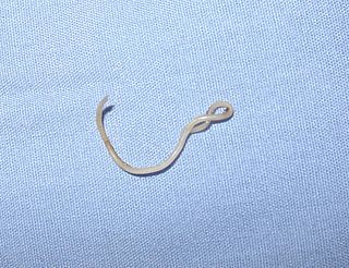
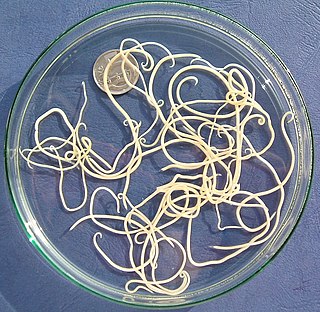
Ascaris lumbricoides is a large parasitic worm that causes ascariasis in humans. A roundworm of genus Ascaris, it is the most common parasitic worm in humans. An estimated one-sixth of the human population is at some point infected by a roundworm such as A. lumbricoides; people living in tropical and subtropical countries are at greater risk of infection.

Toxocariasis is an illness of humans caused by the dog roundworm and, less frequently, the cat roundworm. These are the most common intestinal roundworms of dogs, coyotes, wolves and foxes and domestic cats, respectively. Humans are among the many "accidental" or paratenic hosts of these roundworms.

Gnathostomiasis, also known as larva migrans profundus, is the human infection caused by the nematode Gnathostoma spinigerum and/or Gnathostoma hispidum, which infects vertebrates.

<i>Baylisascaris</i> Genus of worms

Baylisascaris is a genus of roundworms that infect more than fifty animal species.

<i>Anisakis</i> Genus of parasitic nematodes

Anisakis is a genus of parasitic nematodes that have life cycles involving fish and marine mammals. They are infective to humans and cause anisakiasis. People who produce immunoglobulin E in response to this parasite may subsequently have an allergic reaction, including anaphylaxis, after eating fish infected with Anisakis species.

<i>Ascaris suum</i> Species of roundworm

Ascaris suum, also known as the large roundworm of pig, is a parasitic nematode that causes ascariasis in pigs. While roundworms in pigs and humans are today considered as two species with different hosts, cross-infection between humans and pigs is possible; some researchers have thus argued they are the same species. Ascariasis is associated with contact to pigs and pig manure in Denmark.

<span class="mw-page-title-main">Toxocaridae</span> Family of roundworms

The Toxocaridae are a zoonotic family of parasitic nematodes that infect canids and felids and which cause toxocariasis in humans. The worms are unable to reproduce in humans.

<span class="mw-page-title-main">Eucestoda</span> Subclass of flatworms

Eucestoda, commonly referred to as tapeworms, is the larger of the two subclasses of flatworms in the class Cestoda. Larvae have six posterior hooks on the scolex (head), in contrast to the ten-hooked Cestodaria. All tapeworms are endoparasites of vertebrates, living in the digestive tract or related ducts. Examples are the pork tapeworm with a human definitive host, and pigs as the secondary host, and Moniezia expansa, the definitive hosts of which are ruminants.

Spirometra erinaceieuropaei is a parasitic tapeworm that infects domestic animals and humans. The medical term for this infection in humans and other animals is sparganosis. Morphologically, these worms are similar to other worms in the genus Spirometra. They have a long body consisting of three sections: the scolex, the neck, and the strobilia. They have a complex life cycle that consists of three hosts, and can live in varying environments and bodily tissues. Humans can contract this parasite in three main ways. Historically, humans are considered a paratenic host; however, the first case of an adult S. erinaceieuropaei infection in humans was reported in 2017. Spirometra tapeworms exist worldwide and infection is common in animals, but S. erinaceieuropaei infections are rare in humans. Treatment for infection typically includes surgical removal and anti-worm medication.

Gnathostoma spinigerum is a parasitic nematode that causes gnathostomiasis in humans, also known as its clinical manifestations are creeping eruption, larva migrans, Yangtze edema, Choko-Fuschu Tua chid and wandering swelling. Gnathostomiasis in animals can be serious, and even fatal. The first described case of gnathostomiasis was in a young tiger that died in the London Zoo in 1835. The larval nematode is acquired by eating raw or undercooked fish and meat.

<span class="mw-page-title-main">Strongylida</span> Suborder of roundworms

The Strongylida suborder includes many of the important nematodes found in the gastrointestinal tracts of ruminants, horses, and swine, as well as the lungworms of ruminants and the hookworms of dogs and cats.

<i>Toxocara canis</i> Species of roundworm

Toxocara canis is a worldwide-distributed helminth parasite that primarily infects dogs and other canids, but can also infect other animals including humans. The name is derived from the Greek word "toxon," meaning bow or quiver, and the Latin word "caro," meaning flesh. T. canis live in the small intestine of the definitive host. This parasite is very common in puppies and somewhat less common in adult dogs. In adult dogs, infection is usually asymptomatic but may be characterized by diarrhea. By contrast, untreated infection with Toxocara canis can be fatal in puppies, causing diarrhea, vomiting, pneumonia, enlarged abdomen, flatulence, poor growth rate, and other complications.

<span class="mw-page-title-main">Anisakidae</span> Family of roundworms

The Anisakidae are a family of intestinal nematodes (roundworms). The larvae of these worms can cause anisakiasis when ingested by humans, in raw or insufficiently cooked fish.

<i>Baylisascaris procyonis</i> Species of roundworm

Baylisascaris procyonis, also known by the common name of raccoon roundworm, is a roundworm nematode, found ubiquitously in raccoons, the definitive hosts. It is named after H. A. Baylis, who studied them in the 1920s–30s, and Greek askaris. Baylisascaris larvae in paratenic hosts can migrate, causing larva migrans. Baylisascariasis as the zoonotic infection of humans is rare, though extremely dangerous due to the ability of the parasite's larvae to migrate into brain tissue and cause damage. Concern for human infection has been increasing over the years due to urbanization of rural areas resulting in the increase in proximity and potential human interaction with raccoons.

Gnathostoma hispidum is a nematode (roundworm) that infects many vertebrate animals including humans. Infection of Gnathostoma hispidum, like many species of Gnathostoma causes the disease gnathostomiasis due to the migration of immature worms in the tissues.

Eustrongylidosis is a parasitic disease that mainly affects wading birds worldwide; however, the parasite's complex, indirect lifecycle involves other species, such as aquatic worms and fish. Moreover, this disease is zoonotic, which means the parasite can transmit disease from animals to humans. Eustrongylidosis is named after the causative agent Eustrongylides, and typically occurs in eutrophicated waters where concentrations of nutrients and minerals are high enough to provide ideal conditions for the parasite to thrive and persist. Because eutrophication has become a common issue due to agricultural runoff and urban development, cases of eustrongylidosis are becoming prevalent and hard to control. Eustrongylidosis can be diagnosed before or after death by observing behavior and clinical signs, and performing fecal flotations and necropsies. Methods to control it include preventing eutrophication and providing hosts with uninfected food sources in aquaculture farms. Parasites are known to be indicators of environmental health and stability, so should be studied further to better understand the parasite's lifecycle and how it affects predator-prey interactions and improve conservation efforts.

<i>Contracaecum</i> Genus of roundworms

Contracaecum is a genus of parasitic nematodes from the family Anisakidae. These nematodes are parasites of warm-blooded, fish eating animals, i.e. mammals and birds, as sexually mature adults. The eggs and the successive stages of their larvae use invertebrates and increasing size classes of fishes as intermediate hosts. It is the only genus in the family Anisakidae which can infect terrestrial, marine and freshwater animals.

<span class="mw-page-title-main">Cat worm infections</span> Worm infections in cats

Cat worm infections, the infection of cats (Felidae) with parasitic worms, occur frequently. Most worm species occur worldwide in both domestic and other cats, but there are regional, species and lifestyle differences in the frequency of infestation. According to the classification of the corresponding parasites in the zoological system, infections can be divided into those caused by nematode and flatworms - in the case of the latter, mainly cestoda and trematoda - while other strains are of no veterinary significance. While threadworms usually do not require an intermediate host for their reproduction, the development cycle of flatworms always proceeds via alternate hosts.

<span class="mw-page-title-main">Nematode infection in dogs</span> Threadworm infections of dogs are frequent

Nematode infection in dogs - the infection of dogs with parasitic nemamotodes - are, along with tapeworm infections and infections with protozoa, frequent parasitoses in veterinary practice. Nematodes, as so-called endoparasites, colonize various internal organs - most of them the digestive tract - and the skin. To date, about 30 different species of nematode have been identified in domestic dogs; they are essentially also found in wild dog species. However, the majority of them often cause no or only minor symptoms of disease in adult animals. The infection therefore does not necessarily have to manifest itself in a worm disease (helminthosis). For most nematodes, an infection can be detected by examining the feces for eggs or larvae. Roundworm infection in dogs and the hookworm in dogs is of particular health significance in Central Europe, as they can also be transmitted to humans (zoonosis). Regular deworming can significantly reduce the frequency of infection and thus the risk of infection for humans and dogs.

References

  1. Rudolphi, Carolo Asmundo (1809). "Ascaris simplex R.". Animadversiones in generaet species entozoorum. Entozoorum, sive vermium intestinalium: Historia naturalis. 2. Amstelaedami: Taberna Libraria et Artium. p. 170.
  2. 1 2 3 Gang, S., & Hallem, E. (2016). Mechanisms of host seeking by parasitic nematodes. Molecular and Biochemical Parasitology, 208(1), 23–32. doi : 10.1016/j.molbiopara.2016.05.007
  3. 1 2 3 4 5 6 7 8 9 10 11 12 13 14 15 16 17 18 19 20 21 22 23 24 25 26 27 28 29 30 31 32 33 34 35 36 37 38 Audicana, M. T., Del Pozo, M. D., Iglesias, R., & Ubeira, F. M. (2003) Anisakis simplex and Pseudoterranova decipiens. In M. D. Miliotis & J. W. Bier (Ed.), International Handbook of Foodborne Pathogens, 633–656. Florida, United States: CRC Press.
  4. 1 2 3 4 5 6 7 8 9 10 11 12 13 14 15 16 Nagasawa, K. (1990). The Life Cycle of Anisakis simplex: A Review. In H. Ishikura, & K. Kikuchi (Eds.), Intestinal Anisakiasis in Japan, 31–40. Tokyo, Japan: Springer.
  5. 1 2 3 4 5 6 7 8 9 10 11 12 13 14 15 16 Ritter, J. (2001). Anisakis simplex . Retrieved April 17, 2021.
  6. 1 2 3 4 5 6 7 8 9 10 11 12 13 14 Ishii, Y., Fujino, T., & Weerasooriya, M. (1989). Morphology of Anisakine Larvae. In H. Ishikura, & M. Namiki (Eds.), Gastric Anisakiasis in Japan, 19–29. Tokyo, Japan: Springer
  7. 1 2 3 4 5 6 7 8 9 10 11 12 13 14 15 Centre for Disease Control and Prevention [CDC]. (2019). Anisakiasis . Retrieved April 17, 2021.
  8. 1 2 3 4 5 Pravettoni, V., Primavesi, L., & Piantanida, M. (2012). Anisakis simplex: current knowledge. European Annals of Allergy and Clinical Immunology, 44(4), 150–156.
  9. Ángeles-Hernández, J., Gómez-de Anda, F., Reyes-Rodríguez, N., Vega-Sánchez, V., García-Reyna, P., Campos-Montiel, R., Calderón-Apodaca, N., Salgado-Miranda, C., & Zepeda-Velázquez, A. (2020). Genera and Species of the Anisakidae Family and Their Geographical Distribution. Animals (Basel), 10(12), 2374–2397. doi : 10.3390/ani10122374
  10. 1 2 3 Audicana, T. & Kennedy, M. (2008). Anisakis simplex: from Obscure Infectious Worm to Inducer of Immune Hypersensitivity. Clinical Microbiology Reviews, 21(2), 360–379. doi : 10.1128/CMR.00012-07
  11. 1 2 Kuhn, T., Cunze, S., Kochmann, J., & Klimpel, S. (2016). Environmental variables and definitive host distribution: a habitat suitability modelling for endohelminth parasites in the marine realm. Scientific Reports, 6(1), 30246–30246. doi : 10.1038/srep30246
  12. Quiazon, K., Yoshinaga, T., & Ogawa, K. (2011). Distribution of Anisakis species larvae from fishes of the Japanese waters. Parasitology International, 60(2), 223–226. doi : 10.1016/j.parint.2011.03.002
  13. 1 2 3 4 5 Audicana, M., Ansotegui, I., De Corres, L., & Kennedy, M. (2002). Anisakis simplex: dangerous – dead and alive? Trends in Parasitology, 18(1), 20–25. doi : 10.1016/S1471-4922(01)02152-3

Further reading