Aeromonas salmonicida

Last updated

Aeromonas salmonicida
ASalmonicida.png
Scientific classification OOjs UI icon edit-ltr.svg
Domain: Bacteria
Phylum: Pseudomonadota
Class: Gammaproteobacteria
Order: Aeromonadales
Family: Aeromonadaceae
Genus: Aeromonas
Species:
A. salmonicida
Binomial name
Aeromonas salmonicida
(Lehmann and Neumann 1896) Griffin et al. 1953
Synonyms

Bacillus salmonicida (Lehmann and Neumann 1896) Kruse 1896 Bacterium salmonicida Lehmann and Neumann 1896 Proteus salmonicida (Lehmann and Neumann 1896) Pribram 1933

Contents

Aeromonas salmonicida is a pathogenic bacterium that severely impacts salmonid populations and other species. It was first discovered in a Bavarian brown trout hatchery by Emmerich and Weibel in 1894. [1] Aeromonas salmonicida's ability to infect a variety of hosts, multiply, and adapt, make it a prime virulent bacterium. A. salmonicida is an etiological agent for furunculosis, a disease that causes sepsis, haemorrhages, muscle lesions, inflammation of the lower intestine, spleen enlargement, and death in freshwater fish populations. It is found worldwide with the exception of South America. [1] [2] The major route of contamination is poor water quality; however, it can also be associated stress factors such as overcrowding, high temperatures, and trauma. Spawning and smolting fish are prime victims of furunculosis due to their immunocompromised state of being.

Morphology and bacterial characteristics

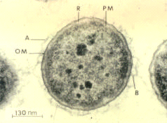
Aeromonas salmonicida is a Gram-negative, facultatively anaerobic, nonmotile bacterium. It is rod-shaped, about 1.3–2.0 by 0.8–1.3 μm in size, and grows optimally at temperatures between 22 and 25 °C. [1] [2] The bacterium readily ferments and oxidizes glucose, and is catalase- and cytochrome oxidase-positive. Its molecular properties include a special surface protein array called the A-layer, which is believed to be responsible for the bacterium's virulent traits, and lipopolysaccharide, the cells' major cell envelope antigen. [3] The A-layer consists of a 50-kD protein, and provides protection to the bacterium. The lipopolysaccharide consists of three moieties: lipid A, a core oligosaccharide, and an O-polysaccharide (O-antigen). The extracellular products of A. salmonicida consist of 25 proteins, enzymes, and toxins, and many more. [3] In addition, the genome is composed of a single circular chromosome (4,702,402 bp), with two large and three small plasmids. The chromosome yields 58.5% of G+C pairs, has 4086 encoding proteins, and totals 4388 genes. [4]

A. salmonicida isolates flourish when grown on blood agar or tyrosine. Large colonies are observed along with a brown diffusible pigment within two to four days. Most typical strains are morphologically and biochemically homogenous with a few exceptions. Some of these exceptions include a distinguishable variation in pigment production, the bacterium's ability to ferment selected sugars, and Voges-Proskauer assay results.

Cell structure and metabolism

A. salmonicida is a facultative anaerobe, which means it is capable of making ATP by aerobic respiration if oxygen is present, but is also capable of switching to fermentation when oxygen is not present. It does not ferment sucrose or lactose, using glucose in this pathway, instead; glucose fermentation creates gas. The bacterium grows optimally at temperatures between 22 and 25 °C. The maximum temperature at which it can grow is 34.5 °C. After about a 24-hour growth period, the bacterial colonies reach about the size of a pin point. The colonies also have a brown pigmented color that appears after it has been growing for 48–72 hours. [5]

Host range

Epizootiology

A. salmonicida, an airborne pathogen, can travel 104 cm from its host into the atmosphere and back to the water, [6] thus making it difficult to control. The bacterium can maintain its pathogenicity in freshwater conditions for 6–9 months, [7] and in saltwater conditions for up to 10 days without a host. Several direct count methods and other detection methods have found the organism does not lose or reduces its titer concentrations. [8]

Transmission of furunculosis mainly occurs through fish-to-fish contact by the skin or by ingestion. Rainbow trout have been found to carry A. salmonicida up to two years after initial infection without re-exposure. Chemically immunosuppressed fish compared with temperature-stressed fish had a 73% mortality as opposed to a 33% mortality rate, respectively. [9] Naturally occurring trout infections consisted of a 5–6% mortality rate per week with an 85% rate in untreated populations. Some clinical furunculosis survivors of an infected trout population became A. salmonicida carriers. [10] When comparing furunculosis epidemics with depressed oxygen levels, when oxygen concentrations were decreased to less than 5 mg/L, A. salmonicida concentrations increased. [11] While observing chum salmon in a density of 14.7 fish per square meter, 12.4% were infected with A. salmonicida, whereas, densities at 4.9 fish per square meter were infection-free. [12] Additionally, A. salmonicida concentrations were considerably elevated in water with low dissolved oxygen (6–7 mg/L), compared to water with higher dissolved oxygen (10 mg/L). High density-low oxygen water resulted in survival rates that were roughly 40% less than in those consisting of low density-high oxygen conditions.

Pathology

The bacterium is pathogenic for fish, and causes the disease known as furunculosis. [13] The symptoms the fish show are external and internal hemorrhaging, swelling of the vents and kidneys, boils, ulcers, liquefaction, and gastroenteritis. Furunculosis is commonly known as tail rot in fish and is common in goldfish and koi. Infected fish with open sores are able to spread the disease to other fish. [5]

It is also one of several bacteria that can cause bald sea urchin disease. [14] Since A. salmonicida cannot grow at 37 °C, it is not pathogenic in humans. [15]

Clinical symptoms and disease diagnosis

Furunculosis is classified into four categories based on severity: acute, subacute, chronic, or latent. When fish are infected, they become listless and weak until they die. Other characteristics observed include anorexia and lethargic movement, and they may exhibit a darkened pigment. Deep or shallow ulcers, exophthalmia, bloody spots, distended abdomen, and petechia at the base of the fin may also occur. Internally, the infected fish may suffer from gastroenteritis, hemorrhagic septicemia, edematous kidney, and an enlarged spleen. The liver may appear pale in color and the spleen may be darkened. The peritoneal cavity may also be bloody and inflamed.

Bacteria must be isolated to positively identify the disease. Isolates are retrieved from muscle lesions, kidney, spleen, or liver, and then grown on trypticase soy agar and brain-heart infusion medium incubated at 20–25 °C. Colonies of A. salmonicida appear hard, friable, smooth, soft, and dark in color.

While cultural procedures produce good results, serological procedures produce more rapid results by using serum agglutination, fluorescent antibody, or enzyme linked immunosorbent assay on infected tissue or cultured bacteria. [16] Mooney et al. [17] developed a DNA probe with polymerase chain reaction to detect A. salmonicida DNA; results were successful in 88% of wild Atlantic salmon.

Detection

A. salmonicida tests negative for indole formation, coagulase, hydrolysis of starch, casein, triglycerides, and phospholipids, hydrogen sulfide production, citrate use, phenylalanine, and the Voges–Proskauer (butanediol fermentation) test. It tests positive for oxidase, lysine decarboxylase, methyl red, gelatin hydrolysis, and catalase. [5]

Related Research Articles

Plesiomonas shigelloides is a species of bacteria and the only member of its genus. It is a Gram-negative, rod-shaped bacterium which has been isolated from freshwater, freshwater fish, shellfish, cattle, goats, swine, cats, dogs, monkeys, vultures, snakes, toads and humans. It is considered a fecal coliform. P. shigelloides is a global distributed species, found globally outside of the polar ice caps.

Aeromonas veronii is a Gram-negative, rod-shaped bacterium found in fresh water and in association with animals. It can be a pathogen of humans and a beneficial symbiont of leeches. In humans A. veronii can cause diseases ranging from wound infections and diarrhea to sepsis in immunocompromised patients. Humans treated with medicinal leeches after vascular surgery can be at risk for infection from A. veronii and are commonly placed on prophylactic antibiotics. Most commonly ciprofloxacin is used but there have been reports of resistant strains leading to infection. In leeches, this bacterium is thought to function in the digestion of blood, provision of nutrients, or preventing other bacteria from growing.

<i>Aeromonas hydrophila</i> Species of heterotrophic, Gram-negative, bacterium

Aeromonas hydrophila is a heterotrophic, Gram-negative, rod-shaped bacterium mainly found in areas with a warm climate. This bacterium can be found in fresh or brackish water. It can survive in aerobic and anaerobic environments, and can digest materials such as gelatin and hemoglobin. A. hydrophila was isolated from humans and animals in the 1950s. It is the best known of the species of Aeromonas. It is resistant to most common antibiotics and cold temperatures and is oxidase- and indole-positive. Aeromonas hydrophila also has a symbiotic relationship as gut flora inside of certain leeches, such as Hirudo medicinalis.

Photobacterium damselaesubsp.piscicida is a gram-negative rod-shaped bacterium that causes disease in fish.

Photobacterium is a genus of gram-negative, oxidase positive and catalase positive bacteria in the family Vibrionaceae. Members of the genus are bioluminescent, that is they have the ability to emit light.

<i>Xanthomonas campestris</i> Species of bacterium

Xanthomonas campestris is a gram-negative, obligate aerobic bacterium that is a member of the Xanthomonas genus, which is a group of bacteria that are commonly known for their association with plant disease. The species is considered to be dominant amongst its genus, as it originally had over 140 identified pathovars and has been found to infect both monocotyledonous and dicotyledonous plants of economical value with various plant diseases. This includes "black rot" in cruciferous vegetables, bacterial wilt of turfgrass, bacterial blight, and leaf spot, for example.

Mycobacterium avium complex is a group of mycobacteria comprising Mycobacterium intracellulare and Mycobacterium avium that are commonly grouped because they infect humans together; this group, in turn, is part of the group of nontuberculous mycobacteria. These bacteria cause Mycobacterium avium-intracellulare infections or Mycobacterium avium complex infections in humans. These bacteria are common and are found in fresh and salt water, in household dust and in soil. MAC bacteria usually cause infection in those who are immunocompromised or those with severe lung disease.

Pseudomonas viridiflava is a fluorescent, Gram-negative, soil bacterium that is pathogenic to plants. It was originally isolated from the dwarf or runner bean, in Switzerland. Based on 16S rRNA analysis, P. viridiflava has been placed in the P. syringae group. Following ribotypical analysis misidentified strains of Pseudomonas syringae pv. ribicola and Pseudomonas syringae pv. primulae were incorporated into this species. This pathogen causes bacterial blight of Kiwifruit.

Shewanella algae is a rod-shaped Gram-negative marine bacterium.

<i>Streptococcus iniae</i> Species of bacterium

Streptococcus iniae is a species of Gram-positive, sphere-shaped bacterium belonging to the genus Streptococcus. Since its isolation from an Amazon freshwater dolphin in the 1970s, S. iniae has emerged as a leading fish pathogen in aquaculture operations worldwide, resulting in over US$100M in annual losses. Since its discovery, S. iniae infections have been reported in at least 27 species of cultured or wild fish from around the world. Freshwater and saltwater fish including tilapia, red drum, hybrid striped bass, and rainbow trout are among those susceptible to infection by S. iniae. Infections in fish manifest as meningoencephalitis, skin lesions, and septicemia.

<span class="mw-page-title-main">Fish disease and parasites</span> Disease that affects fish

Like humans and other animals, fish suffer from diseases and parasites. Fish defences against disease are specific and non-specific. Non-specific defences include skin and scales, as well as the mucus layer secreted by the epidermis that traps microorganisms and inhibits their growth. If pathogens breach these defences, fish can develop inflammatory responses that increase the flow of blood to infected areas and deliver white blood cells that attempt to destroy the pathogens.

Borrelia afzelii is a species of Borrelia a bacterium that can infect various species of vertebrates and invertebrates.

Bacterial cold water disease (BCWD) is a bacterial disease of freshwater fish, specifically salmonid fish. It is caused by the bacterium Flavobacterium psychrophilum, a psychrophilic, gram-negative rod-shaped bacterium of the family Flavobacteriaceae. This bacterium is found in fresh waters with the optimal growth temperature below 13 °C, and it can be seen in any area with water temperatures consistently below 15 °C. Salmon are the most commonly affected species. This disease is not zoonotic.

Edwardsiella tarda is a member of the family Hafniaceae. The bacterium is a facultatively anaerobic, small, motile, gram negative, straight rod with flagella. Infection causes Edwardsiella septicemia in channel catfish, eels, and flounder. Edwardsiella tarda is also found in largemouth bass and freshwater species such as rainbow trout. It is a zoonosis and can infect a variety of animals including fish, amphibians, reptiles, and mammals. Edwardsiella tarda has also been the cause of periodic infections for various animals within zoos. E. tarda has a worldwide distribution and can be found in pond water, mud, and the intestine of fish and other marine animals. It is spread by carrier animal feces.

Lactococcus garvieae is a known fish pathogen affecting saltwater fish in the Far East, specifically in rainbow trout, Japanese yellowtail, Cobia and grey mullet. This bacteria causes lesions in the vascular endothelium, leading to hemorrhages and petechias at the surface of internal organs. As few as 10 bacterial cells per fish can cause an infection. L. garvieae is isolated in saltwater fish in the Far East and specifically in European rainbow trout.

Vibrio coralliilyticus is a Gram-negative, rod-shaped bacterium. It has a polar flagellum that is used for motility and has been shown to be critical for its virulence to corals. It is a versatile pathogen, impacting several marine invertebrates including Pocillopora damicornis corals, both the Pacific and Eastern Oyster's larvae and some vertebrates such as the rainbow trout. It is a bacterium of considerable interest given its direct contribution to temperature dependent coral bleaching as well as its impacts on aquaculture where it can contribute to significant mortalities in larval oyster hatcheries. There are several known virulent strains, which appear on both the Pacific and Atlantic Coasts of the United States. After its initial discovery some strains were incorrectly classified as Vibrio tubiashii including the RE22 and RE98 strains but were later reclassified as Vibrio coralliilyticus.

Yersinia ruckeri is a species of Gram-negative bacteria, known for causing enteric redmouth disease in some species of fish. Strain 2396-61 is its type strain.

Vibrio ordalii is a Gram-negative, rod-shaped bacterium. It causes vibriosis in fish. Its type strain is ATCC 33509.

Aeromonas eucrenophila is a Gram-negative bacterium of the genus Aeromonas isolated from fresh water and infected fish. A. eucrenophila is a pathogen of fish, and it causes diarrhoea in humans.

Aeromonas piscicola is a Gram-negative, catalase and oxidase-positive bacterium of the genus Aeromonas isolated from diseased fish in Spain

References

  1. 1 2 3 "Furunculosis". Merck. Archived from the original on 30 July 2015. Retrieved 2011-06-11.
  2. 1 2 Charette, Steve J. (2021-05-04). "Microbe Profile: Aeromonas salmonicida: an opportunistic pathogen with multiple personalities". Microbiology. 167 (5). doi: 10.1099/mic.0.001052 . hdl: 20.500.11794/106763 . ISSN   1350-0872. PMID   33945463. S2CID   233740911.
  3. 1 2 Chart, H.; Shaw, D.; Ishguro, E.; Trust, T. (April 1984). "Structural and immunochemical homogeneity of Aeromonas salmonicida Lipopolysaccharide". Journal of Bacteriology. 158 (1): 16–22. doi:10.1128/jb.158.1.16-22.1984. PMC   215372 . PMID   6370955.
  4. Reith, M. E.; Singh, R. K.; Curtis, B.; Boyd, J. M.; Bouevitch, A.; Kimball, J.; Munholland, J.; Murphy, C.; Sarty, D.; Williams, J.; Nash, J. H.; Johnson, S. C.; Brown, L. L. (2008). "The genome of Aeromonas salmonicida subsp. salmonicida A449: insights into the evolution of a fish pathogen". BMC Genomics. 9: 427. doi: 10.1186/1471-2164-9-427 . PMC   2556355 . PMID   18801193.
  5. 1 2 3 Staley, James T.; Garrity, George M.; Boone, David R.; Castenholz, Richard W.; Don J. Brenner; Krieg, Noel R. (2001). Bergey's manual of systematic bacteriology . Berlin: Springer. ISBN   978-0-387-24145-6.
  6. Wooster, Gregory A.; Bowser, Paul R. (1996). "The Aerobiological Pathway of a Fish Pathogen: Survival and Dissemination of Aeromonas salmonicida in Aerosols and its Implications in Fish Health Management". Journal of the World Aquaculture Society. 27: 7–14. doi:10.1111/j.1749-7345.1996.tb00588.x.
  7. Michel, C.; Dubois-Darnaudpeys, A. (1980). "Persistence of the virulence of Aeromonas salmonicida strains kept in river sediments". Annales de Recherches Vétérinaires. 11 (4): 375–80. PMID   7337394.
  8. Rose, A; Ellis, E (1990). "The survival of Aeromonas salmonicida subsp. salmonicida in sea water". Journal of Fish Diseases. 13 (3): 205–214. doi:10.1111/j.1365-2761.1990.tb00775.x.
  9. Bullock, G. L.; Stuckey, H. M. (1975). "Aeromonas salmonicida detection of asymptomatically infected trout". The Progressive Fish-Culturist. 37 (4): 237–239. doi:10.1577/1548-8659(1975)37[237:AS]2.0.CO;2. ISSN   1548-8659.
  10. McCarthy, D. H. (1980). "Some ecological aspects of the bacterial fish pathogen Aeromonas salmonicida". Aquatic Microbiology: 299–324.
  11. Kingsbury, Oliver R. (1961). "A Possible Control of Furunculosis". The Progressive Fish-Culturist. 23 (3): 136–137. doi:10.1577/1548-8659(1961)23[136:APCOF]2.0.CO;2.
  12. Kimura, Takahisa; Yoshimizu, Mamoru; Nomura, Tetsuichi (1992). "An Epidemiological Study of Furunculosis in Salmon Propagation": 187–193. hdl:2115/39269.{{cite journal}}: Cite journal requires |journal= (help)
  13. "www.lsc.usgs.gov" (PDF). Archived from the original (PDF) on 2009-05-07. Retrieved 2009-07-03.
  14. Jangoux, M (1987). "Diseases of Echinodermata. I. Agents microorganisms and protistans". Diseases of Aquatic Organisms. 2: 147–162. doi: 10.3354/dao002147 .
  15. Altwegg, M.; Steigerwalt, A. G.; Altwegg-Bissig, R.; Lüthy-Hottenstein, J.; Brenner, D. (1990). "Biochemical identification of Aeromonas genospecies isolated from humans". Journal of Clinical Microbiology. 28 (2): 258–264. doi:10.1128/jcm.28.2.258-264.1990. PMC   269587 . PMID   2312673.
  16. Austin, B.; Bishop, I.; Gray, C.; Watt, B.; Dawes, J. (1986). "Monoclonal antibody- based enzyme-linked immunosorbent assay for the rapid diagnosis of clinical cases of enteric redmouth and furunculosis in fish farms". Journal of Fish Diseases. 9 (5): 469–474. doi:10.1111/j.1365-2761.1986.tb01042.x.
  17. Mooney, J.; Powell, E.; Clabby, C.; Powell, R. (1995). "Detection of Aeromonas salmonicida in wild Atlantic salmon using a specific DNA probe test". Diseases of Aquatic Organisms. 21: 131–135. doi: 10.3354/dao021131 . hdl: 10379/9573 .