Delta-beta thalassemia

Last updated
Delta-beta thalassemia
Delta Beta Thalassemia.jpg
Delta-beta thalassemia
Specialty Hematology   OOjs UI icon edit-ltr-progressive.svg
CausesProduces only gamma-globin and forms HbF(deletes entire delta and beta gene sequence) [1]
Diagnostic method High-performance liquid chromatography
TreatmentBlood transfusions [2]

Delta-beta thalassemia is a rare form of thalassemia in which there is a reduced production of hemoglobin subunit delta and hemoglobin subunit beta and raised levels of hemoglobin subunit gamma. It is an autosomal recessive disorder. [1] [3]

Contents

Signs and symptoms

An individual with delta-beta thalassemia is usually asymptomatic, however microcytosis can occur where the red blood cells are abnormally small. [1] [4]

Mechanism

Structure of hemoglobin the proteins a and b subunits are in red and blue, 1GZX Haemoglobin.png
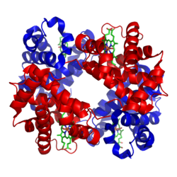
Structure of hemoglobin the proteins α and β subunits are in red and blue,

Delta-beta thalassemia is autosomal recessive disorder, [1] which means both parents are affected and two copies of the gene must be present. [5] A carrier gets a normal gene to produce hemoglobin A, from one parent and the other parent supplies a gene which makes no hemoglobin A. [6] Delta-beta thalassemia is considered rare. [2]

Delta-beta-thalassemia is caused by deletions of the entire delta and beta genes sequences and only gamma-globin and HbF are formed. Rarely, non-deletional forms have been reported. [7] [8]

When two delta0 mutations are inherited, no hemoglobin A2 (alpha2, delta2) are formed. This is innocuous because only 2-3% of normal adult hemoglobin is hemoglobin A2. The individual will have normal hematological parameters (erythrocyte count, total hemoglobin, mean corpuscular volume).[ medical citation needed ] The delta-beta thalassemia demonstrates one mutation is at the +69 position. [9]

Relation to beta thalassemia

Delta-beta thalassemia can mask the diagnosis of beta thalassemia trait. In beta thalassemia, an increase in hemoglobin A2 results, but the co-existence of a delta-beta thalassemia mutation will decrease the value of the hemoglobin A2 into the normal range, thereby obscuring the diagnosis of beta thalassemia trait [10]

Diagnosis

Following the detection of hypochromic microcytic red blood cells, delta-beta thalassemia is confirmed by high-performance liquid chromatography. [11]

Treatment

When needed, treatment for anemia, such as blood transfusions are used. [2]

Stem cell transplant is another option, but the donor and the individual who will receive the bone marrow transplant must be compatible, the risks involved should be evaluated. [2] [12] [13]

See also

Related Research Articles

<span class="mw-page-title-main">Hemoglobinopathy</span> Any of various genetic disorders of blood

Hemoglobinopathy is the medical term for a group of inherited blood disorders and diseases that primarily affect red blood cells. They are single-gene disorders and, in most cases, they are inherited as autosomal co-dominant traits.

<span class="mw-page-title-main">Thalassemia</span> Family of inherited blood disorders

Thalassemias are inherited blood disorders that result in abnormal hemoglobin. Symptoms depend on the type of thalassemia and can vary from none to severe. Often there is mild to severe anemia as thalassemia can affect the production of red blood cells and also affect how long the red blood cells live. Symptoms of anemia include feeling tired and having pale skin. Other symptoms of thalassemia include bone problems, an enlarged spleen, yellowish skin, pulmonary hypertension, and dark urine. Slow growth may occur in children. Symptoms and presentations of thalassemia can change over time.

<span class="mw-page-title-main">Fetal hemoglobin</span> Oxygen carrier protein in the human fetus

Fetal hemoglobin, or foetal haemoglobin is the main oxygen carrier protein in the human fetus. Hemoglobin F is found in fetal red blood cells, and is involved in transporting oxygen from the mother's bloodstream to organs and tissues in the fetus. It is produced at around 6 weeks of pregnancy and the levels remain high after birth until the baby is roughly 2–4 months old. Hemoglobin F has a different composition than adult forms of hemoglobin, allowing it to bind oxygen more strongly; this in turn enables the developing fetus to retrieve oxygen from the mother's bloodstream, which occurs through the placenta found in the mother's uterus.

<span class="mw-page-title-main">Immunodeficiency–centromeric instability–facial anomalies syndrome</span> Medical condition

ICF syndrome is a very rare autosomal recessive immune disorder.

<span class="mw-page-title-main">Hemoglobin A</span> Normal human hemoglobin in adults

Hemoglobin A (HbA), also known as adult hemoglobin, hemoglobin A1 or α2β2, is the most common human hemoglobin tetramer, accounting for over 97% of the total red blood cell hemoglobin. Hemoglobin is an oxygen-binding protein, found in erythrocytes, which transports oxygen from the lungs to the tissues. Hemoglobin A is the most common adult form of hemoglobin and exists as a tetramer containing two alpha subunits and two beta subunits (α2β2). Hemoglobin A2 (HbA2) is a less common adult form of hemoglobin and is composed of two alpha and two delta-globin subunits. This hemoglobin makes up 1-3% of hemoglobin in adults.

Hemoglobin A2 (HbA2) is a normal variant of hemoglobin A that consists of two alpha and two delta chains (α2δ2) and is found at low levels in normal human blood. Hemoglobin A2 may be increased in beta thalassemia or in people who are heterozygous for the beta thalassemia gene.

<span class="mw-page-title-main">Alpha-thalassemia</span> Thalassemia involving the genes HBA1and HBA2 hemoglobin genes

Alpha-thalassemia is a form of thalassemia involving the genes HBA1 and HBA2. Thalassemias are a group of inherited blood conditions which result in the impaired production of hemoglobin, the molecule that carries oxygen in the blood. Normal hemoglobin consists of two alpha chains and two beta chains; in alpha-thalassemia, there is a quantitative decrease in the amount of alpha chains, resulting in fewer normal hemoglobin molecules. Furthermore, alpha-thalassemia leads to the production of unstable beta globin molecules which cause increased red blood cell destruction. The degree of impairment is based on which clinical phenotype is present.

<span class="mw-page-title-main">Beta thalassemia</span> Thalassemia characterized by the reduced or absent synthesis of the beta globin chains of hemoglobin

Beta thalassemias are a group of inherited blood disorders. They are forms of thalassemia caused by reduced or absent synthesis of the beta chains of hemoglobin that result in variable outcomes ranging from severe anemia to clinically asymptomatic individuals. Global annual incidence is estimated at one in 100,000. Beta thalassemias occur due to malfunctions in the hemoglobin subunit beta or HBB. The severity of the disease depends on the nature of the mutation.

<span class="mw-page-title-main">Emery–Dreifuss muscular dystrophy</span> Medical condition

Emery–Dreifuss muscular dystrophy (EDMD) is a type of muscular dystrophy, a group of heritable diseases that cause progressive impairment of muscles. EDMD affects muscles used for movement, causing atrophy, weakness and contractures. It almost always affects the heart, causing abnormal rhythms, heart failure, or sudden cardiac death. It is rare, affecting 0.39 per 100,000 people. It is named after Alan Eglin H. Emery and Fritz E. Dreifuss.

<span class="mw-page-title-main">Hemoglobin subunit beta</span> Mammalian protein found in Homo sapiens

Hemoglobin subunit beta is a globin protein, coded for by the HBB gene, which along with alpha globin (HBA), makes up the most common form of haemoglobin in adult humans, hemoglobin A (HbA). It is 147 amino acids long and has a molecular weight of 15,867 Da. Normal adult human HbA is a heterotetramer consisting of two alpha chains and two beta chains.

<span class="mw-page-title-main">Hemoglobin variants</span> Forms of hemoglobin caused by variations in genetics

Hemoglobin variants are different types of hemoglobin molecules, by different combinations of its subunits and/or mutations thereof. Hemoglobin variants are a part of the normal embryonic and fetal development. They may also be pathologic mutant forms of hemoglobin in a population, caused by variations in genetics. Some well-known hemoglobin variants, such as sickle-cell anemia, are responsible for diseases and are considered hemoglobinopathies. Other variants cause no detectable pathology, and are thus considered non-pathological variants.

In medical genetics, compound heterozygosity is the condition of having two or more heterogeneous recessive alleles at a particular locus that can cause genetic disease in a heterozygous state; that is, an organism is a compound heterozygote when it has two recessive alleles for the same gene, but with those two alleles being different from each other. Compound heterozygosity reflects the diversity of the mutation base for many autosomal recessive genetic disorders; mutations in most disease-causing genes have arisen many times. This means that many cases of disease arise in individuals who have two unrelated alleles, who technically are heterozygotes, but both the alleles are defective.

<span class="mw-page-title-main">KLF1</span> Protein-coding gene in the species Homo sapiens

Krueppel-like factor 1 is a protein that in humans is encoded by the KLF1 gene. The gene for KLF1 is on the human chromosome 19 and on mouse chromosome 8. Krueppel-like factor 1 is a transcription factor that is necessary for the proper maturation of erythroid cells.

<span class="mw-page-title-main">Hemoglobin subunit alpha</span> Human hemoglobin protein

Hemoglobin subunit alpha, Hemoglobin, alpha 1, is a hemoglobin protein that in humans is encoded by the HBA1 gene.

<span class="mw-page-title-main">HBD</span> Mammalian protein found in Homo sapiens

Hemoglobin subunit delta is a protein that in humans is encoded by the HBD gene.

<span class="mw-page-title-main">Hemoglobin E</span> Medical condition

Hemoglobin E (HbE) is an abnormal hemoglobin with a single point mutation in the β chain. At position 26 there is a change in the amino acid, from glutamic acid to lysine (E26K). Hemoglobin E is very common among people of Southeast Asian, Northeast Indian, Sri Lankan and Bangladeshi descent.

<span class="mw-page-title-main">Hemoglobin, alpha 2</span> Mammalian protein found in Homo sapiens

Hemoglobin, alpha 2 also known as HBA2 is a gene that in humans codes for the alpha globin chain of hemoglobin.

<span class="mw-page-title-main">Hemoglobin Lepore syndrome</span> Medical condition

Hemoglobin Lepore syndrome is typically an asymptomatic hemoglobinopathy, which is caused by an autosomal recessive genetic mutation. The Hb Lepore variant, consisting of two normal alpha globin chains (HBA) and two delta-beta globin fusion chains which occurs due to a "crossover" between the delta (HBD) and beta globin (HBB) gene loci during meiosis and was first identified in the Lepore family, an Italian-American family, in 1958. There are three varieties of Hb Lepore, Washington, Baltimore and Hollandia. All three varieties show similar electrophoretic and chromatographic properties and hematological findings bear close resemblance to those of the beta-thalassemia trait; a blood disorder that reduces the production of the iron-containing protein hemoglobin which carries oxygen to cells and which may cause anemia.

<span class="mw-page-title-main">DOCK8 deficiency</span> Medical condition

DOCK8 deficiency, also called DOCK8 immunodeficiency syndrome, is the autosomal recessive form of hyperimmunoglobulin E syndrome, a genetic disorder characterized by elevated immunoglobulin E levels, eosinophilia, and recurrent infections with staphylococcus and viruses. It is caused by a mutation in the DOCK8 gene.

References

  1. 1 2 3 4 "Delta-beta-thalassemia". Orphanet. Retrieved 16 September 2016.
  2. 1 2 3 4 "Thalassaemia | Health | Patient". Patient. Retrieved 17 September 2016.
  3. "HBD - hemoglobin subunit delta". Orphanet. Retrieved 17 September 2016.
  4. Pal, G. K. & (2005). Textbook Of Practical Physiology - 2Nd Edn. Orient Blackswan. p. 53. ISBN   9788125029045 . Retrieved 17 September 2016.
  5. "Autosomal recessive: MedlinePlus Medical Encyclopedia". medlineplus.gov. Retrieved 17 September 2016.
  6. "Delta beta thalassemia carrier" (PDF). Public Health England. Archived from the original (PDF) on 24 September 2016. Retrieved 17 September 2016.
  7. "Transcription and Translation - National Human Genome Research Institute (NHGRI)". www.genome.gov. NIH. Retrieved 17 September 2016.
  8. Proytcheva, Maria, ed. (2010). Diagnostic pediatric hematopathology. Cambridge: Cambridge University Press. p. 61. ISBN   9780521881609 . Retrieved 17 September 2016.
  9. "OMIM Entry - * 142000 - HEMOGLOBIN--DELTA LOCUS; HBD". www.omim.org. Retrieved 17 September 2016.
  10. Galanello, Renzo; Origa, Raffaella (2010). "Beta-thalassemia". Orphanet Journal of Rare Diseases. 5 (1): 11. doi: 10.1186/1750-1172-5-11 . ISSN   1750-1172. PMC   2893117 . PMID   20492708.
  11. Ahmad SQ, Zafar SI, Malik HS, Ahmed S (November 2017). "Delta-Beta Thalassaemia in a Pathan Family". Journal of the College of Physicians and Surgeons (Pakistan). 27 (11): 722–724. PMID   29132487.
  12. Cao, Antonio; Galanello, Renzo (2010-02-01). "Beta-thalassemia". Genetics in Medicine. 12 (2): 61–76. doi: 10.1097/GIM.0b013e3181cd68ed . ISSN   1098-3600. PMID   20098328.
  13. "Risks". nhs.uk. Retrieved 2018-04-28.

Further reading