Nucleoside-phosphate kinase

Last updated
nucleoside phosphate kinase
Identifiers
EC no. 2.7.4.4
CAS no. 9026-50-0
Databases
IntEnz IntEnz view
BRENDA BRENDA entry
ExPASy NiceZyme view
KEGG KEGG entry
MetaCyc metabolic pathway
PRIAM profile
PDB structures RCSB PDB PDBe PDBsum
Gene Ontology AmiGO / QuickGO
Search
PMC articles
PubMed articles
NCBI proteins

In enzymology, a nucleoside-phosphate kinase (EC 2.7.4.4) is an enzyme that catalyzes the chemical reaction [1]

Contents

ATP + nucleoside phosphate ADP + nucleoside diphosphate

Thus, the two substrates of this enzyme are ATP and nucleoside monophosphate, whereas its two products are ADP and nucleoside diphosphate. [2] [3]

This enzyme belongs to the family of transferases, specifically those transferring phosphorus-containing groups (phosphotransferases) with a phosphate group as acceptor. [4] The systematic name of this enzyme class is ATP:nucleoside-phosphate phosphotransferase. This enzyme is also called NMP-kinase, or nucleoside-monophosphate kinase.

Structure

A number of crystal structures have been solved for this class of enzymes, revealing that they share a common ATP binding domain. This section of the enzyme is commonly referred to as the P-loop, [5] in reference to its interaction with the phosphoryl groups on ATP. This binding domain also consists of a β sheet flanked by α helices.

The [P-loop] typically has the amino acid sequence of Gly-X-X-X-X-Gly-Lys. [6] Similar sequences are found in many other nucleotide-binding proteins.

Adenylate kinase, an example nucleoside-phosphate kinase, is shown here in both an open, unbound conformation (left) and with the lid domain closed around Ap5A (right). The P-loop is shown here in green while Ap5A is orange. Adenylate kinase 2.png
Adenylate kinase, an example nucleoside-phosphate kinase, is shown here in both an open, unbound conformation (left) and with the lid domain closed around Ap5A (right). The P-loop is shown here in green while Ap5A is orange.

Mechanism

Metal ion interaction

To allow for interaction with this class of enzymes, ATP must first bind to a metal ion such as magnesium or manganese. [8] The metal ion forms a complex with the phosphoryl-group, as well as several water molecules. [9] These water molecules then form hydrogen bonds to a conserved aspartate residue on the enzyme. [10]

The metal ion interaction facilitates binding by holding the ATP molecule in a position allowing for specific binding to the active site and by providing additional points for binding between the substrate and the enzyme. This increases the binding energy.

Conformational changes

Binding of ATP causes the P-loop to move, in turn making the lid domain lower and secure the ATP in place. [11] [12] Nucleoside monophosphate binding induces further changes that render the enzyme catalytically capable of facilitating a transfer of the phosphoryl group from ATP to nucleoside monophosphate. [13]

The necessity of these conformational changes prevents the wasteful hydrolysis of ATP.

This enzyme mechanism is an example of catalysis by approximation: the nucleoside-phosphate kinase binds the substrates to bring them together in the correct position for the phosphoryl group to be transferred.

Biological function

Similar catalytic domains are present in a variety of proteins, including:

Evolution

When a phylogenetic tree composed of members of the nucleoside-phosphate kinase family was made, [14] it showed that these enzymes had originally diverged from a common ancestor into long and short varieties. This first change was drastic – the three-dimensional structure of the lid domain changed significantly.

Following the evolution of long and short varieties of NMP-kinases, smaller changes in the amino acid sequences resulted in the differentiation of subcellular localization.

Related Research Articles

<span class="mw-page-title-main">Adenylyl cyclase</span> Enzyme with key regulatory roles in most cells

Adenylate cyclase is an enzyme with systematic name ATP diphosphate-lyase . It catalyzes the following reaction:

<span class="mw-page-title-main">Cyclic adenosine monophosphate</span> Cellular second messenger

Cyclic adenosine monophosphate is a second messenger, or cellular signal occurring within cells, that is important in many biological processes. cAMP is a derivative of adenosine triphosphate (ATP) and used for intracellular signal transduction in many different organisms, conveying the cAMP-dependent pathway.

<span class="mw-page-title-main">Kinase</span> Enzyme catalyzing transfer of phosphate groups onto specific substrates

In biochemistry, a kinase is an enzyme that catalyzes the transfer of phosphate groups from high-energy, phosphate-donating molecules to specific substrates. This process is known as phosphorylation, where the high-energy ATP molecule donates a phosphate group to the substrate molecule. This transesterification produces a phosphorylated substrate and ADP. Conversely, it is referred to as dephosphorylation when the phosphorylated substrate donates a phosphate group and ADP gains a phosphate group. These two processes, phosphorylation and dephosphorylation, occur four times during glycolysis.

<span class="mw-page-title-main">Adenosine monophosphate</span> Chemical compound

Adenosine monophosphate (AMP), also known as 5'-adenylic acid, is a nucleotide. AMP consists of a phosphate group, the sugar ribose, and the nucleobase adenine. It is an ester of phosphoric acid and the nucleoside adenosine. As a substituent it takes the form of the prefix adenylyl-.

<span class="mw-page-title-main">Protein kinase A</span> Family of enzymes

In cell biology, protein kinase A (PKA) is a family of serine-threonine kinase whose activity is dependent on cellular levels of cyclic AMP (cAMP). PKA is also known as cAMP-dependent protein kinase. PKA has several functions in the cell, including regulation of glycogen, sugar, and lipid metabolism. It should not be confused with 5'-AMP-activated protein kinase.

<span class="mw-page-title-main">Adenylate kinase</span> Class of enzymes

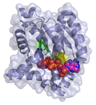
Adenylate kinase is a phosphotransferase enzyme that catalyzes the interconversion of the various adenosine phosphates. By constantly monitoring phosphate nucleotide levels inside the cell, ADK plays an important role in cellular energy homeostasis.

CAMK, also written as CaMK or CCaMK, is an abbreviation for the Ca2+/calmodulin-dependent protein kinase class of enzymes. CAMKs are activated by increases in the concentration of intracellular calcium ions (Ca2+) and calmodulin. When activated, the enzymes transfer phosphates from ATP to defined serine or threonine residues in other proteins, so they are serine/threonine-specific protein kinases. Activated CAMK is involved in the phosphorylation of transcription factors and therefore, in the regulation of expression of responding genes. CAMK also works to regulate the cell life cycle (i.e. programmed cell death), rearrangement of the cell's cytoskeletal network, and mechanisms involved in the learning and memory of an organism.

<span class="mw-page-title-main">Nucleoside-diphosphate kinase</span>

Nucleoside-diphosphate kinases are enzymes that catalyze the exchange of terminal phosphate between different nucleoside diphosphates (NDP) and triphosphates (NTP) in a reversible manner to produce nucleotide triphosphates. Many NDP serve as acceptor while NTP are donors of phosphate group. The general reaction via ping-pong mechanism is as follows: XDP + YTP ←→ XTP + YDP. NDPK activities maintain an equilibrium between the concentrations of different nucleoside triphosphates such as, for example, when guanosine triphosphate (GTP) produced in the citric acid (Krebs) cycle is converted to adenosine triphosphate (ATP). Other activities include cell proliferation, differentiation and development, signal transduction, G protein-coupled receptor, endocytosis, and gene expression.

<span class="mw-page-title-main">GMP synthase</span>

Guanosine monophosphate synthetase, also known as GMPS is an enzyme that converts xanthosine monophosphate to guanosine monophosphate.

<span class="mw-page-title-main">Deoxycytidine kinase</span> Protein-coding gene in the species Homo sapiens

Deoxycytidine kinase (dCK) is an enzyme which is encoded by the DCK gene in humans. dCK predominantly phosphorylates deoxycytidine (dC) and converts dC into deoxycytidine monophosphate. dCK catalyzes one of the initial steps in the nucleoside salvage pathway and has the potential to phosphorylate other preformed nucleosides, specifically deoxyadenosine (dA) and deoxyguanosine (dG), and convert them into their monophosphate forms. There has been recent biomedical research interest in investigating dCK's potential as a therapeutic target for different types of cancer.

<span class="mw-page-title-main">Diphosphomevalonate decarboxylase</span> InterPro Family

Diphosphomevalonate decarboxylase (EC 4.1.1.33), most commonly referred to in scientific literature as mevalonate diphosphate decarboxylase, is an enzyme that catalyzes the chemical reaction

<span class="mw-page-title-main">Guanylate kinase</span>

In enzymology, a guanylate kinase is an enzyme that catalyzes the chemical reaction

In enzymology, a nucleoside-triphosphate-adenylate kinase is an enzyme that catalyzes the chemical reaction

<span class="mw-page-title-main">Phosphoribulokinase</span>

Phosphoribulokinase (PRK) (EC 2.7.1.19) is an essential photosynthetic enzyme that catalyzes the ATP-dependent phosphorylation of ribulose 5-phosphate (RuP) into ribulose 1,5-bisphosphate (RuBP), both intermediates in the Calvin Cycle. Its main function is to regenerate RuBP, which is the initial substrate and CO2-acceptor molecule of the Calvin Cycle. PRK belongs to the family of transferase enzymes, specifically those transferring phosphorus-containing groups (phosphotransferases) to an alcohol group acceptor. Along with ribulose 1,5-bisphosphate carboxylase/oxygenase (RuBisCo), phosphoribulokinase is unique to the Calvin Cycle. Therefore, PRK activity often determines the metabolic rate in organisms for which carbon fixation is key to survival. Much initial work on PRK was done with spinach leaf extracts in the 1950s; subsequent studies of PRK in other photosynthetic prokaryotic and eukaryotic organisms have followed. The possibility that PRK might exist was first recognized by Weissbach et al. in 1954; for example, the group noted that carbon dioxide fixation in crude spinach extracts was enhanced by the addition of ATP. The first purification of PRK was conducted by Hurwitz and colleagues in 1956.

ATP + Mg2+ - D-ribulose 5-phosphate  ADP + D-ribulose 1,5-bisphosphate

In enzymology, a polyphosphate kinase, or polyphosphate polymerase, is an enzyme that catalyzes the formation of polyphosphate from ATP, with chain lengths of up to a thousand or more orthophosphate moieties.

<span class="mw-page-title-main">Pyruvate, phosphate dikinase</span>

Pyruvate, phosphate dikinase, or PPDK is an enzyme in the family of transferases that catalyzes the chemical reaction

<span class="mw-page-title-main">UCK2</span> Protein-coding gene in the species Homo sapiens

Uridine-cytidine kinase 2 (UCK2) is an enzyme that in humans is encoded by the UCK2 gene.

<span class="mw-page-title-main">CMP kinase</span> Protein-coding gene in the species Homo sapiens

UMP-CMP kinase is an enzyme that in humans is encoded by the CMPK1 gene.

The Walker A and Walker B motifs are protein sequence motifs, known to have highly conserved three-dimensional structures. These were first reported in ATP-binding proteins by Walker and co-workers in 1982.

<span class="mw-page-title-main">Prokaryotic riboflavin biosynthesis protein</span> Class of enzymes

The prokaryotic riboflavin biosynthesis protein is a bifunctional enzyme found in bacteria that catalyzes the phosphorylation of riboflavin into flavin mononucleotide (FMN) and the adenylylation of FMN into flavin adenine dinucleotide (FAD). It consists of a C-terminal riboflavin kinase and an N-terminal FMN-adenylyltransferase. This bacterial protein is functionally similar to the monofunctional riboflavin kinases and FMN-adenylyltransferases of eukaryotic organisms, but only the riboflavin kinases are structurally homologous.

References

  1. Boyer PD, Lardy H, Myrback K, eds. (1962). The Enzymes. Vol. 6 (2nd ed.). New York: Academic Press. pp. 139–149.
  2. Ayengar P, Gibson DM, Sanadi DR (July 1956). "Transphosphorylations between nucleoside phosphates". Biochimica et Biophysica Acta. 21 (1): 86–91. doi:10.1016/0006-3002(56)90096-8. PMID   13363863.
  3. Lieberman I, Kornberg A, Simms ES (July 1955). "Enzymatic synthesis of nucleoside diphosphates and triphosphates". The Journal of Biological Chemistry. 215 (1): 429–40. doi: 10.1016/S0021-9258(18)66050-8 . PMID   14392176.
  4. Heppel LA, Strominger JL, Maxwell ES (April 1959). "Nucleoside monophosphate kinases. II. Transphosphorylation between adenosine monophosphate and nucleoside triphosphates". Biochimica et Biophysica Acta. 32: 422–30. doi:10.1016/0006-3002(59)90615-8. PMID   14401179.
  5. Dreusicke D, Schulz GE (November 1986). "The glycine-rich loop of adenylate kinase forms a giant anion hole". FEBS Letters. 208 (2): 301–4. doi:10.1016/0014-5793(86)81037-7. PMID   3023140. S2CID   11786335.
  6. Byeon L, Shi Z, Tsai MD (March 1995). "Mechanism of adenylate kinase. The "essential lysine" helps to orient the phosphates and the active site residues to proper conformations". Biochemistry. 34 (10): 3172–82. doi:10.1021/bi00010a006. PMID   7880812.
  7. Müller CW, Schlauderer GJ, Reinstein J, Schulz GE (February 1996). "Adenylate kinase motions during catalysis: an energetic counterweight balancing substrate binding". Structure. 4 (2): 147–56. doi:10.2210/pdb4ake/pdb. PMID   8805521.
  8. Berg JM, Tymoczko JL, Stryer L (2002). Biochemistry . New York: W H Freeman. ISBN   0-7167-3051-0 . Retrieved 2016-01-08.
  9. Krishnamurthy H, Lou H, Kimple A, Vieille C, Cukier RI (January 2005). "Associative mechanism for phosphoryl transfer: a molecular dynamics simulation of Escherichia coli adenylate kinase complexed with its substrates". Proteins. 58 (1): 88–100. doi:10.1002/prot.20301. PMID   15521058. S2CID   20874015.
  10. Pai EF, Sachsenheimer W, Schirmer RH, Schulz GE (July 1977). "Substrate positions and induced-fit in crystalline adenylate kinase". Journal of Molecular Biology. 114 (1): 37–45. doi:10.1016/0022-2836(77)90281-9. PMID   198550.
  11. Müller CW, Schulz GE (March 1992). "Structure of the complex between adenylate kinase from Escherichia coli and the inhibitor Ap5A refined at 1.9 A resolution. A model for a catalytic transition state". Journal of Molecular Biology. 224 (1): 159–77. doi:10.2210/pdb1ake/pdb. PMID   1548697.
  12. Schlauderer GJ, Proba K, Schulz GE (February 1996). "Structure of a mutant adenylate kinase ligated with an ATP-analogue showing domain closure over ATP". Journal of Molecular Biology. 256 (2): 223–7. doi:10.1006/jmbi.1996.0080. PMID   8594191.
  13. Vonrhein C, Schlauderer GJ, Schulz GE (May 1995). "Movie of the structural changes during a catalytic cycle of nucleoside monophosphate kinases". Structure. 3 (5): 483–90. doi: 10.1016/s0969-2126(01)00181-2 . PMID   7663945.
  14. Fukami-Kobayashi K, Nosaka M, Nakazawa A, Go M (May 1996). "Ancient divergence of long and short isoforms of adenylate kinase: molecular evolution of the nucleoside monophosphate kinase family". FEBS Letters. 385 (3): 214–20. doi: 10.1016/0014-5793(96)00367-5 . PMID   8647254. S2CID   24934783.