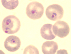
Plasmodium berghei

Last updated

Plasmodium berghei
Berghei 01.png
Blood-borne forms
Scientific classification OOjs UI icon edit-ltr.svg
Domain: Eukaryota
Clade: Sar
Clade: Alveolata
Phylum: Apicomplexa
Class: Aconoidasida
Order: Haemospororida
Family: Plasmodiidae
Genus: Plasmodium
Species:
P. berghei
Binomial name
Plasmodium berghei
Vincke & Lips, 1948
Strains

Plasmodium berghei is a single-celled parasite causing rodent malaria. It is in the Plasmodium subgenus Vinckeia .

Contents

Originally, isolated from thicket rats in Central Africa, [3] P. berghei is one of four Plasmodium species that have been described in African murine rodents, the others are P. chabaudi , P. vinckei , and P. yoelii . Due to its ability to infect rodents and relative ease of genetic engineering, P. berghei is a popular model organism for the study of human malaria. [4]

Biology

Like all malarial parasites of mammals, including the four human malaria parasites, P. berghei is transmitted by Anopheles mosquitoes and it infects the liver after being injected into the bloodstream by a bite of an infected female mosquito. After a short period (a few days) of development and multiplication, these parasites leave the liver and invade erythrocytes (red blood cells). The multiplication of the parasite in the blood causes the pathology such as anaemia and damage of essential organs of the host such as lungs, liver, spleen. P. berghei infections may also affect the brain and can be the cause of cerebral complications in laboratory mice (cerebral murine malaria, CMM). These symptoms are to a certain degree comparable to symptoms of cerebral malaria in patients infected with the human malaria parasite Plasmodium falciparum . [5]

Although sexuality is necessary in vivo in P. berghei as normal for most sexual organisms, it is a stark competitive disadvantage in vitro. Sinha et al., 2014 implement both mechanical passaging and competitive assay to demonstrate the advantage of loss of gametocyte production: During mechanical passage successive generations are found to naturally trend toward lower gametocytaemia; and nonsexuals outcompete sexuals rapidly when placed together in vitro. [6] :575

Immunochemistry

Endothelin 1 has an uncertain role in producing cerebral murine malaria. [2] Martins et al., 2016 find blockade of endothelin-1 prevents CMM and its symptoms and supplementation helps to produce it. [2] Subramaniam et al., 2015 find mice increase production of BTNL2 during infection and so it is probably protective. [2] Chertow et al., 2015 find the asymmetric dimethylarginine-to-arginine ratio is indicative of disease severity in mice with P. berghei ANKA. [7] [8] This ratio is a metric of arginine bioavailability and in this disease they find it predicts degree of endothelial dysfunction. [7] [8]

Pathophysiology

Liver stage

Before progressing to the symptomatic blood stage, P. Berghei infects the hepatocytes (liver cells), in which it undergoes rapid replication and generates thousands of merozoites that eventually burst and enter the bloodstream. The steps for this pathophysiological steps are as follows:

1) Hepatocyte Invasion - After being transmitted by an infected mosquito, Plasmodium sporozoites travel to the liver and invade hepatocytes. [9]

2)PV formation - Upon invasion, they create a parasitophorous vacuole (PV), which acts as a protective barrier against the cells's defense mechanisms in the cytoplasm. The PV is formed when the host cell invaginates (folds inward) around the invading parasite to enclose it within a protective compartment. This compartment provides shielding properties while also remaining permeable enough to allow for nutrient exchange so that the parasite continues to grow. [9] [10]

3) Host manipulation - Inside the parasitophorous vacuole, The P. Berghei parasite alters host processes like the AQP3, UPR and autophagy pathways. This turns the cell defense mechanisms into tools that allow it to grow and expand. [10]

4) Parasite replication - Once the parasite establishes itself within the Hepatocyte by completing the previous steps of invasion, it undergoes rapid nuclear division through schizogony (a type of asexual reproduction utilized by malaria parasites). This causes each single host cell to hold tens of thousands of P. Berghei merozoites. [11]

5) Egress - Once the merozoites within the hepatocyte are fully mature and ready to invade red blood cells, the hepatocyte ruptures and the merozoites are release into the bloodstream. They begin invading circulating red blood cells and thus transition into the blood stage of malaria. [12]

Parasite evasion of the host immune system

Mosquito

P. berghei ookinetes express surface proteins P25 and P28 that protect against complement-like attack in the mosquito midgut by creating a protective coat. [13] Mosquito immune factor TEP1 binds to and destroys pathogens, but the membrane created by the surface proteins shield the ookinetes from the TEP1 mediated lysis. The parasite is also able to suppress encapsulation and melanization, which normally trap and kill invaders. Circumsporozoite proteins (CSP) and thrombosopondin-related anonymous protein (TRAP) mediate sporozoite entry into salivary glands without triggering immune detection. [14] CSP binds heparan sulfate proteoglycans to mosquito salivary gland cells. This allows sporozoites dock specifically where they need to invade, instead of constant circulation throughout the host organism. It also creates a dense coat around the sporozoite shielding other antigens from antibody recognitions. [15]

Liver

The sporozoites are able to traverse liver resident macrophages known as kupffer cells while suppressing reactive oxygen species and inflammatory cytokines allowing the immune response to be minimized and entry into the liver. [16] The parasitophorous vacuole proteins such as UIS3 and UIS4 blocks lysosomal fusion and hijacks host autophagy via LC3 binding. [17] [18] The parasite once inside a hepatocyte is able to activate unfolded protein responses in the cell. In normal circumstances this induces the ER to perform apoptosis but in the case of the plasmodium parasite it is able to skew the pathway forcing it to begin expansion allowing the parasite to have more resources to feed on. [19] In addition to this the parasite also recruits host aquaporin-3 to import glycerol and water, further supporting growth. [20] Once the parasite is ready to leave the liver they are able egress as merosomes, meaning they are able to hide merozoites from the host macrophages by enveloping itself in a host derived vesicle [21] .

Blood

After being released into the blood the parasite has antigenic variation, meaning it is able to switch surface antigens to avoid antibody detection, meaning it can avoid nearby leukocytes [22] . Once the plasmodium is able to infect a red blood cell it is able to undergo cytoadherence. Knobs on infected RBCs anchor to the endothelium, preventing it from circulating throughout the body. This allows the parasites to avoid splenic clearance. [23] They are also able to undergo rosetting, meaning that infected RBCs cluster with uninfected RBCs, physically shielding parasites from immune attack. [24]

Strains

Some strains produce cerebral murine malaria and some do not. [2]

Hosts

Plasmodium berghei was first identified in the thicket rat ( Grammomys surdaster ). It has also been described in Leggada bella , Praomys jacksoni and Thamnomys surdaster.[ citation needed ] In research laboratories, various rodents can be infected, such as mice ( Mus musculus ), rats and gerbils ( Meriones unguiculatus ). [25] In M. musculusP. b. ANKA, downregulation of responses is necessary to prevent self-inflicted damage leading to CMM. [26] [27] :97 Specifically, Sarfo et al., 2011 finds mice produce the cytokine interleukin-10 (cIL-10) to suppress otherwise-potentially-deadly CMM damage from others of their own immune factors. [26] [27]

The natural insect host of P. berghei is likely Anopheles dureni , however in laboratory conditions it has also been shown to infect An. stephensi .[ citation needed ]

Live P. berghei expressing GFP (green) in erythrocytes; visualised using a fluorescence microscope Berghei 02.png
Live P. berghei expressing GFP (green) in erythrocytes; visualised using a fluorescence microscope

Gene interactions

In Mus musculus ⇔ the P. b. ANKA strain various genes affect the incidence of cerebral murine malaria. Kassa et al., 2016 finds several genes to be of no effect:

They find one improves survival probability:

An. gambiae 's hemocytes transcribe a wide array of molecular responses to Plasmodium infections. [28] [29] :138 [30] [31] [32] [33] :221 In response to this species, Baton et al., 2009 find this includes increased expression of the prophenoloxidase gene, cascading to increase phenoloxidase and thereby melanization. [28] [29] :138 [30] [31] [32] [33] :221

Treatment

Infected mouse, with P. berghei in the lungs, spleen and adipose tissue. Transgenic parasites are visualized by their expression of the bioluminescent reporter protein Luciferase Berghei 03.png
Infected mouse, with P. berghei in the lungs, spleen and adipose tissue. Transgenic parasites are visualized by their expression of the bioluminescent reporter protein Luciferase

Some phytochemicals show efficacy against P. berghei. Bankole et al., 2016 find Markhamia tomentosa to be highly effective, comparable to chloroquine, while Monoon longifolium is also significantly effective. They find Trichilia heudelotii to be ineffective. [34]

TMEM33 is an endoplasmic reticulum localized protein that is essential for all life cycle stages of Plasmodium berghei. [4] It is an important regulator of intracellular calcium homeostasis. [35] In humans and other eukaryotes, TMEM33 is a stress-inducible ER transmembrane protein, and is the regulator of UPR response elements. [36] UPR regulators and ER stress response elements play an important role in the blood stage infection and mosquito transmission of Plasmodium berghei. [4] Targeted deletions of TMEM33 show reduced parasitemia and mortality, indicating its potential as a drug target. [4]

The autophagy-related genes of Plasmodium berghei, PbATG5, PbATG8, and PbATG12 respond to 5-fluorouracil and chloroquine treatment, resulting in their upregulation and leading to apoptosis. [37]

History

Plasmodium berghei is found in the forests of Central Africa, where its natural cyclic hosts are the thicket rat ( Grammomys surdaster ) and its vectors are the mosquitos: Anopheles dureni [38] , Praomys jacksoni, and Leggada bella [39] . This species was first discovered by Ignace Vincke and Marcel Lips in blood films of Grammomys surdaster collected in Kisanga in the year 1948 in the Belgian Congo [40] . Plasmodium berghei was characterized by Dr. Richard Carter and David Walliker at the University of Edinburgh in the United Kingdom [41] . The parasite was then named after the director of the Institute for Scientific Research in Central Africa, Dr. Louis van den Berghe. A study conducted in 1950 by Vincke and Lips was the foundation for progressing the understanding of infectious process of P. berghei when infection became evident after injecting the isolated sporozoites into healthy mice [41] . P. berghei was the first in a series of three other malarial plasmodium (there are four total) to be discovered. [38] A trilogy of papers came out describing the discovery of this parasite and early analysis of the biology of P. berghei give an early indication of the natural host range of this parasite, a property that holds potential to underlying successful transmission of the parasite to a variety of laboratory hosts. [42] Though research only suggests that P. berghei is only a threat to rodents, it nevertheless gives us important insight on how to develops different types of medicine or antibiotics for the treatment of malaria.

A liver cell with P. berghei (a schizont with daughter parasites) expressing mCherry (red). Here the parasite membrane is stained green with an antibody, while the nuclei of liver cells and parasites are stained with DAPI (blue) Liver stage malaria parasite.jpg
A liver cell with P. berghei (a schizont with daughter parasites) expressing mCherry (red). Here the parasite membrane is stained green with an antibody, while the nuclei of liver cells and parasites are stained with DAPI (blue)

Research

Plasmodium berghei infection of laboratory mouse strains is frequently used in research as a model for human malaria. [43] In the laboratory the natural hosts have been replaced by a number of commercially available laboratory mouse strains, and the mosquito Anopheles stephensi , which is comparatively easily reared and maintained under defined laboratory conditions.

P. berghei is used as a model organism for the investigation of human malaria because of its similarity to the Plasmodium species which cause human malaria. P. berghei has a very similar life-cycle to the species that infect humans, and it causes disease in mice which has signs similar to those seen in human malaria. Importantly, P. berghei can be genetically manipulated more easily than the species which infect humans, making it a useful model for research into Plasmodium genetics.

In several aspects the pathology caused by P. berghei in mice differs from malaria caused by P. falciparum in humans. In particular, while death from P. falciparum malaria in humans is most frequently caused by the accumulation of red blood cells in the blood vessels of the brain, it is unclear to what extent this occurs in mice infected with P. berghei. [43] Instead, in P. berghei infection, mice are found to have an accumulation of immune cells in brain blood vessels. [43] This has led some to question the use of P. berghei infections in mice as an appropriate model of cerebral malaria in humans. [43]

P. berghei can be genetically manipulated in the laboratory using standard genetic engineering technologies. Consequently, this parasite is often used for the analysis of the function of malaria genes using the technology of genetic modification. [44] [45] [46] Additionally, the genome of P. berghei has been sequenced and it shows a high similarity, both in structure and gene content, with the genome of the primate malaria parasite Plasmodium falciparum . [47] [48] [49]

A number of genetically modified P. berghei lines have been generated which express fluorescent reporter proteins such as Green Fluorescent Protein (GFP) and mCherry (red) or bioluminescent reporters such as Luciferase. These transgenic parasites are important tools to study and visualize the parasites in the living host. [50] [51]

P. berghei is used in research programs for development and screening of anti-malarial drugs and for the development of an effective vaccine against malaria. [52]

Significance to One Health

Malaria is regarded as one of the most pervasive diseases in the world with the number of cases surpassing the hundreds of millions every year. [53] P. berghei zoonotic disease has a major impact on rodents within many different ecosystems and food chains around the world. While this parasite is not a threat to humans, it is used as a research tool for studying the life cycles, cellular biology, and pathogenicity of malaria within its animal hosts, which is then used for understanding its effects on human hosts. Understanding the intricacies of this parasite and how it operates allows for the development and improvement of vaccines and therapies that can then be distributed to areas where the risk for malaria within the human population is high. One strategy for combating this disease is by developing transmission blocking strategies (TBS) to stop the development of P. berghei within the mosquito stage, so that transmission may be halted all together. [53]

References

Reference: R. Sinden et al. "Maintenance of the Plasmodium berghei life cycle.." Methods in molecular medicine, 72 (2002): 25-40 . https://doi.org/10.1385/1-59259-271-6:25.

  1. 1 2 3 4 5 6 7 Torre S, Langlais D, Gros P (August 2018). "Genetic analysis of cerebral malaria in the mouse model infected with Plasmodium berghei". Mammalian Genome. 29 (7–8). Springer Science+Business Media: 488–506. doi:10.1007/s00335-018-9752-9. PMID   29922917. S2CID   49309005. International Mammalian Genome Society.
  2. 1 2 3 4 5 6 7 8 9 Moxon CA, Gibbins MP, McGuinness D, Milner DA, Marti M (January 2020). "New Insights into Malaria Pathogenesis". Annual Review of Pathology. 15 (1). Annual Reviews: 315–343. doi:10.1146/annurev-pathmechdis-012419-032640. PMID   31648610. S2CID   204882296.
  3. Conteh S, Anderson C, Lambert L, Orr-Gonzalez S, Herrod J, Robbins YL, et al. (April 2017). "Grammomys surdaster, the Natural Host for Plasmodium berghei Parasites, as a Model to Study Whole-Organism Vaccines Against Malaria". The American Journal of Tropical Medicine and Hygiene. 96 (4): 835–841. doi:10.4269/ajtmh.16-0745. PMC   5392629 . PMID   28115674.
  4. 1 2 3 4 Kamil M, Kina UY, Atmaca HN, Unal S, Deveci G, Burak P, et al. (April 2024). "Endoplasmic reticulum localized TMEM33 domain-containing protein is crucial for all life cycle stages of the malaria parasite". Molecular Microbiology. 121 (4): 767–780. doi: 10.1111/mmi.15228 . PMID   38238886.
  5. Franke-Fayard B, Fonager J, Braks A, Khan SM, Janse CJ (September 2010). "Sequestration and tissue accumulation of human malaria parasites: can we learn anything from rodent models of malaria?". PLOS Pathogens. 6 (9) e1001032. doi: 10.1371/journal.ppat.1001032 . PMC   2947991 . PMID   20941396.
  6. Josling GA, Llinás M (September 2015). "Sexual development in Plasmodium parasites: knowing when it's time to commit". Nature Reviews. Microbiology. 13 (9). Nature Portfolio: 573–587. doi:10.1038/nrmicro3519. PMID   26272409. S2CID   2182486.
  7. 1 2 3 Ngai M, Weckman AM, Erice C, McDonald CR, Cahill LS, Sled JG, et al. (February 2020). "Malaria in Pregnancy and Adverse Birth Outcomes: New Mechanisms and Therapeutic Opportunities". Trends in Parasitology. 36 (2). Cell Press: 127–137. doi:10.1016/j.pt.2019.12.005. PMID   31864896. S2CID   209446589.
  8. 1 2 3 Kayano AC, Dos-Santos JC, Bastos MF, Carvalho LJ, Aliberti J, Costa FT (April 2016). Andrews-Polymenis HL (ed.). "Pathophysiological Mechanisms in Gaseous Therapies for Severe Malaria". Infection and Immunity. 84 (4). American Society for Microbiology: 874–882. doi:10.1128/iai.01404-15. PMC   4807480 . PMID   26831465. S2CID   29927044.
  9. 1 2 Hildebrandt F, Urrutia Iturritza M, Zwicker C (2024). "Host-pathogen interactions in the Plasmodium-infected mouse liver at spatial and single-cell resolution". Nature Communications. 15 (1): 7105. Bibcode:2024NatCo..15.7105H. doi:10.1038/s41467-024-51418-2. PMC   11333755 . PMID   39160174.
  10. 1 2 De Niz M, Caldelari R, Kaiser G, Zuber B, Heo WD, Heussler VT, et al. (2021). "Hijacking of the host cell Golgi by Plasmodium berghei liver stage parasites". Journal of Cell Science. 134 (10) jcs252213. doi:10.1242/jcs.252213. PMC   8186485 . PMID   34013963.
  11. Hildebrandt F, Iturritza MU, Zwicker C, Vanneste B, Van Hul N, Semle E, et al. (August 19, 2024). "Host-pathogen interactions in the Plasmodium-infected mouse liver at spatial and single-cell resolution". Nature Communications. 15 (1) 7105. Bibcode:2024NatCo..15.7105H. doi:10.1038/s41467-024-51418-2. PMC   11333755 . PMID   39160174.
  12. Dewi Putrianti E (September 21, 2020). "A Plasmodium cysteine protease required for efficient transition from the liver infection stage". PLOS Pathogens.
  13. Baton LA, Ranford-Cartwright LC (2005-02-25). "Do malaria ookinete surface proteins P25 and P28 mediate parasite entry into mosquito midgut epithelial cells?". Malaria Journal. 4 (1) 15. doi: 10.1186/1475-2875-4-15 . ISSN   1475-2875.
  14. Blandin S, Shiao SH, Moita LF, Janse CJ, Waters AP, Kafatos FC, et al. (March 2004). "Complement-like protein TEP1 is a determinant of vectorial capacity in the malaria vector Anopheles gambiae". Cell. 116 (5): 661–70. doi:10.1016/s0092-8674(04)00173-4. PMID   15006349.
  15. Sultan AA, Thathy V, Frevert U, Robson KJ, Crisanti A, Nussenzweig V, et al. (August 1997). "TRAP is necessary for gliding motility and infectivity of plasmodium sporozoites". Cell. 90 (3): 511–22. doi:10.1016/s0092-8674(00)80511-5. PMID   9267031.
  16. Risco-Castillo V, Topçu S, Marinach C, Manzoni G, Bigorgne AE, Briquet S, et al. (November 2015). "Malaria Sporozoites Traverse Host Cells within Transient Vacuoles". Cell Host & Microbe. 18 (5): 593–603. doi:10.1016/j.chom.2015.10.006. PMID   26607162.
  17. Mueller AK, Camargo N, Kaiser K, Andorfer C, Frevert U, Matuschewski K, et al. (2005-02-07). "Plasmodiumliver stage developmental arrest by depletion of a protein at the parasite–host interface". Proceedings of the National Academy of Sciences. 102 (8): 3022–3027. Bibcode:2005PNAS..102.3022M. doi: 10.1073/pnas.0408442102 . ISSN   0027-8424.
  18. Real E, Rodrigues L, Cabal GG, Enguita FJ, Mancio-Silva L, Mello-Vieira J, et al. (2017-11-06). "Plasmodium UIS3 sequesters host LC3 to avoid elimination by autophagy in hepatocytes". Nature Microbiology. 3 (1): 17–25. doi:10.1038/s41564-017-0054-x. ISSN   2058-5276. PMC   5739284 . PMID   29109477.
  19. Inácio P, Zuzarte-Luís V, Ruivo MT, Falkard B, Nagaraj N, Rooijers K, et al. (August 2015). "Parasite-induced ER stress response in hepatocytes facilitates Plasmodium liver stage infection". EMBO Reports. 16 (8): 955–64. doi:10.15252/embr.201439979. PMC   4552488 . PMID   26113366.
  20. Posfai D, Sylvester K, Reddy A, Ganley JG, Wirth J, Cullen QE, et al. (2018-05-18). "Plasmodium parasite exploits host aquaporin-3 during liver stage malaria infection". PLOS Pathogens. 14 (5) e1007057. doi: 10.1371/journal.ppat.1007057 . ISSN   1553-7374.
  21. Roepe P (2006-09-01). "Faculty Opinions recommendation of Manipulation of host hepatocytes by the malaria parasite for delivery into liver sinusoids". doi: 10.3410/f.1033777.388108 .{{cite web}}: Missing or empty |url= (help)
  22. Scherf A, Lopez-Rubio JJ, Riviere L (2008-10-01). "Antigenic Variation in Plasmodium falciparum". Annual Review of Microbiology. 62 (1): 445–470. doi:10.1146/annurev.micro.61.080706.093134. ISSN   0066-4227. PMID   18785843.
  23. Miller LH, Baruch DI, Marsh K, Doumbo OK (February 2002). "The pathogenic basis of malaria". Nature. 415 (6872): 673–9. doi:10.1038/415673a. PMID   11832955.
  24. Rowe A, Obeiro J, Newbold CI, Marsh K (June 1995). "Plasmodium falciparum rosetting is associated with malaria severity in Kenya". Infection and Immunity. 63 (6): 2323–6. doi:10.1128/iai.63.6.2323-2326.1995. PMC   173304 . PMID   7768616.
  25. Junaid QO, Khaw LT, Mahmud R, Ong KC, Lau YL, Borade PU, et al. (2017). "Pathogenesis of Plasmodium berghei ANKA infection in the gerbil (Meriones unguiculatus) as an experimental model for severe malaria". Parasite. 24: 38. doi:10.1051/parasite/2017040. PMC   5642054 . PMID   29034874.
  26. 1 2 Xu Z, Zhang X, Lau J, Yu J (September 2016). "C-X-C motif chemokine 10 in non-alcoholic steatohepatitis: role as a pro-inflammatory factor and clinical implication". Expert Reviews in Molecular Medicine. 18 e16. Cambridge University Press (CUP). doi:10.1017/erm.2016.16. PMID   27669973. S2CID   28322523.
  27. 1 2 Fillatreau S, O'Garra A (2014). Interleukin-10 in Health and Disease. Current Topics in Microbiology and Immunology. Vol. 20. Springer. p. 649. doi:10.1007/978-3-662-43492-5. ISBN   978-3-662-43491-8.
  28. 1 2 Smith RC, Vega-Rodríguez J, Jacobs-Lorena M (August 2014). "The Plasmodium bottleneck: malaria parasite losses in the mosquito vector". Memórias do Instituto Oswaldo Cruz. 109 (5). FapUNIFESP (SciELO): 644–661. doi:10.1590/0074-0276130597. PMC   4156458 . PMID   25185005.
  29. 1 2 Willis JH, Papandreou NC, Iconomidou VA, Hamodrakas SJ (2012). "5 Cuticular Proteins". In Gilbert LI (ed.). Insect Molecular Biology and Biochemistry. Elsevier. ISBN   978-0-12-384747-8. OCLC   742299021.
  30. 1 2 Clayton AM, Dong Y, Dimopoulos G (2014). "The Anopheles innate immune system in the defense against malaria infection". Journal of Innate Immunity. 6 (2). Karger Publishers: 169–181. doi:10.1159/000353602. PMC   3939431 . PMID   23988482.
  31. 1 2 Hillyer JF, Strand MR (September 2014). "Mosquito hemocyte-mediated immune responses". Current Opinion in Insect Science. 3. Elsevier: 14–21. Bibcode:2014COIS....3...14H. doi:10.1016/j.cois.2014.07.002. PMC   4190037 . PMID   25309850. NIHMSID 615755.
  32. 1 2 Cheng G, Liu Y, Wang P, Xiao X (March 2016). "Mosquito Defense Strategies against Viral Infection". Trends in Parasitology. 32 (3). Cell Press: 177–186. doi:10.1016/j.pt.2015.09.009. PMC   4767563 . PMID   26626596.
  33. 1 2 Hillyer JF (2010). "Mosquito Immunity". In Söderhäll K (ed.). Invertebrate Immunity. Advances in Experimental Medicine and Biology. Vol. 708. Boston, MA: Springer Nature. pp. 218–238. doi:10.1007/978-1-4419-8059-5_12. ISBN   978-1-4419-8058-8. ISSN   0065-2598. PMID   21528701.
  34. Dkhil MA, Al-Quraishy S, Al-Shaebi EM, Abdel-Gaber R, Thagfan FA, Qasem MA (March 2021). "Medicinal plants as a fight against murine blood-stage malaria". Saudi Journal of Biological Sciences. 28 (3). Elsevier: 1723–1738. Bibcode:2021SJBS...28.1723D. doi:10.1016/j.sjbs.2020.12.014. PMC   7938113 . PMID   33732056. S2CID   232241302. Saudi Biological Society.
  35. Arhatte M, Gunaratne GS, El Boustany C, Kuo IY, Moro C, Duprat F, et al. (May 2019). "TMEM33 regulates intracellular calcium homeostasis in renal tubular epithelial cells". Nature Communications. 10 (1) 2024. Bibcode:2019NatCo..10.2024A. doi:10.1038/s41467-019-10045-y. PMC   6497644 . PMID   31048699.
  36. Sakabe I, Hu R, Jin L, Clarke R, Kasid UN (September 2015). "TMEM33: a new stress-inducible endoplasmic reticulum transmembrane protein and modulator of the unfolded protein response signaling". Breast Cancer Research and Treatment. 153 (2): 285–297. doi:10.1007/s10549-015-3536-7. PMC   4559576 . PMID   26268696.
  37. Unal S, Kina UY, Kamil M, Aly AS, Palabiyik B (July 2024). "Drug-induced ER stress leads to induction of programmed cell death pathways of the malaria parasite". Parasitology Research. 123 (7) 263. doi:10.1007/s00436-024-08281-3. PMC   11230985 . PMID   38976068.
  38. 1 2 Otun SO, Graca R, Onisuru O, Achilonu I (2024-09-12). "Evaluation of Plasmodium berghei Models in Malaria Research". Journal of Cellular Signaling. 5 (3): 96–113. doi:10.33696/Signaling.5.117. ISSN   2692-0638.
  39. "Introduction to Plasmodium berghei – P.BERGHEI" . Retrieved 2025-10-02.
  40. Vincke IH, Lips M (1948). "Un nouveau plasmodium d'un rongeur sauvage du Congo: Plasmodium berghei n.sp". Annales de la Société Belge de Médecine Tropicale. 28: 97–104.
  41. 1 2 Pattaradilokrat S, Wu J, Xu F, Su XZ (December 2022). "The origins, isolation, and biological characterization of rodent malaria parasites". Parasitology International. 91 102636. doi:10.1016/j.parint.2022.102636. ISSN   1873-0329. PMC   9465976 . PMID   35926694.
  42. Sinden RE, Butcher GA, Beetsma AL (2002-06-28), "Maintenance of the Plasmodium berghei Life Cycle", Malaria Methods and Protocols, vol. 72, New Jersey: Humana Press, pp. 25–40, doi:10.1385/1-59259-271-6:25, ISBN   978-1-59259-271-5, PMID   12125122 , retrieved 2025-10-02
  43. 1 2 3 4 Craig AG, Grau GE, Janse C, Kazura JW, Milner D, Barnwell JW, et al. (February 2012). "The role of animal models for research on severe malaria". PLOS Pathogens. 8 (2) e1002401. doi: 10.1371/journal.ppat.1002401 . PMC   3271056 . PMID   22319438.
  44. Janse CJ, Ramesar J, Waters AP (2006). "High-efficiency transfection and drug selection of genetically transformed blood stages of the rodent malaria parasite Plasmodium berghei". Nature Protocols. 1 (1): 346–356. doi:10.1038/nprot.2006.53. PMID   17406255. S2CID   20096737.
  45. Janse CJ, Kroeze H, van Wigcheren A, Mededovic S, Fonager J, Franke-Fayard B, et al. (January 2011). "A genotype and phenotype database of genetically modified malaria-parasites". Trends in Parasitology. 27 (1): 31–39. doi:10.1016/j.pt.2010.06.016. PMID   20663715.
  46. Khan SM, Kroeze H, Franke-Fayard B, Janse CJ (2013). "Standardization in Generating and Reporting Genetically Modified Rodent Malaria Parasites: The RMGMDB Database". Malaria. Methods in Molecular Biology. Vol. 923. pp. 139–50. doi:10.1007/978-1-62703-026-7_9. ISBN   978-1-62703-025-0. PMID   22990775.
  47. Hall N, Karras M, Raine JD, Carlton JM, Kooij TW, Berriman M, et al. (January 2005). "A comprehensive survey of the Plasmodium life cycle by genomic, transcriptomic, and proteomic analyses". Science. 307 (5706): 82–86. Bibcode:2005Sci...307...82H. doi:10.1126/science.1103717. PMID   15637271. S2CID   7230793.
  48. Kooij TW, Janse CJ, Waters AP (May 2006). "Plasmodium post-genomics: better the bug you know?". Nature Reviews. Microbiology. 4 (5): 344–357. doi: 10.1038/nrmicro1392 . PMID   16582929. S2CID   38403613.
  49. Otto TD, Böhme U, Jackson AP, Hunt M, Franke-Fayard B, Hoeijmakers WA, et al. (October 2014). "A comprehensive evaluation of rodent malaria parasite genomes and gene expression". BMC Biology. 12 86. doi: 10.1186/s12915-014-0086-0 . PMC   4242472 . PMID   25359557.
  50. Amino R, Ménard R, Frischknecht F (August 2005). "In vivo imaging of malaria parasites--recent advances and future directions". Current Opinion in Microbiology. 8 (4): 407–414. doi:10.1016/j.mib.2005.06.019. PMID   16019254.
  51. Franke-Fayard B, Waters AP, Janse CJ (2006). "Real-time in vivo imaging of transgenic bioluminescent blood stages of rodent malaria parasites in mice". Nature Protocols. 1 (1): 476–485. doi:10.1038/nprot.2006.69. PMID   17406270. S2CID   20812965.
  52. Khan SM, Janse CJ, Kappe SH, Mikolajczak SA (December 2012). "Genetic engineering of attenuated malaria parasites for vaccination". Current Opinion in Biotechnology. 23 (6): 908–916. doi:10.1016/j.copbio.2012.04.003. PMID   22560204.
  53. 1 2 Dehghan H, Oshaghi MA, Mosa-Kazemi SH, Abai MR, Rafie F, Nateghpour M, et al. (2018). "Experimental Study on Plasmodium berghei, Anopheles Stephensi, and BALB/c Mouse System: Implications for Malaria Transmission Blocking Assays". Iranian Journal of Parasitology. 13 (4): 549–559. ISSN   1735-7020. PMC   6348208 . PMID   30697308.
General information about (the biology of) P. berghei
Information about the genome and genes of P. berghei