HslVU

Last updated
HslU—HslV peptidase
Hslvu ecoli.png
Top view of the hslV/hslU complex isolated from E. coli (PDB ID 1G4A).
Identifiers
EC no. 3.4.25.2
Databases
IntEnz IntEnz view
BRENDA BRENDA entry
ExPASy NiceZyme view
KEGG KEGG entry
MetaCyc metabolic pathway
PRIAM profile
PDB structures RCSB PDB PDBe PDBsum
Search
PMC articles
PubMed articles
NCBI proteins
Heat shock protein HslU
Identifiers
SymbolHslU
InterPro IPR004491
ATP-dependent protease, HslV subunit
Identifiers
SymbolHslV
InterPro IPR022281

The heat shock proteins HslV and HslU (HslVU complex; also known as ClpQ and ClpY respectively, or ClpQY) are expressed in many bacteria such as E. coli in response to cell stress. [1] The hslV protein is a protease and the hslU protein is an ATPase; the two form a symmetric assembly of four stacked rings, consisting of an hslV dodecamer bound to an hslU hexamer, with a central pore in which the protease and ATPase active sites reside. The hslV protein degrades unneeded or damaged proteins only when in complex with the hslU protein in the ATP-bound state. HslV is thought to resemble the hypothetical ancestor of the proteasome, a large protein complex specialized for regulated degradation of unneeded proteins in eukaryotes, many archaea, and a few bacteria. HslV bears high similarity to core subunits of proteasomes. [2]

Contents

Genetics

Both proteins are encoded on the same operon within the bacterial genome. Unlike many eukaryotic proteasomes, which have several different peptide substrate specificities, hslV has a specificity similar to that of chymotrypsin; hence it is inhibited by proteasome inhibitors that specifically target the chymotrypsin site in eukaryotic proteasomes. [3] Although the HslVU complex is stable on its own, some evidence suggests that the complex is formed in vivo in a substrate-induced manner due to a conformational change in the hslU-substrate complex that promotes hslV binding. [4]

HslV and hslU genes have also been identified in some eukaryotes, although these also require the constitutively expressed proteasome for survival. These eukaryotic HslVU complexes assemble to apparently functional units, suggesting that these eukaryotes have both functional proteasomes and functional hslVU systems. [5]

Regulation

The promoter region of the operon encoding HslU and HslV contains a stem-loop structure which is necessary for gene expression. This structure contributes to mRNA stability. [6]

Motifs in peptide unfolding

A four-amino acid sequence motif - GYVG, glycine-tyrosine-valine-glycine - conserved in hslU ATPases and located on the inner surface of the assembled pore dramatically accelerates the degradation of some proteins, and is required for the degradation of others. However, these motifs are not necessary for the degradation of short peptides and play no direct role in hydrolysis, suggesting that their major role is in unfolding the native state structure of the substrate and transferring the resulting disordered polypeptide chain to the hslV subunits for degradation. These motifs also influence the assembly of the complex. [7] Translocation is also facilitated by the C-terminal tails of the HslU subunits, which form a gate closing off the proteolytic active sites in the central pore until a substrate has been bound and unfolded. [8]

Mechanism

The basic mechanism by which the hslVU complex undertakes proteolytic substrate degradation is essentially the same as that observed in the eukaryotic proteasome, catalyzed by Nactive-site threonine residues. Both are members of the T1 family. [9] It is inhibited by enzyme inhibitors that covalently bind the threonine. [10] Like the proteasome, hslU must bind ATP in a magnesium-dependent manner before substrate binding and unfolding can occur. [11]

Related Research Articles

<span class="mw-page-title-main">Proteasome</span> Protein complexes which degrade unnecessary or damaged proteins by proteolysis

Proteasomes are protein complexes which degrade unneeded or damaged proteins by proteolysis, a chemical reaction that breaks peptide bonds. Enzymes that help such reactions are called proteases.

<span class="mw-page-title-main">Chaperone (protein)</span> Proteins assisting in protein folding

In molecular biology, molecular chaperones are proteins that assist the conformational folding or unfolding of large proteins or macromolecular protein complexes. There are a number of classes of molecular chaperones, all of which function to assist large proteins in proper protein folding during or after synthesis, and after partial denaturation. Chaperones are also involved in the translocation of proteins for proteolysis.

<span class="mw-page-title-main">Hsp90</span> Heat shock proteins with a molecular mass around 90kDa

Hsp90 is a chaperone protein that assists other proteins to fold properly, stabilizes proteins against heat stress, and aids in protein degradation. It also stabilizes a number of proteins required for tumor growth, which is why Hsp90 inhibitors are investigated as anti-cancer drugs.

<span class="mw-page-title-main">AAA proteins</span> Protein family

AAA proteins or ATPases Associated with diverse cellular Activities are a protein family sharing a common conserved module of approximately 230 amino acid residues. This is a large, functionally diverse protein family belonging to the AAA+ protein superfamily of ring-shaped P-loop NTPases, which exert their activity through the energy-dependent remodeling or translocation of macromolecules.

<span class="mw-page-title-main">Endopeptidase Clp</span>

Endopeptidase Clp (EC 3.4.21.92, endopeptidase Ti, caseinolytic protease, protease Ti, ATP-dependent Clp protease, ClpP, Clp protease). This enzyme catalyses the following chemical reaction

Non-chaperonin molecular chaperone ATPase (EC 3.6.4.10, molecular chaperone Hsc70 ATPase) is an enzyme with systematic name ATP phosphohydrolase (polypeptide-polymerizing). This enzyme catalyses the following chemical reaction

<span class="mw-page-title-main">PSMC3</span> Enzyme found in humans

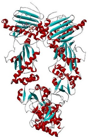
26S protease regulatory subunit 6A, also known as 26S proteasome AAA-ATPase subunit Rpt5, is an enzyme that in humans is encoded by the PSMC3 gene. This protein is one of the 19 essential subunits of a complete assembled 19S proteasome complex Six 26S proteasome AAA-ATPase subunits together with four non-ATPase subunits form the base sub complex of 19S regulatory particle for proteasome complex.

<span class="mw-page-title-main">PSMC5</span> Enzyme found in humans

26S protease regulatory subunit 8, also known as 26S proteasome AAA-ATPase subunit Rpt6, is an enzyme that in humans is encoded by the PSMC5 gene. This protein is one of the 19 essential subunits of a complete assembled 19S proteasome complex Six 26S proteasome AAA-ATPase subunits together with four non-ATPase subunits form the base sub complex of 19S regulatory particle for proteasome complex.

<span class="mw-page-title-main">PSMD4</span> Enzyme found in humans

26S proteasome non-ATPase regulatory subunit 4, also as known as 26S Proteasome Regulatory Subunit Rpn10, is an enzyme that in humans is encoded by the PSMD4 gene. This protein is one of the 19 essential subunits that contributes to the complete assembly of 19S proteasome complex.

<span class="mw-page-title-main">PSMC2</span> Enzyme found in humans

26S protease regulatory subunit 7, also known as 26S proteasome AAA-ATPase subunit Rpt1, is an enzyme that in humans is encoded by the PSMC2 gene This protein is one of the 19 essential subunits of a complete assembled 19S proteasome complex. Six 26S proteasome AAA-ATPase subunits together with four non-ATPase subunits form the base sub complex of 19S regulatory particle for proteasome complex.

<span class="mw-page-title-main">PSMC1</span> Enzyme found in humans

26S protease regulatory subunit 4, also known as 26S proteasome AAA-ATPase subunit Rpt2, is an enzyme that in humans is encoded by the PSMC1 gene. This protein is one of the 19 essential subunits of a complete assembled 19S proteasome complex. Six 26S proteasome AAA-ATPase subunits together with four non-ATPase subunits form the base sub complex of 19S regulatory particle for proteasome complex.

<span class="mw-page-title-main">PSMC4</span> Enzyme found in humans

26S protease regulatory subunit 6B, also known as 26S proteasome AAA-ATPase subunit Rpt3, is an enzyme that in humans is encoded by the PSMC4 gene. This protein is one of the 19 essential subunits of a complete assembled 19S proteasome complex Six 26S proteasome AAA-ATPase subunits together with four non-ATPase subunits form the base sub complex of 19S regulatory particle for proteasome complex.

<span class="mw-page-title-main">PSME2</span> Protein found in humans

Proteasome activator complex subunit 2 is a protein that in humans is encoded by the PSME2 gene.

<span class="mw-page-title-main">PSMC6</span> Enzyme found in humans

26S protease regulatory subunit S10B, also known as 26S proteasome AAA-ATPase subunit Rpt4, is an enzyme that in humans is encoded by the PSMC6 gene. This protein is one of the 19 essential subunits of a complete assembled 19S proteasome complex Six 26S proteasome AAA-ATPase subunits together with four non-ATPase subunits form the base sub complex of 19S regulatory particle for proteasome complex.

<span class="mw-page-title-main">PSMD2</span> Enzyme found in humans

26S proteasome non-ATPase regulatory subunit 2, also as known as 26S Proteasome Regulatory Subunit Rpn1, is an enzyme that in humans is encoded by the PSMD2 gene.

<span class="mw-page-title-main">ATP-dependent Clp protease proteolytic subunit</span> Protein-coding gene in the species Homo sapiens

ATP-dependent Clp protease proteolytic subunit (ClpP) is an enzyme that in humans is encoded by the CLPP gene. This protein is an essential component to form the protein complex of Clp protease.

The N-end rule is a rule that governs the rate of protein degradation through recognition of the N-terminal residue of proteins. The rule states that the N-terminal amino acid of a protein determines its half-life. The rule applies to both eukaryotic and prokaryotic organisms, but with different strength, rules, and outcome. In eukaryotic cells, these N-terminal residues are recognized and targeted by ubiquitin ligases, mediating ubiquitination thereby marking the protein for degradation. The rule was initially discovered by Alexander Varshavsky and co-workers in 1986. However, only rough estimations of protein half-life can be deduced from this 'rule', as N-terminal amino acid modification can lead to variability and anomalies, whilst amino acid impact can also change from organism to organism. Other degradation signals, known as degrons, can also be found in sequence.

<span class="mw-page-title-main">ATP-dependent Clp protease adaptor protein ClpS</span>

ClpS is an N-recognin in the N-end rule pathway. ClpS interacts with protein substrates that have a bulky hydrophobic residue at the N-terminus. The protein substrate is then degraded by the ClpAP protease.

<span class="mw-page-title-main">ClpX</span> Mammalian protein found in Homo sapiens

ATP-dependent Clp protease ATP-binding subunit clpX-like, mitochondrial is an enzyme that in humans is encoded by the CLPX gene. This protein is a member of the family of AAA Proteins and is to form the protein complex of Clp protease.

Alfred Lewis Goldberg was an American cell biologist-biochemist and professor at Harvard University. His major discoveries have concerned the mechanisms and physiological importance of protein degradation in cells. Of wide impact have been his lab's demonstration that all cells contain a pathway for selectively eliminating misfolded proteins, his discoveries about the role of proteasomes in this process and of the enzyme systems catalyzing protein breakdown in bacteria, his elucidating the mechanisms for muscle atrophy and the role of proteasomes in antigen presentation to the immune system, and his introduction of proteasome inhibitors now widely used as research tools and in the treatment of blood cancers.

References

  1. Ramachandran R, Hartmann C, Song HK, Huber R, Bochtler M. (2002). Functional interactions of HslV (ClpQ) with the ATPase HslU (ClpY). Proc Natl Acad Sci USA 99(11):7396-401.
  2. Gille C, Goedel A, Schloetelburg C, Preißner R, Kloetzell PM, Gobel UB, Frommell C. (2003). A Comprehensive View on Proteasomal Sequences: Implications for the Evolution of the Proteasome. J Mol Biol 326: 1437–1448.
  3. Rohrwild M, Coux O, Huang HC, R P Moerschell RP, Yoo SJ, Seol JH, Chung CH, Goldberg AL. (1996). HslV-HslU: A novel ATP-dependent protease complex in Escherichia coli related to the eukaryotic proteasome. Proc Natl Acad Sci USA 93(12): 5808–5813
  4. Azim MK, Goehring W, Song HK, Ramachandran R, Bochtler M, Goettig P. (2005). Characterization of the HslU chaperone affinity for HslV protease. Protein Sci 14(5):1357-62.
  5. Ruiz-Gonzalez MX, Marin I. (2006). Proteasome-related HslU and HslV genes typical of eubacteria are widespread in eukaryotes. J Mol Evol 63(4):504-12.
  6. Lien, HY; Yu, CH; Liou, CM; Wu, WF (2009). "Regulation of clpQ⁺Y⁺ (hslV⁺U⁺) gene expression in Escherichia coli". The Open Microbiology Journal. 3: 29–39. doi: 10.2174/1874285800903010029 . PMC   2681174 . PMID   19440251.
  7. Park E, Rho YM, Koh OJ, Ahn SW, Seong IS, Song JJ, Bang O, Seol JH, Wang J, Eom SH, Chung CH. (2005). Role of the GYVG pore motif of HslU ATPase in protein unfolding and translocation for degradation by HslV peptidase. J Biol Chem 280(24):22892-8.
  8. Seong IS, Kang MS, Choi MK, Lee JW, Koh OJ, Wang J, Eom SH, Chung CH. (2002). The C-terminal tails of HslU ATPase act as a molecular switch for activation of HslV peptidase. J Biol Chem 277(29):25976-82.
  9. Bogyo M, McMaster JS, Gaczynska M, Tortorella D, Goldberg AL, Ploegh H. (1997). Covalent modification of the active site threonine of proteasomal beta subunits and the Escherichia coli homolog HslV by a new class of inhibitors. Proc Natl Acad Sci USA 94(13):6629-34.
  10. Sousa MC, Kessler BM, Overkleeft HS, McKay DB. (2002). Crystal structure of HslUV complexed with a vinyl sulfone inhibitor: corroboration of a proposed mechanism of allosteric activation of HslV by HslU. J Mol Biol 318(3):779-85.
  11. Burton RE, Baker TA, Sauer RT. (2005). Nucleotide-dependent substrate recognition by the AAA+ HslUV protease. Nat Struct Mol Biol 12(3):245-51.

Further reading