Dipeptidase 1

Last updated
DPEP1
Protein DPEP1 PDB 1itq.png
Available structures
PDB Ortholog search: PDBe RCSB
Identifiers
Aliases DPEP1 , MBD1, MDP, RDP, dipeptidase 1 (renal), dipeptidase 1
External IDs OMIM: 179780 MGI: 94917 HomoloGene: 80192 GeneCards: DPEP1
Orthologs
SpeciesHumanMouse
Entrez
Ensembl
UniProt
RefSeq (mRNA)

NM_001128141
NM_004413

NM_007876

RefSeq (protein)

NP_001121613
NP_004404

NP_031902

Location (UCSC) Chr 16: 89.61 – 89.64 Mb Chr 8: 123.91 – 123.93 Mb
PubMed search [3] [4]
Wikidata
View/Edit Human View/Edit Mouse

Dipeptidase 1 (DPEP1), or renal dipeptidase, is a membrane-bound glycoprotein responsible for hydrolyzing dipeptides. It is found in the microsomal fraction of the porcine kidney cortex. [5] It exists as a disulfide-linked homodimer that is glygosylphosphatidylinositol (GPI)-anchored to the renal brush border of the kidney. [6] The active site on each homodimer is made up of a barrel subunit with binuclear zinc ions that are bridged by the Gly125 side-chain located at the bottom of the barrel. [7]

Contents

Structure

The gene coding for DPEP1 is 6 kb long and consists of ten exons and nine introns. The protein itself is made of 411 amino acid residues and is only transcribed in kidney cells. [8] Although disulfide linkages in DPEP1 do not contribute to the enzyme’s activity, they are essential for the enzyme’s proper function because they keep the enzyme’s subunits together and attached to the renal brush border. Cysteine 261 is involved in disulfide linkage between the enzyme’s subunits, and is also located very close to both the site of the GPI-anchor and the membrane, suggesting that it is also involved in the enzyme’s linkage to the membrane. [9]

DPEP1 is also a metalloenzyme that specifically uses zinc as its cofactor. [10] The enzyme’s typical zinc content is 1.42 ug/mg. [11] The addition of cobalt or manganese ions cause the enzyme to take on different conformations, which suggests that the enzyme may be able to hydrolyze different dipeptides depending on which metal ions are present—aka the metal-content of one’s micronutrient intake could affect their renal dipeptidase’s ability to metabolize various dipeptides. [12]

Function

The primary function of DPEP1 is to hydrolyze various dipeptides in renal metabolism. Specifically, it has been found to hydrolyze glutathione and its conjugates such as leukotriene D (Kozak and Tate, 1982).

Several pieces of evidence suggest that DPEP1 is also responsible for the hydrolysis of the beta-lactam ring of various THM-class antibiotics, such as penem and carbapenem (Campbell et al., 1984). First, the metabolism of these THM-class antibiotics is known to be localized in the kidney, specifically by a membrane-bound protein. Second, the metabolism of these antibiotics is significantly hindered when the zinc concentration is altered, suggesting the enzyme responsible for the drugs’ metabolism is a zinc-metalloenzyme. Finally, when DPEP1 was experimentally added to penem and carbapenem antibiotics in vitro, the resulting products were structurally identical to their respective metabolites found in an organism's urine (8). The hydrolysis of these antibiotics hinders their antibacterial capabilities, so information on the specific structure of DPEPI is highly sought after in order to find viable inhibitors that could be taken along with these antibiotics to make them more effective. [13]

Earlier, beta-lactamase enzymes were thought to occur only in bacteria, where their probable function was in protecting the organisms against the action of beta-lactam antibiotics. These antibiotics exhibit selective toxicity against bacteria but virtual inertness against many eukaryotic cells (Adachi et al., 1990).[supplied by OMIM] [14]

Reaction Mechanism

When hydrolyzing a substrate, DPEP1 goes through a tetrahedral intermediate, after which the bridging solvent attacks the face of the carbonyl carbon of the scissile peptide bond. [15] Although DPEP1 shows preference for dipeptide substrates with D amino acids at the carboxy positions, it has been shown that DPEP1 can accommodate substrates with both D and L amino acids. [16]

Interactions

Dipeptidase 1 has been shown to interact with KIAA1279. [17]

Cancer

DPEP1 has been found to be highly expressed in colon tumor cells compared to normal colon cells—one study even found a ≥2 fold over-expression of DPEP1. Increased levels of DPEP1 have also been detected in colorectal cancer patients, suggesting that DPEP1 is a viable marker for disseminated colon tumor cells. [18]

Related Research Articles

<span class="mw-page-title-main">Beta-lactamase</span> Class of enzymes

Beta-lactamases, (β-lactamases) are enzymes produced by bacteria that provide multi-resistance to beta-lactam antibiotics such as penicillins, cephalosporins, cephamycins, monobactams and carbapenems (ertapenem), although carbapenems are relatively resistant to beta-lactamase. Beta-lactamase provides antibiotic resistance by breaking the antibiotics' structure. These antibiotics all have a common element in their molecular structure: a four-atom ring known as a beta-lactam (β-lactam) ring. Through hydrolysis, the enzyme lactamase breaks the β-lactam ring open, deactivating the molecule's antibacterial properties.

<span class="mw-page-title-main">Angiotensin-converting enzyme</span> Mammalian protein found in Homo sapiens

Angiotensin-converting enzyme, or ACE, is a central component of the renin–angiotensin system (RAS), which controls blood pressure by regulating the volume of fluids in the body. It converts the hormone angiotensin I to the active vasoconstrictor angiotensin II. Therefore, ACE indirectly increases blood pressure by causing blood vessels to constrict. ACE inhibitors are widely used as pharmaceutical drugs for treatment of cardiovascular diseases.

<span class="mw-page-title-main">DD-transpeptidase</span>

DD-transpeptidase is a bacterial enzyme that catalyzes the transfer of the R-L-αα-D-alanyl moiety of R-L-αα-D-alanyl-D-alanine carbonyl donors to the γ-OH of their active-site serine and from this to a final acceptor. It is involved in bacterial cell wall biosynthesis, namely, the transpeptidation that crosslinks the peptide side chains of peptidoglycan strands.

<span class="mw-page-title-main">Piperacillin</span> Chemical compound

Piperacillin is a broad-spectrum β-lactam antibiotic of the ureidopenicillin class. The chemical structure of piperacillin and other ureidopenicillins incorporates a polar side chain that enhances penetration into Gram-negative bacteria and reduces susceptibility to cleavage by Gram-negative beta lactamase enzymes. These properties confer activity against the important hospital pathogen Pseudomonas aeruginosa. Thus piperacillin is sometimes referred to as an "anti-pseudomonal penicillin".

<span class="mw-page-title-main">Penicillin-binding proteins</span> Class of proteins

Penicillin-binding proteins (PBPs) are a group of proteins that are characterized by their affinity for and binding of penicillin. They are a normal constituent of many bacteria; the name just reflects the way by which the protein was discovered. All β-lactam antibiotics bind to PBPs, which are essential for bacterial cell wall synthesis. PBPs are members of a subgroup of enzymes called transpeptidases. Specifically, PBPs are DD-transpeptidases.

<span class="mw-page-title-main">Leucyl/cystinyl aminopeptidase</span> Protein-coding gene in the species Homo sapiens

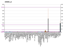
Leucyl/cystinyl aminopeptidase, also known as cystinyl aminopeptidase (CAP), insulin-regulated aminopeptidase (IRAP), human placental leucine aminopeptidase (PLAP), oxytocinase, and vasopressinase, is an enzyme of the aminopeptidase group that in humans is encoded by the LNPEP gene.

<span class="mw-page-title-main">Doripenem</span> Chemical compound

Doripenem is an antibiotic drug in the carbapenem class. It is a beta-lactam antibiotic drug able to kill Pseudomonas aeruginosa.

<span class="mw-page-title-main">Thienamycin</span> Chemical compound

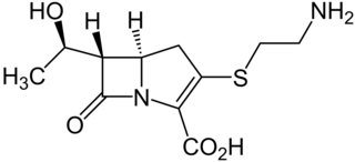
Thienamycin is one of the most potent naturally produced antibiotics known thus far, discovered in Streptomyces cattleya in 1976. Thienamycin has excellent activity against both Gram-positive and Gram-negative bacteria and is resistant to bacterial β-lactamase enzymes. Thienamycin is a zwitterion at pH 7.

<span class="mw-page-title-main">Cephaloridine</span> Chemical compound

Cephaloridine is a first-generation semisynthetic derivative of antibiotic cephalosporin C. It is a Beta lactam antibiotic, like penicillin. Its chemical structure contains 3 cephems, 4 carboxyl groups and three pyridinium methyl groups.

<span class="mw-page-title-main">Gastric lipase</span> Class of enzymes

Gastric lipase, also known as LIPF, is an enzymatic protein that, in humans, is encoded by the LIPF gene.

<span class="mw-page-title-main">PEPD</span> Protein-coding gene in the species Homo sapiens

Xaa-Pro dipeptidase, also known as prolidase, is an enzyme that in humans is encoded by the PEPD gene.

<span class="mw-page-title-main">Carnosinemia</span> Disease

Carnosinemia is a rare autosomal recessive metabolic disorder caused by a deficiency of carnosinase, a dipeptidase.

<span class="mw-page-title-main">Dipeptidase 2</span> Mammalian protein found in Homo sapiens

Dipeptidase 2 (DPEP2) is a protein which in humans is encoded by the DPEP2 gene.

<span class="mw-page-title-main">Dipeptidase 3</span> Protein-coding gene in the species Homo sapiens

Dipeptidase 3 (DPEP3) is a protein that in humans is encoded by the DPEP3 gene.

<span class="mw-page-title-main">Ubenimex</span> Chemical compound

Ubenimex (INN), also known more commonly as bestatin, is a competitive, reversible protease inhibitor. It is an inhibitor of arginyl aminopeptidase (aminopeptidase B), leukotriene A4 hydrolase (a zinc metalloprotease that displays both epoxide hydrolase and aminopeptidase activities), alanyl aminopeptidase (aminopeptidase M/N), leucyl/cystinyl aminopeptidase (oxytocinase/vasopressinase), and membrane dipeptidase (leukotriene D4 hydrolase). It is being studied for use in the treatment of acute myelocytic leukemia and lymphedema. It is derived from Streptomyces olivoreticuli. Ubenimex has been found to inhibit the enzymatic degradation of oxytocin, vasopressin, enkephalins, and various other peptides and compounds.

<span class="mw-page-title-main">Plasmid-mediated resistance</span> Antibiotic resistance caused by a plasmid

Plasmid-mediated resistance is the transfer of antibiotic resistance genes which are carried on plasmids. Plasmids possess mechanisms that ensure their independent replication as well as those that regulate their replication number and guarantee stable inheritance during cell division. By the conjugation process, they can stimulate lateral transfer between bacteria from various genera and kingdoms. Numerous plasmids contain addiction-inducing systems that are typically based on toxin-antitoxin factors and capable of killing daughter cells that don't inherit the plasmid during cell division. Plasmids often carry multiple antibiotic resistance genes, contributing to the spread of multidrug-resistance (MDR). Antibiotic resistance mediated by MDR plasmids severely limits the treatment options for the infections caused by Gram-negative bacteria, especially family Enterobacteriaceae. The global spread of MDR plasmids has been enhanced by selective pressure from antimicrobial medications used in medical facilities and when raising animals for food.

Membrane dipeptidase (EC 3.4.13.19, renal dipeptidase, dehydropeptidase I (DPH I), dipeptidase, aminodipeptidase, dipeptide hydrolase, dipeptidyl hydrolase, nonspecific dipeptidase, glycosyl-phosphatidylinositol-anchored renal dipeptidase, MBD, MDP, leukotriene D4 hydrolase) is an enzyme. This enzyme catalyses the following chemical reaction

<span class="mw-page-title-main">Peptidyl-dipeptidase Dcp</span> Class of enzymes

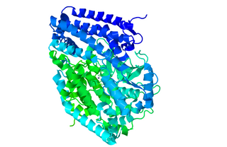
Peptidyl-dipeptidase Dcp (EC 3.4.15.5, dipeptidyl carboxypeptidase (Dcp), dipeptidyl carboxypeptidase) is a metalloenzyme found in the cytoplasm of bacterium E. Coli responsible for the C-terminal cleavage of a variety of dipeptides and unprotected larger peptide chains. The enzyme does not hydrolyze bonds in which P1' is Proline, or both P1 and P1' are Glycine. Dcp consists of 680 amino acid residues that form into a single active monomer which aids in the intracellular degradation of peptides. Dcp coordinates to divalent zinc which sits in the pocket of the active site and is composed of four subsites: S1’, S1, S2, and S3, each subsite attracts certain amino acids at a specific position on the substrate enhancing the selectivity of the enzyme. The four subsites detect and bind different amino acid types on the substrate peptide in the P1 and P2 positions. Some metallic divalent cations such as Ni+2, Cu+2, and Zn+2 inhibit the function of the enzyme around 90%, whereas other cations such as Mn+2, Ca+2, Mg+2, and Co+2 have slight catalyzing properties, and increase the function by around 20%. Basic amino acids such as Arginine bind preferably at the S1 site, the S2 site sits deeper in the enzyme therefore is restricted to bind hydrophobic amino acids with phenylalanine in the P2 position. Dcp is divided into two subdomains (I, and II), which are the two sides of the clam shell-like structure and has a deep inner cavity where a pair of histidine residues bind to the catalytic zinc ion in the active site. Peptidyl-Dipeptidase Dcp is classified like Angiotensin-I converting enzyme (ACE) which is also a carboxypeptidase involved in blood pressure regulation, but due to structural differences and peptidase activity between these two enzymes they had to be examined separately. ACE has endopeptidase activity, whereas Dcp strictly has exopeptidase activity based on its cytoplasmic location and therefore their mechanisms of action are differentiated. Another difference between these enzymes is that the activity of Peptidyl-Dipeptidase Dcp is not enhanced in the presence of chloride anions, whereas chloride enhances ACE activity.

<span class="mw-page-title-main">Natalie Strynadka</span>

Natalie C. J. Strynadka FRS is a professor of Biochemistry in the Department of Biochemistry and Molecular Biology at the University of British Columbia.

<span class="mw-page-title-main">OBPgp279</span>

OBPgp279 is an endolysin that hydrolyzes peptidoglycan, a major constituent in bacterial membrane. OBPgp279 is found in Pseudomonas fluorescens phage OBP, which belongs in the Myoviridae family of bacteriophages. Because of its role in hydrolyzing the peptidoglycan layer, OBPgp279 is a key enzyme in the lytic cycle of the OBP bacteriophage; it allows the bacteriophage to lyse its host internally to escape. Unlike other endolysins, OBPgp279 does not rely on holins to perforate the inner bacterial membrane in order to reach the peptidoglycan layer. Although OBPgp279 is not a well-studied enzyme, it has garnered interest as a potential antibacterial protein due to its activity against multidrug-resistant gram-negative bacteria.

References

  1. 1 2 3 GRCh38: Ensembl release 89: ENSG00000015413 - Ensembl, May 2017
  2. 1 2 3 GRCm38: Ensembl release 89: ENSMUSG00000019278 - Ensembl, May 2017
  3. "Human PubMed Reference:". National Center for Biotechnology Information, U.S. National Library of Medicine.
  4. "Mouse PubMed Reference:". National Center for Biotechnology Information, U.S. National Library of Medicine.
  5. Armstrong, David J., Sunil K. Mukhopadhyay, and Benedict J. Campbell. "Physicochemical characterization of renal dipeptidase." Biochemistry 13.8 (1974): 1745-750. Web.
  6. Keynan, Shoshana, Nicolette T. Habgood, Nigel M. Hooper, and Anthony J. Turner. "Site-Directed Mutagenesis of Conserved Cysteine Residues in Porcine Membrane Dipeptidase. Cys 361 Alone Is Involved in Disulfide-Linked Dimerization†." Biochemistry 35.38 (1996): 12511-2517. Web.
  7. Nitanai, Yasushi, Yoshinori Satow, Hideki Adachi, and Masafumi Tsujimoto. "Crystal Structure of Human Renal Dipeptidase Involved in β-Lactam Hydrolysis." Journal of Molecular Biology 321.2 (2002): 177-84. Web.
  8. Satoh, Susumu, Kazuyuki Ohtsuka, Yuriko Keida, Chihiro Kusunoki, Yoshiyuki Konta, Mineo Niwa, and Masanobu Kohsaka. "Gene structural analysis and expression of human renal dipeptidase." Biotechnology Progress 10.2 (1994): 134-40. Web.
  9. Thoden, James B., Ricardo Marti-Arbona, Frank M. Raushel, and Hazel M. Holden. "High-Resolution X-Ray Structure of Isoaspartyl Dipeptidase from Escherichia coli†,‡." Biochemistry 42.17 (2003): 4874-882. Web.
  10. Armstrong, David J., Sunil K. Mukhopadhyay, and Benedict J. Campbell. "Physicochemical characterization of renal dipeptidase." Biochemistry 13.8 (1974): 1745-750. Web.
  11. Wu, Yong Qian, and Shahriar Mobashery. "Targeting renal dipeptidase (dehydropeptidase I) for inactivation by mechanism-based inactivators." Journal of Medicinal Chemistry 34.6 (1991): 1914-916. Web.
  12. Hayman, Selma, Joselina S. Gatmaitan, and Elizabeth K. Patterson. "Relation of extrinsic and intrinsic metal ions to the specificity of a dipeptidase from Escherichia coli B." Biochemistry 13.22 (1974): 4486-494. Web.
  13. Nitanai, Yasushi, Yoshinori Satow, Hideki Adachi, and Masafumi Tsujimoto. "Crystal Structure of Human Renal Dipeptidase Involved in β-Lactam Hydrolysis." Journal of Molecular Biology 321.2 (2002): 177-84. Web.
  14. "Entrez Gene: DPEP1 dipeptidase 1 (renal)".
  15. Thoden, James B., Ricardo Marti-Arbona, Frank M. Raushel, and Hazel M. Holden. "High-Resolution X-Ray Structure of Isoaspartyl Dipeptidase from Escherichia coli†,‡." Biochemistry 42.17 (2003): 4874-882. Web.
  16. Wu, Yong Qian, and Shahriar Mobashery. "Targeting renal dipeptidase (dehydropeptidase I) for inactivation by mechanism-based inactivators." Journal of Medicinal Chemistry 34.6 (1991): 1914-916. Web.
  17. Rual JF, Venkatesan K, Hao T, Hirozane-Kishikawa T, Dricot A, Li N, Berriz GF, Gibbons FD, Dreze M, Ayivi-Guedehoussou N, Klitgord N, Simon C, Boxem M, Milstein S, Rosenberg J, Goldberg DS, Zhang LV, Wong SL, Franklin G, Li S, Albala JS, Lim J, Fraughton C, Llamosas E, Cevik S, Bex C, Lamesch P, Sikorski RS, Vandenhaute J, Zoghbi HY, Smolyar A, Bosak S, Sequerra R, Doucette-Stamm L, Cusick ME, Hill DE, Roth FP, Vidal M (Oct 2005). "Towards a proteome-scale map of the human protein-protein interaction network". Nature. 437 (7062): 1173–8. Bibcode:2005Natur.437.1173R. doi:10.1038/nature04209. PMID   16189514. S2CID   4427026.
  18. Mciver, C.m, J.m Lloyd, P.j Hewett, and J.e Hardingham. "Dipeptidase 1: a candidate tumor-specific molecular marker in colorectal carcinoma." Cancer Letters 209.1 (2004): 67-74. Web.

Further reading