Pulmonary venoocclusive disease

Last updated
Pulmonary veno-occlusive disease
Other namesObstructive disease of the pulmonary veins [1]
Pulmonary veno-occlusive disease (3627471184).jpg
Pulmonary veno-occlusive disease-Intimal fibrosis with marked narrowing of lumen of a large pulmonary vein
Specialty Pulmonology, cardiology   OOjs UI icon edit-ltr-progressive.svg
Symptoms Shortness of breath, fatigue [2]
CausesNarrow pulmonary vein, Pulmonary artery hypertension [2]
Diagnostic method Chest x-ray, Chest CT [2]
TreatmentVasodilators can be used [2]

Pulmonary veno-occlusive disease (PVOD) is a rare form of pulmonary hypertension caused by progressive blockage of the small veins in the lungs. [2] The blockage leads to high blood pressures in the arteries of the lungs, which, in turn, leads to heart failure. The disease is progressive and fatal, with median survival of about 2 years from the time of diagnosis to death. [3] The definitive therapy is lung transplantation. [4]

Contents

Signs and symptoms

Cyanosis Cynosis.JPG
Cyanosis

The symptoms for pulmonary veno-occlusive disease are the following: [2] [5]

Cause

The genetic cause of pulmonary veno-occlusive disease is mutations in EIF2AK4 gene. Though this does not mean other possible causes do not exist, such as viral infection and risk of toxic chemicals (chemotherapy drugs). [6]

Pathophysiology

Pulmonary veno-occlusive disease may have a genetic basis. Published reports have indicated fatal occurrences that appeared to possess a familial pattern, more to the point, a germline mutation. [7] The pathophysiology of veno-occlusive disease culminates in occlusion of the pulmonary blood vessels. This could be due to edematous tissue (sclerotic fibrous tissue). Thickening is identified in lobular septal veins, also dilatation of lymphatics happens. Furthermore, alveolar capillaries become dilated (due to back-pressure). [8]

Diagnosis

Pulmonary veno-occlusive disease can only be well diagnosed with a lung biopsy. CT scans may show characteristic findings such as ground-glass opacities in centrilobular distribution, and mediastinal lymphadenopathy, but these findings are non-specific and may be seen in other conditions. However, pulmonary hypertension (revealed via physical examination), in the presence of pleural effusion (done via CT scan) usually indicates a diagnosis of pulmonary veno-occlusive disease. The prognosis indicates usually a 2-year (24 month) life expectancy after diagnosis. [9] [10]

Treatment

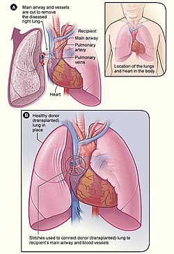
The technique of Lung transplant, the definitive therapy of PVOD. Lung transplant.jpg
The technique of Lung transplant, the definitive therapy of PVOD.

Treatments for primary pulmonary hypertension such as prostacyclins and endothelin receptor antagonists can be fatal in people with PVOD due to the development of severe pulmonary edema, and worsening symptoms after initiation of these medications may be a clue to the diagnosis of pulmonary veno-occlusive disease. [7]

The definitive therapy is lung transplantation, though transplant rejection is always a possibility, in this measures must be taken in terms of appropriate treatment and medication. [11] [12]

Epidemiology

Pulmonary venoocclusive disease is rare, difficult to diagnose, and probably frequently misdiagnosed as idiopathic pulmonary arterial hypertension. Prevalence in parts of Europe is estimated to be 0.1-0.2 cases per million. [13]

PVOD appears to occur as frequently in men as in women, and age at diagnosis ranges from 7–74 years with a median of 39 years. [13] PVOD may occur in patients with associated diseases such as HIV, bone marrow transplantation, and connective tissue diseases. [13] PVOD has also been associated with several chemotherapy regimens such as bleomycin, BCNU, and mitomycin. [13]

Society

Related Research Articles

<span class="mw-page-title-main">Pulmonary hypertension</span> Increased blood pressure in lung arteries

Pulmonary hypertension is a condition of increased blood pressure in the arteries of the lungs. Symptoms include shortness of breath, fainting, tiredness, chest pain, swelling of the legs, and a fast heartbeat. The condition may make it difficult to exercise. Onset is typically gradual. According to the definition at the 6th World Symposium of Pulmonary Hypertension in 2018, a patient is deemed to have pulmonary hypertension if the pulmonary mean arterial pressure is greater than 20mmHg at rest, revised down from a purely arbitrary 25mmHg, and pulmonary vascular resistance (PVR) greater than 3 Wood units.

<span class="mw-page-title-main">Eisenmenger syndrome</span> Medical condition

Eisenmenger syndrome or Eisenmenger's syndrome is defined as the process in which a long-standing left-to-right cardiac shunt caused by a congenital heart defect causes pulmonary hypertension and eventual reversal of the shunt into a cyanotic right-to-left shunt. Because of the advent of fetal screening with echocardiography early in life, the incidence of heart defects progressing to Eisenmenger syndrome has decreased.

<span class="mw-page-title-main">Interstitial lung disease</span> Diseases of the space or tissue between the alveoli of the lungs

Interstitial lung disease (ILD), or diffuse parenchymal lung disease (DPLD), is a group of respiratory diseases affecting the interstitium and space around the alveoli of the lungs. It concerns alveolar epithelium, pulmonary capillary endothelium, basement membrane, and perivascular and perilymphatic tissues. It may occur when an injury to the lungs triggers an abnormal healing response. Ordinarily, the body generates just the right amount of tissue to repair damage, but in interstitial lung disease, the repair process is disrupted, and the tissue around the air sacs (alveoli) becomes scarred and thickened. This makes it more difficult for oxygen to pass into the bloodstream. The disease presents itself with the following symptoms: shortness of breath, nonproductive coughing, fatigue, and weight loss, which tend to develop slowly, over several months. The average rate of survival for someone with this disease is between three and five years. The term ILD is used to distinguish these diseases from obstructive airways diseases.

<span class="mw-page-title-main">Lymphangioleiomyomatosis</span> Progressive cystic lung disease

Lymphangioleiomyomatosis (LAM) is a rare, progressive and systemic disease that typically results in cystic lung destruction. It predominantly affects women, especially during childbearing years. The term sporadic LAM is used for patients with LAM not associated with tuberous sclerosis complex (TSC), while TSC-LAM refers to LAM that is associated with TSC.

<span class="mw-page-title-main">Pulmonary fibrosis</span> Disease that causes scarring of the lungs

Pulmonary fibrosis is a condition in which the lungs become scarred over time. Symptoms include shortness of breath, a dry cough, feeling tired, weight loss, and nail clubbing. Complications may include pulmonary hypertension, respiratory failure, pneumothorax, and lung cancer.

<span class="mw-page-title-main">Respiratory disease</span> Disease of the respiratory system

Respiratory diseases, or lung diseases, are pathological conditions affecting the organs and tissues that make gas exchange difficult in air-breathing animals. They include conditions of the respiratory tract including the trachea, bronchi, bronchioles, alveoli, pleurae, pleural cavity, the nerves and muscles of respiration. Respiratory diseases range from mild and self-limiting, such as the common cold, influenza, and pharyngitis to life-threatening diseases such as bacterial pneumonia, pulmonary embolism, tuberculosis, acute asthma, lung cancer, and severe acute respiratory syndromes, such as COVID-19. Respiratory diseases can be classified in many different ways, including by the organ or tissue involved, by the type and pattern of associated signs and symptoms, or by the cause of the disease.

Portopulmonary hypertension (PPH) is defined by the coexistence of portal and pulmonary hypertension. PPH is a serious complication of liver disease, present in 0.25 to 4% of all patients with cirrhosis. Once an absolute contraindication to liver transplantation, it is no longer, thanks to rapid advances in the treatment of this condition. Today, PPH is comorbid in 4-6% of those referred for a liver transplant.

<span class="mw-page-title-main">Idiopathic pulmonary fibrosis</span> Fibrosis of Lungs due to unknown causes

Idiopathic pulmonary fibrosis (IPF) synonymous with cryptogenic fibrosing alveolitis is a rare, progressive illness of the respiratory system, characterized by the thickening and stiffening of lung tissue, associated with the formation of scar tissue. It is a type of chronic pulmonary fibrosis characterized by a progressive and irreversible decline in lung function.

<span class="mw-page-title-main">Usual interstitial pneumonia</span> Scarring of the lungs

Usual interstitial pneumonia (UIP) is a form of lung disease characterized by progressive scarring of both lungs. The scarring involves the pulmonary interstitium. UIP is thus classified as a form of interstitial lung disease.

Malignant pleural effusion is a condition in which cancer causes an abnormal amount of fluid to collect between the thin layers of tissue (pleura) lining the outside of the lung and the wall of the chest cavity. Lung cancer and breast cancer account for about 50-65% of malignant pleural effusions. Other common causes include pleural mesothelioma and lymphoma.

Hepatic veno-occlusive disease (VOD) or veno-occlusive disease with immunodeficiency is a potentially life-threatening condition in which some of the small veins in the liver are obstructed. It is a complication of high-dose chemotherapy given before a bone marrow transplant or excessive exposure to hepatotoxic pyrrolizidine alkaloids. It is classically marked by weight gain due to fluid retention, increased liver size, and raised levels of bilirubin in the blood. The name sinusoidal obstruction syndrome (SOS) is preferred if hepatic veno-occlusive disease happens as a result of chemotherapy or bone marrow transplantation.

Combined pulmonary fibrosis and emphysema (CPFE), describes a medical syndrome involving both pulmonary fibrosis and emphysema. The combination is most commonly found in male smokers. Pulmonary function tests typically show preserved lung volume with very low transfer factor.

Alveolar capillary dysplasia (ACD) is a rare, congenital diffuse lung disease characterized by abnormal blood vessels in the lungs that cause highly elevated pulmonary blood pressure and an inability to effectively oxygenate and remove carbon dioxide from the blood. ACD typically presents in newborn babies within hours of birth as rapid and labored breathing, blue-colored lips or skin, quickly leading to respiratory failure and death. Atypical forms of ACD have been reported with initially milder symptoms and survival of many months before the onset of respiratory failure or lung transplantation.

<span class="mw-page-title-main">Pulmonary capillary hemangiomatosis</span> Medical condition

Pulmonary capillary hemangiomatosis (PCH) is a disease affecting the blood vessels of the lungs, where abnormal capillary proliferation and venous fibrous intimal thickening result in progressive increase in vascular resistance. It is a rare cause of pulmonary hypertension, and occurs predominantly in young adults. Together with pulmonary veno-occlusive disease, PCH comprises WHO Group I' causes for pulmonary hypertension. Indeed, there is some evidence to suggest that PCH and pulmonary veno-occlusive disease are different forms of a similar disease process.

<span class="mw-page-title-main">Asbestos-related diseases</span> Disorders of the lung and pleura caused by the inhalation of asbestos fibres

Asbestos-related diseases are disorders of the lung and pleura caused by the inhalation of asbestos fibres. Asbestos-related diseases include non-malignant disorders such as asbestosis, diffuse pleural thickening, pleural plaques, pleural effusion, rounded atelectasis and malignancies such as lung cancer and malignant mesothelioma.

<span class="mw-page-title-main">Pulmonary alveolar microlithiasis</span> Medical condition

Pulmonary alveolar microlithiasis (PAM) is a rare, inherited disorder of lung phosphate balance that is associated with small stone formation in the airspaces of the lung. Mutations in the gene SLC34A2 result in loss of a key sodium, phosphate co-transporter, known to be expressed in distal alveolar type II cells, as well as in the mammary gland, and to a lesser extent in intestine, kidney, skin, prostate and testes. As the disease progresses, the lung fields become progressively more dense (white) on the chest xray, and low oxygen level, lung inflammation and fibrosis, elevated pressures in the lung blood vessels, and respiratory failure ensue, usually in middle age. The clinical course of PAM can be highly variable, with some patients remaining asymptomatic for decades, and others progressing more rapidly. There is no effective treatment, and the mechanisms of stone formation, inflammation and scarring are not known.

Chronic thromboembolic pulmonary hypertension (CTEPH) is a long-term disease caused by a blockage in the blood vessels that deliver blood from the heart to the lungs. These blockages cause increased resistance to flow in the pulmonary arterial tree which in turn leads to rise in pressure in these arteries. The blockages either result from organised blood clots that usually originate from the deep veins of the lower limbs of the body (thromboembolism) and lodge in the pulmonary arterial tree after passing through the right side of the heart. The blockages may also result from scar tissue that forms at the site where the clot has damaged the endothelial lining of the pulmonary arteries, causing permanent fibrous obstruction. Most patients have a combination of microvascular and macrovascular obstruction. Some patients may present with normal or near-normal pulmonary pressures at rest despite symptomatic disease. These patients are labelled as having chronic thromboembolic disease (CTED).

<span class="mw-page-title-main">Hepatic hydrothorax</span> Medical condition

Hepatic hydrothorax is a rare form of pleural effusion that occurs in people with liver cirrhosis. It is defined as an effusion of over 500 mL in people with liver cirrhosis that is not caused by heart, lung, or pleural disease. It is found in 5–10% of people with liver cirrhosis and 2–3% of people with pleural effusions. In cases of decompensated liver cirrhosis, prevalence rises significantly up to 90%. Over 85% of cases occurring on the right, 13% on the left, and 2% on both. Although it is most common in people with severe ascites, it can also occur in people with mild or no ascites. Symptoms are not specific and mostly involve the respiratory system.

<span class="mw-page-title-main">Pulmonary arterial hypertension</span> Elevated pressure in the pulmonary circulation limited to the pre-capillary blood vessels

Pulmonary Arterial Hypertension (PAH) is a syndrome in which the blood pressure in the pulmonary arteries and pulmonary arterioles is elevated. This pre-capillary pulmonary artery pressure being elevated is essential, and by definition a mean pulmonary artery pressure greater than 20 mmHg as measured by a right heart catheterization is required for the diagnosis. This pre-capillary pulmonary hypertension is confirmed with measuring pulmonary vascular resistance being greater than 3 Woods Units. A pulmonary artery wedge pressure being less than 15 mmHg excludes post-capillary bed pulmonary hypertension. Pulmonary arterial hypertension is a subgroup of pulmonary hypertension and is categorized as World Health Organization as group 1. PAH is further subdivided into various categories based on the cause, including idiopathic, heritable, drug and toxin induced, PAH associated with specific diseases, PAH that is responsive to vasodilators, PAH with venous or capillary involvement, and persistent PAH in the newborn period.

References

  1. "Pulmonary venoocclusive disease | Genetic and Rare Diseases Information Center (GARD) – an NCATS Program". rarediseases.info.nih.gov. Retrieved 31 July 2019.
  2. 1 2 3 4 5 6 "Pulmonary veno-occlusive disease: MedlinePlus Medical Encyclopedia". www.nlm.nih.gov. Retrieved 2015-11-07.
  3. Escribano (2012). "Survival in pulmonary hypertension in Spain: insights from the Spanish registry". Eur Respir J. 40 (3): 596–603. doi: 10.1183/09031936.00101211 . PMID   22362843.
  4. Ye (2011). "Lengthy Diagnostic Challenge in a Rare Case of Pulmonary Veno-Occlusive Disease: Case Report and Review of the Literature". Intern Med. 50 (12): 1323–7. doi: 10.2169/internalmedicine.50.5035 . PMID   21673470.
  5. Zander, Dani S.; Farver, Carol F. (2008-01-01). Pulmonary Pathology. Elsevier Health Sciences. p. 121. ISBN   978-0-443-06741-9.
  6. "Pulmonary veno-occlusive disease". Genetics Home Reference. 2015-11-02. Retrieved 2015-11-07.
  7. 1 2 Montani, David (2010). "Pulmonary veno-occlusive disease: Recent progress and current challenges". Respiratory Medicine. 104: S23 –S32. doi: 10.1016/j.rmed.2010.03.014 . PMID   20456932.
  8. Ali, Hakim Azfar (2024-05-28). "Pulmonary Veno-Occlusive Disease: Practice Essentials, Pathophysiology, Etiology". Medscape Reference. Retrieved 2024-08-09.
  9. Light, Richard W. (2007-01-01). Pleural Diseases. Lippincott Williams & Wilkins. p. 128. ISBN   9780781769570.
  10. Grosse, Claudia; Grosse, Alexandra (2010-11-01). "CT Findings in Diseases Associated with Pulmonary Hypertension: A Current Review". RadioGraphics. 30 (7): 1753–1777. doi:10.1148/rg.307105710. ISSN   0271-5333. PMID   21057119.
  11. Ali, Hakim Azfar (2024-05-28). "Pulmonary Veno-Occlusive Disease Treatment & Management: Approach Considerations, Specific PAH Therapies, Immunosuppressants, Steroids, and Antithrombotic Agents". Medscape Reference. Retrieved 2024-08-09.
  12. "Lung Transplantation: MedlinePlus". www.nlm.nih.gov. Retrieved 2015-11-07.
  13. 1 2 3 4 Montani (2009). "Pulmonary veno-occlusive disease". Eur Respir J. 33 (1): 189–200. doi: 10.1183/09031936.00090608 . PMID   19118230.
  14. Phull, Hardeep (July 26, 2016). "She's tethered to an oxygen tank, but her singing career is soaring". New York Post.
  15. Insdorf, Annette (November 10, 2013). "The Challenges of Chloe Temtchine". The Huffington Post.

Further reading