Coomassie Brilliant Blue

Last updated
Coomassie Brilliant Blue R-250
Coomassie Brilliant Blue R-250.svg
Coomassie Brilliant Blue zwitterion 3D spacefill.png
Names
Other names
C.I. 42660, C.I. Acid Blue 83
Brilliant indocyanine 6B, Brillantindocyanin 6B
Brilliant Cyanine 6B, Serva Blue R
Identifiers
  • 6104-59-2 Yes check.svgY
3D model (JSmol)
ECHA InfoCard 100.025.509 OOjs UI icon edit-ltr-progressive.svg
PubChem CID
  • CCN(CC1=CC(=CC=C1)S(=O)(=O)O)C2=CC=C(C=C2)C(=C3C=CC(=[N+](CC)CC4=CC(=CC=C4)S(=O)(=O)[O-])C=C3)C5=CC=C(C=C5)NC6=CC=C(C=C6)OCC.[Na+]
Properties
C45H44N3NaO7S2 (Sodium salt)
Molar mass 825.97 g/mol
Insoluble in cold, slightly soluble in hot (bright red blue)
Solubility in ethanolSlightly soluble
Except where otherwise noted, data are given for materials in their standard state (at 25 °C [77 °F], 100 kPa).
Yes check.svgY  verify  (what is  Yes check.svgYX mark.svgN ?)
Infobox references
Coomassie Brilliant Blue G-250
Coomassie Brilliant Blue G-250.svg
Sample of Coomassie Brilliant Blue G.jpg
Solid Coomassie Brilliant Blue G
Coomassie Brilliant Blue G in Isopropanol.jpg
Brilliant Blue G in Isopropanol solution
Names
Other names
C.I. 42655, C.I. Acid Blue 90
Brilliant indocyanine G, Brillantindocyanin G
Xylene Brilliant Cyanine G, Serva Blue G
Identifiers
  • 6104-58-1
3D model (JSmol)
ECHA InfoCard 100.025.509 OOjs UI icon edit-ltr-progressive.svg
KEGG
PubChem CID
  • CCN(CC1=CC(=CC=C1)S(=O)(=O)O)C2=CC(=C(C=C2)/C(=C\3/C=CC(=[N+](CC)CC4=CC(=CC=C4)S(=O)(=O)O)C=C3C)/C5=CC=C(C=C5)NC6=CC=C(C=C6)OCC)C
Properties
C47H50N3NaO7S2 (Sodium salt)
Molar mass 856.03 g/mol
Slightly soluble in cold, soluble in hot (bright blue)
Solubility in ethanolSoluble
Except where otherwise noted, data are given for materials in their standard state (at 25 °C [77 °F], 100 kPa).
Infobox references

Coomassie Brilliant Blue is the name of two similar triphenylmethane dyes that were developed for use in the textile industry but are now commonly used for staining proteins in analytical biochemistry. Coomassie Brilliant Blue G-250 differs from Coomassie Brilliant Blue R-250 by the addition of two methyl groups. The name "Coomassie" is a registered trademark of Imperial Chemical Industries.

Contents

Name and discovery

The name Coomassie was adopted at the end of the 19th century as a trade name by the Blackley-based dye manufacturer Levinstein Ltd, in marketing a range of acid wool dyes. [1] In 1896 during the Fourth Anglo-Ashanti War, British forces had occupied the town of Coomassie (modern-day Kumasi in Ghana). In 1918 Levinstein Ltd became part of British Dyestuffs which in 1926 became part of Imperial Chemical Industries. [2] Although ICI still owns the Coomassie trademark, the company no longer manufactures the dyes.

The blue disulfonated triphenylmethane dyes were first produced in 1913 by Max Weiler who was based in Elberfeld, Germany. [3] Various patents were subsequently taken out on the organic synthesis. [4] [5] [6]

Papers published in biochemistry journals frequently refer to these dyes simply as "Coomassie" without specifying which dye was actually used. In fact the Colour Index lists over 40 dyes with "Coomassie" in their name. There are also other Coomassie "blue" dyes. For example, the Merck Index (10th edition) lists Coomassie Blue RL (Acid Blue 92, C.I. 13390) which has a completely different structure.

Dye colour

The suffix "R" in the name of Coomassie Brilliant Blue R-250 is an abbreviation for Red as the blue colour of the dye has a slight reddish tint. For the "G" variant the blue colour has a more greenish tint. The "250" originally denoted the purity of the dye.

The colour of the two dyes depends on the acidity of the solution. The "G" form of the dye has been studied in detail. [7] At a pH of less than 0 the dye has a red colour with an absorption maximum at a wavelength of 465 nm. At a pH of around 1 the dye is green with an absorption maximum at 620 nm while above pH 2 the dye is bright blue with a maximum at 595 nm. At pH 7 the dye has an extinction coefficient of 43,000 M−1 cm−1. [7]

The different colours are a result of the different charged states of the dye molecule. In the red form, all three nitrogen atoms carry a positive charge. The two sulfonic acid groups have extremely low pKa and will normally be negatively charged, thus at a pH of around zero the dye will be a cation with an overall charge of +1. The green colour corresponds to a form of the dye with no net overall charge. In neutral media (pH 7), only the nitrogen atom of the diphenylamine moiety carries a positive charge and the blue dye molecule is an anion with an overall charge of −1. The pKa for the loss of the two protons are 1.15 and 1.82 respectively. The final proton is lost under alkaline conditions and the dye becomes pink in colour (pKa 12.4). [7]

The dye interacts electrostatically but noncovalently with the amino and carboxyl groups of proteins. The dye molecules bind to proteins including wool (keratin) to form a protein–dye complex. The formation of the complex stabilises the negatively charged anionic form of the dye producing the blue colour, even under acid conditions when most of the molecules in solution are in the cationic form. [7] This is the basis of the Bradford assay which is a protein determination method that involves the Coomassie Brilliant Blue dye binding to proteins. The binding of the dye to a protein causes a shift in the absorbance maximum of the dye from 465 to 595 nm. The increase of absorption at 595 nm is monitored to determine protein concentration. [8]

The dye also forms a complex with the anionic detergent sodium dodecylsulfate (SDS). [9] The formation of this complex stabilizes the neutral green form of the dye. This effect can interfere with the estimation of protein concentration using the Bradford assay. It is also likely that the anionic detergent competes with the dye for binding to the protein.

Applications in biochemistry

Coomassie Brilliant Blue R-250 was first used to visualise proteins in 1964 by Fazekas de St. Groth and colleagues. Protein samples were separated electrophoretically on a cellulose acetate sheet. The sheet was then soaked in sulfosalicylic acid to fix the protein bands and then transferred to a solution of the dye. [10]

Two years later in 1965 Meyer and Lambert used Coomassie Brilliant Blue R-250 to stain protein samples after electrophoretic separation in a polyacrylamide gel. They soaked the gel in a dye solution containing methanol, acetic acid and water. As the dye stained the polyacrylamide gel as well as the protein, in order to visualise the protein bands they needed to destain the gel which they did electrophoretically. [11] Subsequent publications reported that polyacrylamide gels could be successfully destained using an acetic acid solution.

The first report of the use of the "G" form of the dye to visualise protein bands in polyacrylamide gels came in 1967, where the dye was dissolved in an acetic acid solution containing methanol. [12] It was subsequently discovered that the protein bands could be stained without staining the polyacrylamide by using a colloid of the "G" form of the dye in a trichloroacetic acid solution containing no methanol. Using this procedure it was no longer necessary to destain the gel. [13] Modern formulations typically use a colloid of the "G" form of dye in a solution containing phosphoric acid, ethanol (or methanol) and ammonium sulfate (or aluminium sulfate). [14] [15] [16] [17]

The Bradford assay uses the spectral properties of Coomassie Brilliant Blue G-250 to estimate the amount of protein in a solution. [18] A protein sample is added to a solution of the dye in phosphoric acid and ethanol. Under the acid conditions the dye is normally a brownish colour but on binding to the protein the blue form of the dye is produced. The optical absorbance of the solution is measured at a wavelength of 595 nm. The dye is notable due to its high level of sensitivity, 5 μg of protein is enough to distinguish the difference. However, among the disadvantages of the method is its variability of color development with different proteins: the absorbance change per unit mass of proteins varies with the type of the protein. [19]

On binding to a protein the negatively charged Coomassie Brilliant Blue G-250 dye molecule will give an overall negative charge to the protein. This property can be used to separate proteins or protein complexes using polyacrylamide gel electrophoresis under non-denaturing conditions in a technique called Blue Native PAGE. [20] [21] The mobility of the complex in the polyacrylamide gel will depend on both the size of the protein complex (i.e. the molecular weight) and on the amount of dye bound to the protein.

Coomassie Blue staining can also be used as a loading control staining method in western blot analysis. [22] It is applied as an anionic pre-antibody stain.

Medical uses

In 2009, Brilliant Blue G was used in scientific experiments to treat spinal injuries in laboratory rats. [23] It acts by reducing the body's natural swelling response, which can cause neurons in the area to die of metabolic stress. Testing on the rats proved effective. Two groups of injured rats were tested, with one group given the dye as a treatment for the spinal injuries and the other group was not. The results of the test proved that in comparison to the rats that had not received the dye, the rats that were treated with the dye were able to move around better and able to out perform the rats without the dye treatment on motion tests. [24] Testing is still in progress to determine whether this treatment can be used effectively in humans. The recent tests have administered the dye within 15 minutes of injury, but to be effective in a real-life setting, where it may take time for a patient to reach the emergency room, the treatment needs to be effective even when administered up to two hours after injury. The only reported side effect was that the rats temporarily turned blue. [23] [25] [26]

Under the trade names ILM Blue and Brilliant Peel, Brilliant Blue G is used as a stain to assist surgeons in retinal surgery. [27]

In December 2019, Brilliant Blue G (under the trade name TissueBlue, DORC International, Netherlands) was approved for use in the United States. [28] [29] [30]

Application in forensic science

Through a study done at the University of Albany, it was shown that the ability of the Coomassie dye to target amino acids with aromatic groups (phenylalanine, tyrosine, tryptophan) and basic side chains (lysine, arginine and histidine), allows Bradford assay to be used for fingerprint analysis. Bradford assay was successfully used to identify the biological sex of the fingerprint. Female samples were shown to have a higher absorbance compared to male samples when tested at similar wavelengths. This provides a simpler method for fingerprint analysis by reducing the number of amino acids needed to be analyzed from 23 to 6, and having little to no assay preparation, in comparison to the ninhydrin chemical assay which requires assay preparation such as heating and enzyme cascade. [31]

Related Research Articles

Agarose gel electrophoresis

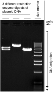
Agarose gel electrophoresis is a method of gel electrophoresis used in biochemistry, molecular biology, genetics, and clinical chemistry to separate a mixed population of macromolecules such as DNA or proteins in a matrix of agarose, one of the two main components of agar. The proteins may be separated by charge and/or size, and the DNA and RNA fragments by length. Biomolecules are separated by applying an electric field to move the charged molecules through an agarose matrix, and the biomolecules are separated by size in the agarose gel matrix.

Gel electrophoresis Method for separation and analysis of macromolecules

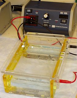
Gel electrophoresis is a method for separation and analysis of macromolecules and their fragments, based on their size and charge. It is used in clinical chemistry to separate proteins by charge or size and in biochemistry and molecular biology to separate a mixed population of DNA and RNA fragments by length, to estimate the size of DNA and RNA fragments or to separate proteins by charge.

Southern blot DNA analysis technique

A Southern blot is a method used in molecular biology for detection of a specific DNA sequence in DNA samples. Southern blotting combines transfer of electrophoresis-separated DNA fragments to a filter membrane and subsequent fragment detection by probe hybridization.

Polyacrylamide gel electrophoresis

Polyacrylamide gel electrophoresis (PAGE) is a technique widely used in biochemistry, forensic chemistry, genetics, molecular biology and biotechnology to separate biological macromolecules, usually proteins or nucleic acids, according to their electrophoretic mobility. Electrophoretic mobility is a function of the length, conformation and charge of the molecule. Polyacrylamide gel electrophoresis is a powerful tool used to analyze RNA samples. When polyacrylamide gel is denatured after electrophoresis, it provides information on the sample composition of the RNA species.

Western blot Analytical technique used in molecular biology

The western blot, or western blotting, is a widely used analytical technique in molecular biology and immunogenetics to detect specific proteins in a sample of tissue homogenate or extract.

Two-dimensional gel electrophoresis

Two-dimensional gel electrophoresis, abbreviated as 2-DE or 2-D electrophoresis, is a form of gel electrophoresis commonly used to analyze proteins. Mixtures of proteins are separated by two properties in two dimensions on 2D gels. 2-DE was first independently introduced by O'Farrell and Klose in 1975.

Protein purification is a series of processes intended to isolate one or a few proteins from a complex mixture, usually cells, tissues or whole organisms. Protein purification is vital for the specification of the function, structure and interactions of the protein of interest. The purification process may separate the protein and non-protein parts of the mixture, and finally separate the desired protein from all other proteins. Separation of one protein from all others is typically the most laborious aspect of protein purification. Separation steps usually exploit differences in protein size, physico-chemical properties, binding affinity and biological activity. The pure result may be termed protein isolate.

Gel electrophoresis of proteins

Protein electrophoresis is a method for analysing the proteins in a fluid or an extract. The electrophoresis may be performed with a small volume of sample in a number of alternative ways with or without a supporting medium: SDS polyacrylamide gel electrophoresis, free-flow electrophoresis, electrofocusing, isotachophoresis, affinity electrophoresis, immunoelectrophoresis, counterelectrophoresis, and capillary electrophoresis. Each method has many variations with individual advantages and limitations. Gel electrophoresis is often performed in combination with electroblotting immunoblotting to give additional information about a specific protein. Because of practical limitations, protein electrophoresis is generally not suited as a preparative method.

The Bradford protein assay was developed by Marion M. Bradford in 1976. It is a quick and accurate spectroscopic analytical procedure used to measure the concentration of protein in a solution. The reaction is dependent on the amino acid composition of the measured proteins.

In pathology, silver staining is the use of silver to selectively alter the appearance of a target in microscopy of histological sections; in temperature gradient gel electrophoresis; and in polyacrylamide gels.

Protein methods are the techniques used to study proteins. There are experimental methods for studying proteins. Computational methods typically use computer programs to analyze proteins. However, many experimental methods require computational analysis of the raw data.

Electrophoretic mobility shift assay

An electrophoretic mobility shift assay (EMSA) or mobility shift electrophoresis, also referred as a gel shift assay, gel mobility shift assay, band shift assay, or gel retardation assay, is a common affinity electrophoresis technique used to study protein–DNA or protein–RNA interactions. This procedure can determine if a protein or mixture of proteins is capable of binding to a given DNA or RNA sequence, and can sometimes indicate if more than one protein molecule is involved in the binding complex. Gel shift assays are often performed in vitro concurrently with DNase footprinting, primer extension, and promoter-probe experiments when studying transcription initiation, DNA gang replication, DNA repair or RNA processing and maturation, as well as pre-mRNA splicing. Although precursors can be found in earlier literature, most current assays are based on methods described by Garner and Revzin and Fried and Crothers.

QPNC-PAGE, or quantitative preparative native continuous polyacrylamide gel electrophoresis, is a bioanalytical, high-resolution and highly accurate technique applied in biochemistry and bioinorganic chemistry to separate proteins quantitatively by isoelectric point. This standardized variant of native gel electrophoresis is used by biologists to isolate biomacromolecules in solution, for example, active or native metalloproteins in biological samples or properly and improperly folded metal cofactor-containing proteins or protein isoforms in complex protein mixtures.

Electroblotting

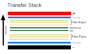
Electroblotting is a method in molecular biology/biochemistry/immunogenetics to transfer proteins or nucleic acids onto a membrane by using PVDF or nitrocellulose, after gel electrophoresis. The protein or nucleic acid can then be further analyzed using probes such as specific antibodies, ligands like lectins, or stains. This method can be used with all polyacrylamide and agarose gels. An alternative technique for transferring proteins from a gel is capillary blotting.

Affinity electrophoresis

Affinity electrophoresis is a general name for many analytical methods used in biochemistry and biotechnology. Both qualitative and quantitative information may be obtained through affinity electrophoresis. The methods include the so-called electrophoretic mobility shift assay, charge shift electrophoresis and affinity capillary electrophoresis. The methods are based on changes in the electrophoretic pattern of molecules through biospecific interaction or complex formation. The interaction or binding of a molecule, charged or uncharged, will normally change the electrophoretic properties of a molecule. Membrane proteins may be identified by a shift in mobility induced by a charged detergent. Nucleic acids or nucleic acid fragments may be characterized by their affinity to other molecules. The methods have been used for estimation of binding constants, as for instance in lectin affinity electrophoresis or characterization of molecules with specific features like glycan content or ligand binding. For enzymes and other ligand-binding proteins, one-dimensional electrophoresis similar to counter electrophoresis or to "rocket immunoelectrophoresis", affinity electrophoresis may be used as an alternative quantification of the protein. Some of the methods are similar to affinity chromatography by use of immobilized ligands.

Marion Mckinley Bradford was an American scientist who developed and patented the Bradford protein assay, a method to quickly quantify the amount of protein in a sample. His paper describing the method is among the most cited scholarly articles of all time.

Normalization of Western blot data is an analytical step that is performed to compare the relative abundance of a specific protein across the lanes of a blot or gel under diverse experimental treatments, or across tissues or developmental stages. The overall goal of normalization is to minimize effects arising from variations in experimental errors, such as inconsistent sample preparation, unequal sample loading across gel lanes, or uneven protein transfer, which can compromise the conclusions that can be obtained from Western blot data. Currently, there are two methods for normalizing Western blot data: (i) housekeeping protein normalization and (ii) total protein normalization.

SDS-PAGE biochemical technique

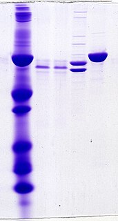
SDS-PAGE, is a discontinuous electrophoretic system developed by Ulrich K. Laemmli which is commonly used as a method to separate proteins with molecular masses between 5 and 250 kDa. The combined use of sodium dodecyl sulfate and polyacrylamide gel allows to eliminate the influence of structure and charge, and proteins are separated solely on the basis of differences in their molecular weight.

Stains-all Dye

Stains-all is a carbocyanine dye, which stains anionic proteins, nucleic acids, anionic polysaccharides and other anionic molecules.

SYBR Gold Chemical compound

SYBR Gold is an asymmetrical cyanine dye. It can be used as a stain for double-stranded DNA, single-stranded DNA, and RNA. SYBR Gold is the most sensitive fluorescent stain of the SYBR family of dyes for the detection of nucleic acids. The SYBR family of dyes is produced by Molecular Probes Inc., now owned by Thermo Fisher Scientific

References

  1. Fox, M. R. (1987). Dye-makers of Great Britain 1856-1976 : A History of Chemists, Companies, Products and Changes. Manchester: Imperial Chemical Industries. p. 38.
  2. Fox, M. R. (1987). Dye-makers of Great Britain 1856-1976 : A History of Chemists, Companies, Products and Changes. Manchester: Imperial Chemical Industries. p. 259.
  3. Colour Index (PDF). 4 (3rd ed.). Bradford: Society of Dyers and Colourists. 1971. pp. 4397–4398. Archived from the original (PDF) on 2011-07-19. Retrieved 2010-07-20.
  4. FRpatent 474260,"Procédé de production de colorants de la série du triarylméthane",issued 1915-02-16, assigned to Bayer
  5. USpatent 1218232,Weiler, Max,"Blue Triphenylmethane Dye",issued 1917-03-06
  6. GBpatent 275609,"Manufacture of Triarylmethane-dyestuffs",issued 1927-11-03, assigned to IG Farbenindustrie
  7. 1 2 3 4 Chial, H. J.; Thompson, H. B.; Splittgerber, A. G. (1993). "A spectral study of the charge forms of Coomassie Blue G". Analytical Biochemistry. 209 (2): 258–266. doi:10.1006/abio.1993.1117. PMID   7682385.
  8. Bradford, Marion M. (1976). "A rapid and sensitive method for the quantitation of microgram quantities of protein utilizing the principle of protein-dye binding" (PDF). Analytical Biochemistry. 72 (1–2): 248–254. doi:10.1016/0003-2697(76)90527-3. PMID   942051.
  9. Compton, S. J.; Jones, C. G. (1985). "Mechanism of dye response and interference in the Bradford protein assay". Analytical Biochemistry. 151 (2): 369–374. doi:10.1016/0003-2697(85)90190-3. PMID   4096375.
  10. Fazekas de St. Groth, S.; Webster, R. G.; Datyner, A. (1963). "Two new staining procedures for quantitative estimation of proteins on electrophoretic strips". Biochimica et Biophysica Acta. 71: 377–391. doi:10.1016/0006-3002(63)91092-8. PMID   18421828.
  11. Meyer, T. S.; Lambert, B. L. (1965). "Use of Coomassie brilliant blue R250 for the electrophoresis of microgram quantities of parotid saliva proteins on acrylamide-gel strips". Biochimica et Biophysica Acta (BBA) - General Subjects. 107 (1): 144–145. doi:10.1016/0304-4165(65)90403-4. PMID   4159310.
  12. Altschul, A. M.; Evans, W. J. (1967). Zone electrophoresis with polyacrylamide gel. Methods in Enzymology. 11. pp. 179–186. doi:10.1016/S0076-6879(67)11019-7. ISBN   9780121818609.. Page 184 personal communication from W. J. Saphonov.
  13. Diezel, W.; Kopperschläger, G.; Hofmann, E. (1972). "An improved procedure for protein staining in polyacrylamide gels with a new type of Coomassie Brilliant Blue". Analytical Biochemistry. 48 (2): 617–620. doi:10.1016/0003-2697(72)90117-0. PMID   4115985.
  14. Neuhoff, V.; Stamm, R.; Eibl, H. (1985). "Clear background and highly sensitive protein staining with Coomassie Blue dyes in polyacrylamide gels: a systematic analysis". Electrophoresis. 6 (9): 427–448. doi:10.1002/elps.1150060905. S2CID   94797941.
  15. Candiano, G.; Bruschi, M.; Musante, L.; Santucci, L.; Ghiggeri, G. M.; Carnemolla, B.; Orecchia, P.; Zardi, L.; Righetti, P. G. (2004). "Blue silver: a very sensitive colloidal Coomassie G-250 staining for proteome analysis". Electrophoresis. 25 (9): 1327–1333. doi:10.1002/elps.200305844. PMID   15174055. S2CID   25960150.
  16. Steinberg, T. H. (2009). Protein gel staining methods: an introduction and overview. Methods in Enzymology. 463. pp. 541–563. doi:10.1016/S0076-6879(09)63031-7. ISBN   9780123745361. PMID   19892191.
  17. Pink, M.; Verma, N.; Rettenmeier, A. W.; Schmitz-Spanke, S. (2010). "CBB staining protocol with higher sensitivity and mass spectrometric compatibility". Electrophoresis. 31 (4): 593–598. doi:10.1002/elps.200900481. PMID   20162584. S2CID   39038371.
  18. Bradford, M. M. (1976). "Rapid and sensitive method for the quantitation of microgram quantities of protein utilizing the principle of protein-dye binding". Analytical Biochemistry. 72 (1–2): 248–254. doi:10.1016/0003-2697(76)90527-3. PMID   942051.
  19. Congdon, Robert W.; Muth, Gregory W.; Splittgerber, Allan G. (September 1993). "The binding interaction of Coomassie blue with proteins". Analytical Biochemistry. 213 (2): 407–413. doi:10.1006/abio.1993.1439. PMID   7694523.
  20. Schägger, H.; Jagow, G. (1991). "Blue native electrophoresis for isolation of membrane protein complexes in enzymatically active form". Analytical Biochemistry. 199 (2): 223–231. doi:10.1016/0003-2697(91)90094-A. PMID   1812789.
  21. Wittig, I.; Braun, H. P.; Schägger, H. (2006). "Blue native PAGE". Nature Protocols. 1 (1): 418–428. doi:10.1038/nprot.2006.62. PMID   17406264. S2CID   19715017.
  22. Welinder, Charlotte; Ekblad, Lars (2011). "Coomassie Staining as Loading Control in Western Blot Analysis". Journal of Proteome Research. 10 (3): 1416–1419. doi:10.1021/pr1011476. PMID   21186791.
  23. 1 2 Peng, W.; Cotrina, M. L.; Han, X.; Yu, H.; Bekar, L.; Blum, L.; Takano, T.; Tian, G. F.; et al. (2009). "Systemic administration of an antagonist of the ATP-sensitive receptor P2X7 improves recovery after spinal cord injury". Proceedings of the National Academy of Sciences of the United States of America. 106 (30): 12489–12493. doi: 10.1073/pnas.0902531106 . PMC   2718350 . PMID   19666625.
  24. Ehrenberg, Rachel (23 September 2013). "Brilliant blue for the spine". Science News. Society for Science & the Public. Retrieved 9 December 2017.
  25. "Blue M&Ms 'mend spinal injuries'". Telegraph. 2009-07-28. Retrieved 2010-01-19.
  26. "Blue Food Dye Treats Spine Injury in Rats". Wired.com. 2009-07-27. Retrieved 2010-01-19.
  27. Mennel, S.; Meyer, C. H.; Schmidt, J. C.; Kaempf, S.; Thumann, G. (2008). Trityl dyes patent blue V and brilliant blue G - clinical relevance and in vitro analysis of the function of the outer blood-retinal barrier. Developments in Ophthalmology. 42. pp. 101–114. doi:10.1159/000138988. ISBN   978-3-8055-8551-4. PMID   18535384.
  28. "Drug Trials Snapshots: TissueBlue". U.S. Food and Drug Administration (FDA). 20 December 2019. Retrieved 17 March 2020.
  29. "TissueBlue- brilliant blue g injection, solution". DailyMed. 29 December 2019. Retrieved 17 March 2020.
  30. "Drug Approval Package: TissueBlue". U.S. Food and Drug Administration (FDA). 10 January 2020. Retrieved 17 March 2020.
  31. Brunelle, Erica; Le, Anh Minh; Huynh, Crystal; Wingfield, Kelly; Halámková, Lenka; Agudelo, Juliana; Halámek, Jan (2017-04-04). "Coomassie Brilliant Blue G-250 Dye: An Application for Forensic Fingerprint Analysis". Analytical Chemistry. 89 (7): 4314–4319. doi:10.1021/acs.analchem.7b00510. ISSN   0003-2700. PMID   28293949.

Further reading