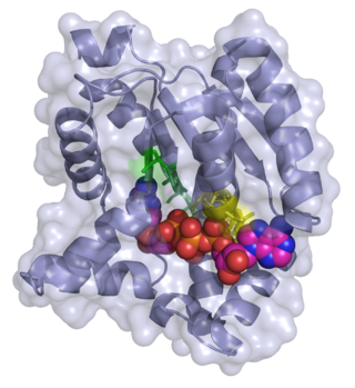
Kanamycin kinase

Last updated
kanamycin kinase
APHStructure.png
Structure of APH(3'), taken from 1L8T [1]
Identifiers
EC no. 2.7.1.95
CAS no. 62213-36-9
Databases
IntEnz IntEnz view
BRENDA BRENDA entry
ExPASy NiceZyme view
KEGG KEGG entry
MetaCyc metabolic pathway
PRIAM profile
PDB structures RCSB PDB PDBe PDBsum
Gene Ontology AmiGO / QuickGO
Search
PMC articles
PubMed articles
NCBI proteins

Aminoglycoside-3'-phosphotransferase (APH(3')), also known as aminoglycoside kinase, is an enzyme that primarily catalyzes the addition of phosphate from ATP to the 3'-hydroxyl group of a 4,6-disubstituted aminoglycoside, such as kanamycin. [2] However, APH(3') has also been found to phosphorylate at the 5'-hydroxyl group in 4,5-disubstituted aminoglycosides, which lack a 3'-hydroxyl group, and to diphosphorylate hydroxyl groups in aminoglycosides that have both 3'- and 5'-hydroxyl groups. [2] [3] Primarily positively charged at biological conditions, aminoglycosides bind to the negatively charged backbone of nucleic acids to disrupt protein synthesis, effectively inhibiting bacterial cell growth. [4] APH(3') mediated phosphorylation of aminoglycosides effectively disrupts their mechanism of action, introducing a phosphate group that reduces their binding affinity due to steric hindrances and unfavorable electrostatic interactions. [5] APH(3') is primarily found in certain species of gram-positive bacteria. [6] [7] [8]

Contents

APH(3') catalyzes the phosphorylation of kanamycin A, a 4,6-disubstituted aminoglycoside, at the 3'-hydroxyl group. APHRxnScheme.png
APH(3') catalyzes the phosphorylation of kanamycin A, a 4,6-disubstituted aminoglycoside, at the 3'-hydroxyl group.

This enzyme belongs to the family of transferases, specifically those transferring phosphorus-containing groups (phosphotransferases) with an alcohol group as acceptor. The systematic name of this enzyme class is ATP:kanamycin 3'-O-phosphotransferase. This enzyme is also called neomycin-kanamycin phosphotransferase. [9]

Structure

APH(3') thermodynamically favors a dimer form of two identical APH(3') monomers that are connected by two disulfide bonds between Cys19 and Cys156, with the active sites facing each other. [2] [10] However, the large distance between the two monomers' active sites suggests that they are independent of each other, and do not operate in a cooperative fashion. Additionally, dimerization of APH(3') does not affect the activity of the enzyme. [2] [10] [11]

Interactions of negatively charged residues and kanamycin A in APH(3') binding pocket. APHNegativeBindinPocket.png
Interactions of negatively charged residues and kanamycin A in APH(3') binding pocket.

Each monomer consists of two lobes, the beta-sheet rich N-terminus and alpha-helix rich C-terminus, with a twelve amino acid region connecting the two. The N-terminal lobe is composed of 5 antiparallel ß-sheets, with an α-helix between sheets 3 and 4. The C-terminal lobe is divided into a central core region (two α-helices and a hairpin-loop followed by four ß-sheets), an insert region (two α-helices connected by a loop structure), and a C-terminal region (two α-helices). [11] The resulting pocket that is encapsulated by the two lobes make up the enzyme active site. [2] This pocket is largely composed of negatively charged amino acid residues, which stabilize the positive charge of and orient the substrate in the active site. Additionally, this pocket is thought to contribute to the promiscuity of the enzyme, allowing it to take in and stabilize several different kinds of aminoglycosides. [6]

Mechanism

ADP and Kanamycin A in the active site of APH(3'). Two magnesium ions are coordinated by Asn195 and Asp208 residues, which in turn facilitate the binding of ATP in the active site. The NPL, in conjunction with magnesium ions, mediate the phosphorylation of aminoglycosides. APHActiveSite.png
ADP and Kanamycin A in the active site of APH(3'). Two magnesium ions are coordinated by Asn195 and Asp208 residues, which in turn facilitate the binding of ATP in the active site. The NPL, in conjunction with magnesium ions, mediate the phosphorylation of aminoglycosides.

While earlier studies of APH(3') supported a mechanism involving the nucleophilic attack of γ-phosphate by the 3'-hydroxyl, more recent studies suggest that APH(3') catalyzes the transfer of the γ-phosphate from ATP to an aminoglycoside through a dissociative mechanism, where deprotonation of the substrate is not critical to phosphate transfer, but instead the stabilization of a metaphosphate transition state. [8] [12] Additionally, APH(3') has a nucleotide positioning loop (NPL) that closes down on the enzyme active site after binding ATP, facilitating the phosphorylation of the 3'-hydroxyl group. Key to correctly positioning the phosphate group are Ser27 and Met26 residues. Initially, two magnesium ions stabilized by Asn195 and Asp208 facilitate the binding of ATP in the active site and orient the ß- and γ-phosphate groups. The NPL then undergoes a conformational change to form a hydrogen bond between Ser27 and the ß-phosphate group. Upon binding of substrate, APH(3') undergoes another conformational change to orient Ser27 such that its amide backbone disrupts the alignment of ß-phosphate and γ-phosphate, weakening the γ-phosphate bond. The amide backbone of Met26 forms a hydrogen bond with the metaphosphate to stabilize the transition state, as a magnesium ion (designated Mg1) then lengthens the γ-phosphate bond, breaking it and effectively phosphorylating the hydroxyl group. [8]

Reaction mechanism of APH(3'). Magnesium ions coordinate ATP into place, and the addition of the substrate induces a conformational change that engages the amide backbone of Ser27 in hydrogen bonding with the ss-phosphate, disrupting the g-PO bond and facilitating the phosphorylation of a 4,6-disubstituted aminoglycoside. APHReactionMechanism.png
Reaction mechanism of APH(3'). Magnesium ions coordinate ATP into place, and the addition of the substrate induces a conformational change that engages the amide backbone of Ser27 in hydrogen bonding with the ß-phosphate, disrupting the γ-PO bond and facilitating the phosphorylation of a 4,6-disubstituted aminoglycoside.

Evolution and biological function

The central core region of APH(3') has a high degree of conformational similarity to regions of serine/tyrosine and threonine protein kinases, functionally equivalent enzymes found in eukaryotes. Additionally, X-ray crystallography and mutagenesis of key active site residues supports claims that APH(3') and eukaryotic protein kinases are related, despite sharing less than 10% of total residue content. [2] [8] [11] Several studies have suggested that serine/tyrosine/threonine protein kinases, once thought to only occur in eukaryotes, are also found in the prokaryotes. [13] [14] Additionally, it has been found that aminoglycoside biosynthesis requires phosphorylation of hydroxyl groups during certain steps of synthesis. Thus, it has been speculated that APH(3') and other protein kinases originate from enzymes from the metabolic pathway for aminoglycosides, and developed in order to counteract the toxic effects of these antibiotics in the host bacterial cell. [11] [15]

Use in research

Aminoglycoside resistance genes are commonly used in the realm of genetic engineering in order to select for correctly transformed bacterial organisms. When constructing a vector plasmid, including antibiotic resistance in the vector is crucial to effectively expressing the gene of interest. Antibiotics, such as the aminoglycosides kanamycin or neomycin, are added to the cultures during growth phases in order to selectively destroy the cells that did not effectively take up the plasmid.

Related Research Articles

<span class="mw-page-title-main">Kinase</span> Enzyme catalyzing transfer of phosphate groups onto specific substrates

In biochemistry, a kinase is an enzyme that catalyzes the transfer of phosphate groups from high-energy, phosphate-donating molecules to specific substrates. This process is known as phosphorylation, where the high-energy ATP molecule donates a phosphate group to the substrate molecule. This transesterification produces a phosphorylated substrate and ADP. Conversely, it is referred to as dephosphorylation when the phosphorylated substrate donates a phosphate group and ADP gains a phosphate group. These two processes, phosphorylation and dephosphorylation, occur four times during glycolysis.

<span class="mw-page-title-main">Neomycin</span> Type of antibiotic

Neomycin is an aminoglycoside antibiotic that displays bactericidal activity against gram-negative aerobic bacilli and some anaerobic bacilli where resistance has not yet arisen. It is generally not effective against gram-positive bacilli and anaerobic gram-negative bacilli. Neomycin comes in oral and topical formulations, including creams, ointments, and eyedrops. Neomycin belongs to the aminoglycoside class of antibiotics that contain two or more amino sugars connected by glycosidic bonds.

<span class="mw-page-title-main">Phosphofructokinase 1</span> Class of enzymes

Phosphofructokinase-1 (PFK-1) is one of the most important regulatory enzymes of glycolysis. It is an allosteric enzyme made of 4 subunits and controlled by many activators and inhibitors. PFK-1 catalyzes the important "committed" step of glycolysis, the conversion of fructose 6-phosphate and ATP to fructose 1,6-bisphosphate and ADP. Glycolysis is the foundation for respiration, both anaerobic and aerobic. Because phosphofructokinase (PFK) catalyzes the ATP-dependent phosphorylation to convert fructose-6-phosphate into fructose 1,6-bisphosphate and ADP, it is one of the key regulatory steps of glycolysis. PFK is able to regulate glycolysis through allosteric inhibition, and in this way, the cell can increase or decrease the rate of glycolysis in response to the cell's energy requirements. For example, a high ratio of ATP to ADP will inhibit PFK and glycolysis. The key difference between the regulation of PFK in eukaryotes and prokaryotes is that in eukaryotes PFK is activated by fructose 2,6-bisphosphate. The purpose of fructose 2,6-bisphosphate is to supersede ATP inhibition, thus allowing eukaryotes to have greater sensitivity to regulation by hormones like glucagon and insulin.

<span class="mw-page-title-main">Phosphatase</span> Enzyme which catalyzes the removal of a phosphate group from a molecule

In biochemistry, a phosphatase is an enzyme that uses water to cleave a phosphoric acid monoester into a phosphate ion and an alcohol. Because a phosphatase enzyme catalyzes the hydrolysis of its substrate, it is a subcategory of hydrolases. Phosphatase enzymes are essential to many biological functions, because phosphorylation and dephosphorylation serve diverse roles in cellular regulation and signaling. Whereas phosphatases remove phosphate groups from molecules, kinases catalyze the transfer of phosphate groups to molecules from ATP. Together, kinases and phosphatases direct a form of post-translational modification that is essential to the cell's regulatory network.

<span class="mw-page-title-main">Aminoglycoside</span> Antibacterial drug

Aminoglycoside is a medicinal and bacteriologic category of traditional Gram-negative antibacterial medications that inhibit protein synthesis and contain as a portion of the molecule an amino-modified glycoside (sugar). The term can also refer more generally to any organic molecule that contains amino sugar substructures. Aminoglycoside antibiotics display bactericidal activity against Gram-negative aerobes and some anaerobic bacilli where resistance has not yet arisen but generally not against Gram-positive and anaerobic Gram-negative bacteria.

<span class="mw-page-title-main">Adenylate kinase</span> Class of enzymes

Adenylate kinase is a phosphotransferase enzyme that catalyzes the interconversion of the various adenosine phosphates. By constantly monitoring phosphate nucleotide levels inside the cell, ADK plays an important role in cellular energy homeostasis.

<span class="mw-page-title-main">Kanamycin A</span> Antibiotic

Kanamycin A, often referred to simply as kanamycin, is an antibiotic used to treat severe bacterial infections and tuberculosis. It is not a first line treatment. It is used by mouth, injection into a vein, or injection into a muscle. Kanamycin is recommended for short-term use only, usually from 7 to 10 days. As with most antibiotics, it is ineffective in viral infections.

Pantothenate kinase (EC 2.7.1.33, PanK; CoaA) is the first enzyme in the Coenzyme A (CoA) biosynthetic pathway. It phosphorylates pantothenate (vitamin B5) to form 4'-phosphopantothenate at the expense of a molecule of adenosine triphosphate (ATP). It is the rate-limiting step in the biosynthesis of CoA.

<span class="mw-page-title-main">Diphosphomevalonate decarboxylase</span> InterPro Family

Diphosphomevalonate decarboxylase (EC 4.1.1.33), most commonly referred to in scientific literature as mevalonate diphosphate decarboxylase, is an enzyme that catalyzes the chemical reaction

In enzymology, an aminoglycoside N6'-acetyltransferase (EC 2.3.1.82) is an enzyme that catalyzes the chemical reaction

Choline kinase is an enzyme which catalyzes the first reaction in the choline pathway for phosphatidylcholine (PC) biosynthesis. This reaction involves the transfer of a phosphate group from adenosine triphosphate (ATP) to choline in order to form phosphocholine.

In enzymology, a dephospho-[reductase kinase] kinase is an enzyme that catalyzes the chemical reaction

In enzymology, a low-density-lipoprotein receptor kinase is an enzyme that catalyzes the chemical reaction

In enzymology, a nucleoside-phosphate kinase is an enzyme that catalyzes the chemical reaction

<span class="mw-page-title-main">Phosphoribulokinase</span>

Phosphoribulokinase (PRK) (EC 2.7.1.19) is an essential photosynthetic enzyme that catalyzes the ATP-dependent phosphorylation of ribulose 5-phosphate (RuP) into ribulose 1,5-bisphosphate (RuBP), both intermediates in the Calvin Cycle. Its main function is to regenerate RuBP, which is the initial substrate and CO2-acceptor molecule of the Calvin Cycle. PRK belongs to the family of transferase enzymes, specifically those transferring phosphorus-containing groups (phosphotransferases) to an alcohol group acceptor. Along with ribulose 1,5-bisphosphate carboxylase/oxygenase (RuBisCo), phosphoribulokinase is unique to the Calvin Cycle. Therefore, PRK activity often determines the metabolic rate in organisms for which carbon fixation is key to survival. Much initial work on PRK was done with spinach leaf extracts in the 1950s; subsequent studies of PRK in other photosynthetic prokaryotic and eukaryotic organisms have followed. The possibility that PRK might exist was first recognized by Weissbach et al. in 1954; for example, the group noted that carbon dioxide fixation in crude spinach extracts was enhanced by the addition of ATP. The first purification of PRK was conducted by Hurwitz and colleagues in 1956.

ATP + Mg2+ - D-ribulose 5-phosphate  ADP + D-ribulose 1,5-bisphosphate

In enzymology, a polo kinase is a kinase enzyme i.e. one that catalyzes the chemical reaction

In enzymology, a protein-histidine tele-kinase is an enzyme that catalyzes the chemical reaction

In enzymology, an [RNA-polymerase]-subunit kinase is an enzyme that catalyzes the chemical reaction

The Walker A and Walker B motifs are protein sequence motifs, known to have highly conserved three-dimensional structures. These were first reported in ATP-binding proteins by Walker and co-workers in 1982.

<span class="mw-page-title-main">Kanamycin nucleotidyltransferase</span>

In molecular biology, kanamycin nucleotidyltransferase EC 2.7.7.- (KNTase) is an enzyme which is involved in conferring resistance to aminoglycoside antibiotics. It catalyses the transfer of a nucleoside monophosphate group from a nucleotide to kanamycin. This enzyme is dimeric with each subunit being composed of two domains. The C-terminal domain contains five alpha helices, four of which are organised into an up-and-down alpha helical bundle. Residues found in this domain may contribute to this enzyme's active site.

References

  1. Fong DH, Berghuis AM (2002). "Crystal Structure Of 3',5"-Aminoglycoside Phosphotransferase Type IIIa ADP Kanamycin A Complex". World-wide Protein Data Bank. doi:10.2210/pdb1l8t/pdb.
  2. 1 2 3 4 5 6 7 Wright GD, Thompson PR (1999). "Aminoglycoside phosphotransferases: proteins, structure, and mechanism". Front Biosci. 4 (1–3): D9–21. doi: 10.2741/wright . PMID   9872733.
  3. Thompson PR, Hughes DW, Wright GD (1996). "Regiospecificity of aminoglycoside phosphotransferase from Enterococci and Staphylococci (APH(3')-IIIa)". Biochemistry. 35 (26): 8686–95. doi:10.1021/bi960389w. PMID   8679631.
  4. Cavallo G, Martinetto P (1981). "The mechanism of action of aminoglycosides". G Batteriol Virol Immunol. 74 (7–12): 335–46. PMID   6182050.
  5. Kotra LP, Haddad J, Mobashery S (2000). "Aminoglycosides: Perspectives on Mechanisms of Action and Resistance and Strategies to Counter Resistance". Antimicrobial Agents and Chemotherapy. 44 (12): 3249–56. doi:10.1128/aac.44.12.3249-3256.2000. PMC   90188 . PMID   11083623.
  6. 1 2 Fong DH, Berghuis AM (2002). "Substrate promiscuity of an aminoglycoside antibiotic resistance enzyme via target mimicry". The EMBO Journal. 21 (10): 2323–31. doi:10.1093/emboj/21.10.2323. PMC   126009 . PMID   12006485.
  7. Gray GS, Fitch WM (1983). "Evolution of antibiotic resistance genes: the DNA sequence of a kanamycin resistance gene from Staphylococcus aureus". Mol Biol Evol. 1 (1): 57–66. doi: 10.1093/oxfordjournals.molbev.a040298 . PMID   6100986.
  8. 1 2 3 4 5 Thompson PR, Boehr DD, Berghuis AM, Wright GD (2002). "Mechanism of Aminoglycoside Antibiotic Kinase APH(3')-IIIa: Role of the Nucleotide Positioning Loop". Biochemistry. 41 (22): 7001–7. doi:10.1021/bi0256680. PMID   12033933.
  9. McKay GA, Wright GD (1996). "Catalytic mechanism of enterococcal kanamycin kinase (APH(3')-IIIa): viscosity, thio, and solvent isotope effects support a Theorell-Chance mechanism". Biochemistry. 35 (26): 8680–5. doi:10.1021/bi9603884. PMID   8679630.
  10. 1 2 McKay GA, Thompson PR, Wright GD (1994). "Broad spectrum aminoglycoside phosphotransferase type III from Enterococcus: overexpression, purification, and substrate specificity". Biochemistry. 33 (22): 6936–44. doi:10.1021/bi00188a024. PMID   8204627.
  11. 1 2 3 4 Hon WC, McKay GA, Thompson PR, Sweet RM, Yang DS, Wright GD, Berhuis AM (1997). "Structure of an Enzyme Required for Aminoglycoside Antibiotic Resistance Reveals Homology to Eukaryotic Protein Kinases". Cell. 89 (6): 887–95. doi: 10.1016/s0092-8674(00)80274-3 . PMID   9200607. S2CID   13251696.
  12. Boehr DD, Thompson PR, Wright GD (2001). "Molecular mechanism of aminoglycoside antibiotic kinase APH(3')-IIIa: roles of conserved active site residues". J Biol Chem. 276 (26): 23929–36. doi: 10.1074/jbc.m100540200 . PMID   11279088.
  13. Kennelly PJ (1996). "Fancy meeting you here! A fresh look at "prokaryotic" protein phosphorylation". J Bacteriol. 178 (16): 4759–64. doi:10.1128/jb.178.16.4759-4764.1996. PMC   178254 . PMID   8759835.
  14. Zhang CC (1996). "Bacterial signalling involving eukaryotic-type protein kinases". Mol Microbiol. 20 (1): 9–15. doi:10.1111/j.1365-2958.1996.tb02483.x. PMID   8861199. S2CID   33493179.
  15. Pierpersberg, W; Distler, J; Heinzel, P; Perez-Gonzalaez, JA (1988). "Antibiotic resistance by modification: Many resistance genes could be derived from cellular control genes in actinomycetes - a hypothesis". Actinomycetologica. 2 (2): 83–98. doi: 10.3209/saj.2_83 .

Further reading