Rheumatoid arthritis (RA) is a long-term autoimmune disorder that primarily affects joints. It typically results in warm, swollen, and painful joints. Pain and stiffness often worsen following rest. Most commonly, the wrist and hands are involved, with the same joints typically involved on both sides of the body. The disease may also affect other parts of the body, including skin, eyes, lungs, heart, nerves and blood. This may result in a low red blood cell count, inflammation around the lungs, and inflammation around the heart. Fever and low energy may also be present. Often, symptoms come on gradually over weeks to months.

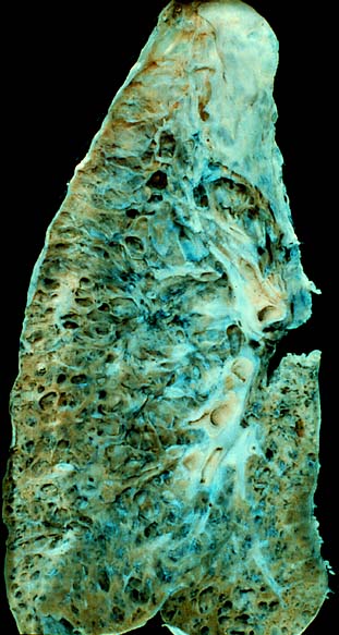
Pneumoconiosis is the general term for a class of interstitial lung disease where inhalation of dust has caused interstitial fibrosis. The three most common types are asbestosis, silicosis, and coal miner's lung. Pneumoconiosis often causes restrictive impairment, although diagnosable pneumoconiosis can occur without measurable impairment of lung function. Depending on extent and severity, it may cause death within months or years, or it may never produce symptoms. It is usually an occupational lung disease, typically from years of dust exposure during work in mining; textile milling; shipbuilding, ship repairing, and/or shipbreaking; sandblasting; industrial tasks; rock drilling ; or agriculture. It is one of the most common occupational diseases in the world.

Silicosis is a form of occupational lung disease caused by inhalation of crystalline silica dust. It is marked by inflammation and scarring in the form of nodular lesions in the upper lobes of the lungs. It is a type of pneumoconiosis. Silicosis is characterized by shortness of breath, cough, fever, and cyanosis. It may often be misdiagnosed as pulmonary edema, pneumonia, or tuberculosis. Using workplace controls, silicosis is almost always a preventable disease.

Interstitial lung disease (ILD), or diffuse parenchymal lung disease (DPLD), is a group of respiratory diseases affecting the interstitium (the tissue and space around the alveoli of the lungs. It concerns alveolar epithelium, pulmonary capillary endothelium, basement membrane, and perivascular and perilymphatic tissues. It may occur when an injury to the lungs triggers an abnormal healing response. Ordinarily, the body generates just the right amount of tissue to repair damage, but in interstitial lung disease, the repair process is disrupted, and the tissue around the air sacs becomes scarred and thickened. This makes it more difficult for oxygen to pass into the bloodstream. The disease presents itself with the following symptoms: shortness of breath, nonproductive coughing, fatigue, and weight loss, which tend to develop slowly, over several months. The average rate of survival for someone with this disease is between three and five years. The term ILD is used to distinguish these diseases from obstructive airways diseases.

A chest radiograph, called a chest X-ray (CXR), or chest film, is a projection radiograph of the chest used to diagnose conditions affecting the chest, its contents, and nearby structures. Chest radiographs are the most common film taken in medicine.
Siderosis is the deposition of excess iron in body tissue. When used without qualification, it usually refers to an environmental disease of the lung, also known more specifically as pulmonary siderosis or Welder's disease, which is a form of pneumoconiosis.

Cryptogenic organizing pneumonia (COP), formerly known as bronchiolitis obliterans organizing pneumonia (BOOP), is an inflammation of the bronchioles (bronchiolitis) and surrounding tissue in the lungs. It is a form of idiopathic interstitial pneumonia.

Black lung disease (BLD), also known as coal workers' pneumoconiosis, or simply black lung, is an occupational type of pneumoconiosis caused by long-term inhalation and deposition of coal dust in the lungs and the consequent lung tissue's reaction to its presence. It is common in coal miners and others who work with coal. It is similar to both silicosis from inhaling silica dust and asbestosis from inhaling asbestos dust. Inhaled coal dust progressively builds up in the lungs and leads to inflammation, fibrosis, and in worse cases, necrosis.
Progressive massive fibrosis (PMF), characterized by the development of large conglomerate masses of dense fibrosis, can complicate silicosis and coal worker's pneumoconiosis. Conglomerate masses may also occur in other pneumoconioses, such as talcosis, berylliosis (CBD), kaolin pneumoconiosis, and pneumoconiosis from carbon compounds, such as carbon black, graphite, and oil shale. Conglomerate masses can also develop in sarcoidosis, but usually near the hilae and with surrounding paracicatricial emphysema.
Occupational lung diseases are work-related, lung conditions that have been caused or made worse by the materials a person is exposed to within the workplace. It includes a broad group of diseases, including occupational asthma, industrial bronchitis, chronic obstructive pulmonary disease (COPD), bronchiolitis obliterans, inhalation injury, interstitial lung diseases, infections, lung cancer and mesothelioma. These diseases can be caused directly or due to immunological response to an exposure to a variety of dusts, chemicals, proteins or organisms.
Baritosis is a benign type of pneumoconiosis, which is caused by long-term exposure to barium dust.

Usual interstitial pneumonia (UIP) is a form of lung disease characterized by progressive scarring of both lungs. The scarring (fibrosis) involves the pulmonary interstitium. UIP is thus classified as a form of interstitial lung disease.

High-resolution computed tomography (HRCT) is a type of computed tomography (CT) with specific techniques to enhance image resolution. It is used in the diagnosis of various health problems, though most commonly for lung disease, by assessing the lung parenchyma. On the other hand, HRCT of the temporal bone is used to diagnose various middle ear diseases such as otitis media, cholesteatoma, and evaluations after ear operations.
Restrictive lung diseases are a category of extrapulmonary, pleural, or parenchymal respiratory diseases that restrict lung expansion, resulting in a decreased lung volume, an increased work of breathing, and inadequate ventilation and/or oxygenation. Pulmonary function test demonstrates a decrease in the forced vital capacity.

A rheumatoid nodule is a lump of tissue, or an area of swelling, that appears on the exterior of the skin usually around the olecranon or the interphalangeal joints, but can appear in other areas. There are four different types of rheumatoid nodules: subcutaneous rheumatoid nodules, cardiac nodules, pulmonary nodules, and central nervous systems nodules. These nodules occur almost exclusively in association with rheumatoid arthritis. Very rarely do rheumatoid nodules occur as rheumatoid nodulosis in the absence of rheumatoid arthritis. Rheumatoid nodules can also appear in areas of the body other than the skin. Less commonly they occur in the lining of the lungs or other internal organs. The occurrence of nodules in the lungs of miners exposed to silica dust was known as Caplan’s syndrome. Rarely, the nodules occur at diverse sites on body.
The ILO International Classification of Radiographs of Pneumoconioses is a system of classifying chest radiographs (X-rays) for persons with a form of pneumoconiosis. The intent is to provide a standardized, uniform method of interpreting and describing abnormalities in chest x-rays that are thought to be caused by prolonged dust inhalation. In use, it provides a system for both epidemiological comparisons of many individuals exposed to dust and evaluation of an individual's potential disease relative to established standards.
Rheumatoid lung disease is a disease of the lung associated with RA, rheumatoid arthritis. Rheumatoid lung disease is characterized by pleural effusion, pulmonary fibrosis, lung nodules and pulmonary hypertension. Common symptoms associated with the disease include shortness of breath, cough, chest pain and fever. It is estimated that about one quarter of people with rheumatoid arthritis develop this disease, which are more likely to develop among elderly men with a history of smoking.
Mineral dust airway disease is a general term used to describe complications due to inhaled mineral dust causing fibrosis and narrowing of primarily the respiratory bronchioles. It is a part of a group of disorders known as pneumoconioses which is characterized by inhaled mineral dust and the effects on the lungs.

Philip Hugh-Jones FRCP was a British respiratory physician and Medical Research Council (MRC) researcher who during the Second World War investigated the effects of gun fumes on tank operators in Dorset and the effect of coal dust on Welsh coal miners with particular relevance to pneumoconiosis. This work led to future post-war pioneering research in lung physiology, the effect of asbestos on the lungs and lung diseases including emphysema.

A focal lung pneumatosis, is an enclosed pocket of air or gas in the lung and includes blebs, bullae, pulmonary cysts, and lung cavities. Blebs and bullae can be classified by their wall thickness.


