Viroporin

Last updated
The transmembrane helical tetramer of the influenza A virus M2 protein, which functions as a proton channel, in complex with the channel-blocking drug amantadine (shown in red). Highly conserved tryptophan and histidine residues known to play key roles in mediating proton transport are shown as sticks. From PDB: 3C9J . M2 influenza A proton channel amantadine 3C9J.png
The transmembrane helical tetramer of the influenza A virus M2 protein, which functions as a proton channel, in complex with the channel-blocking drug amantadine (shown in red). Highly conserved tryptophan and histidine residues known to play key roles in mediating proton transport are shown as sticks. From PDB: 3C9J .

Viroporins are small and usually hydrophobic multifunctional viral proteins that modify cellular membranes, thereby facilitating virus release from infected cells. [2] [3] Viroporins are capable of assembling into oligomeric ion channels or pores in the host cell's membrane, rendering it more permeable and thus facilitating the exit of virions from the cell. Many viroporins also have additional effects on cellular metabolism and homeostasis mediated by protein-protein interactions with host cell proteins. [3] Viroporins are not necessarily essential for viral replication, but do enhance growth rates. They are found in a variety of viral genomes but are particularly common in RNA viruses. Many viruses that cause human disease express viroporins. These viruses include hepatitis C virus, HIV-1, influenza A virus, poliovirus, respiratory syncytial virus, and SARS-CoV. [3] [4] [5]

Contents

Structure

Viroporins are usually small - under 100 or 120 amino acid residues - and contain at least one region capable of folding into an amphipathic transmembrane helix. Some examples also contain stretches of basic amino acids, or stretches of aromatic amino acids thought to reside in the interfacial region of the membrane. [3] Oligomers of these proteins, most often tetramers, [6] form ion channels or pores of usually weak ion selectivity that permit diffusion of ions across the cell membrane. The molecular architecture of the pore, its degree of selectivity, the extent to which it incorporates lipids from the surrounding membrane, and the presence of portions of the protein that extend beyond the membrane all vary among viroporins and indicate that these proteins have a diverse array of functional roles. [4] [5]

Classification

A proposed classification scheme sorts viroporins into four classes based on their topology and orientation in the membrane. Class I viroporins possess a single transmembrane helix; in class IA the C-terminus is oriented into the cytosol and in class IB the N-terminus is so oriented. Class II viroporins possess a helix-turn-helix motif with both helices crossing the membrane; in class IIA both termini are oriented externally (extracellularly or toward the lumen of the endoplasmic reticulum) and in class IIB the termini are oriented toward the cytosol. [5] Likely exceptions to this scheme exist, such as the rotavirus protein non-structural protein 4. [7] [8]

Function

Essentiality

Most viroporins are not essential, but their absence significantly reduces the efficiency of viral propagation. There is significant variation in the consequences of viroporin depletion: while hepatitis C virus is incapable of propagation without its p7 protein viroporin, influenza A virus and HIV-1 see decreases in in vitro viral titer of 10- to 100-fold in the absence of their respective viroporins, but remain capable of propagation. [4] [9] In most cases absence of viroporin in the viral genome can be rescued by the presence of viroporin in trans, and sometimes viral replication can be partially rescued in the presence of another virus' viroporin. [5]

Membrane permeabilization

The most well-studied and well-established function of viroporins is the permeabilization of the cell membrane to ions and small solutes. [10] Before viroporins themselves were understood as a class, it was well known that many viruses induce membrane permeabilization in infected cells; viroporins are at least partially responsible for this effect, particularly when it occurs late in the viral replication cycle. [2] [3] [11] Viroporins expressed transgenically, in the absence of their virus of origin, induce the same effect, a feature that has facilitated viroporin discovery. [5] [12]

Solid-state NMR-based model of the pentameric pore formed by the transmembrane helices of the SARS-CoV-2 E protein, which forms a viroporin permeable to cations. Rendered from PDB: 7K3G . 7k3g.png
Solid-state NMR-based model of the pentameric pore formed by the transmembrane helices of the SARS-CoV-2 E protein, which forms a viroporin permeable to cations. Rendered from PDB: 7K3G .

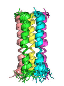
In most cases, pores formed by viroporins are nonselective or only weakly selective for particular ions or small molecules. [9] However, some examples do show strong selectivity; examples include the influenza A virus M2 proton channel protein, which is highly selective for protons and is active at low pH, and the Chlorella virus Kcv protein, which is selective for potassium ions. An alternative mechanism is illustrated by the SARS-CoV E protein, which forms a pore that integrates membrane lipids whose polar head groups influence ion selectivity. [4] The homologous E protein of SARS-CoV-2 has been structurally characterized by solid-state NMR and found to form a pentamer permeable to cations. [13] [14]

Loss of membrane polarization can promote viral yields through a variety of mechanisms that operate throughout the viral life cycle. In enveloped viruses, viroporins are not highly concentrated in the viral envelope, but nevertheless their presence may promote viral entry into the cell; the influenza A virus provides a well-studied example. Viroporins in the membranes of organelles such as the Golgi apparatus can influence those organelles' internal environments, which can modulate protein trafficking of viral proteins or protect the proteins from the low pH they would otherwise encounter in these cellular compartments. In non-enveloped viruses, the membrane permeability changes may be sufficient to induce cell lysis, thereby permitting the new virions to exit the cell. In enveloped viruses, viroporins' depolarization effect is thought to promote viral budding. [4] [5] Abrogating the ion channel or pore function of viroporins, either through mutations that block conductance without disrupting other functions or through channel-blocking drugs, usually reduces or eliminates viral propagation. [4]

Genome replication

Most viruses encoding viroporins can replicate their genomes in the absence of the viroporin, even if they are impaired in propagation. Rotaviruses and picornaviruses, however, rely on their viroporins to facilitate the formation of viroplasm, or specialized intracellular compartments remodeled from the membrane of the endoplasmic reticulum in which genome replication occurs. [5]

Protein-protein interactions

Some viroporins have established functional effects exerted through protein-protein interactions. For example, the HIV-1 viroporin Vpu promotes viral budding through interactions with CD4 and tetherin, though the precise molecular mechanism of this interaction is not known. [6] [7] [9] The JC polyomavirus agnoprotein functions as a viroporin in addition to other roles mediated through interactions with viral proteins such as major capsid protein VP1. [15]

Role in disease

Virulence factors

Viroporins can also be considered virulence factors; in viruses in which viroporins are not essential, their pathogenicity is attenuated in the absence of viroporin beyond the level expected by the effects on viral propagation. In some cases the membrane permeabilization effects of viroporins activate the inflammasome, a protein complex associated with activation of innate immunity which, when overactive, can cause disease symptoms. [4]

Oncoproteins

The human papillomavirus 16 E5 protein, the least well-studied of the three known oncogenic HPV proteins, was reported in 2012 to be a viroporin. [16] This was the first known example of an oncogenic viroporin. [7]

Drug targets

Because some viroporins are essential for viral propagation, they are often considered to be appealing drug targets for development of antiviral drugs. [3] [9] Although many chemical compounds have been reported to interfere with the ion channel functions of various viroporins, clinical usage is relatively rare. Amantadine, which was discovered in the 1960s and has been in clinical use against influenza A for some time, is an example of a viroporin-targeting drug; [4] [17] [18] however, a 2014 Cochrane review did not find benefit for its use in children or elderly people [19] and the US CDC does not recommend drugs of this class due to widespread resistance mutations. [20]

Examples

Viroporins can be found in a large number of viruses with distinct genomic organizations and replication mechanisms.

Known viroporins [3] [5] [6]
FamilyVirusTypeViroporin protein
Coronaviridae SARS coronavirus (+)ssRNA E protein, 3A protein
Coronaviridae Murine hepatitis virus (+)ssRNA E protein
Flaviviridae Hepatitis C virus (+)ssRNA p7 protein
Orthomyxoviridae Influenza A virus (-)ssRNA M2 protein
Orthomyxoviridae Influenza B virus (-)ssRNA NB protein, BM2 protein
Orthomyxoviridae Influenza C virus (-)ssRNA CM2 protein
Papillomaviridae Human papillomavirus 16 dsDNA E protein
Phycodnaviridae Paramecium bursaria chlorella virus 1 dsDNA Kcv protein
Phycodnaviridae Acanthocystis turfacea chlorella virus 1 dsDNA Kcv protein
Picornaviridae Coxsackievirus (+)ssRNA Protein 2B
Picornaviridae Enterovirus 71 (+)ssRNA Protein 2B
Picornaviridae Poliovirus (+)ssRNA Protein 2B, Protein 3A
Pneumoviridae Human respiratory syncytial virus (-)ssRNA Small hydrophobic (SH) protein
Polyomaviridae JC polyomavirus dsDNA Agnoprotein
Polyomaviridae SV40 dsDNA Viral protein 4
Reoviridae Avian reovirus dsRNA p10 protein
Retroviridae Human immunodeficiency virus 1 ssRNA-RT Vpu
Rhabdoviridae Bovine ephemeral fever virus (-)ssRNA Alpha 10p protein
Togaviridae Semliki Forest virus (+)ssRNA Protein 6K
Togaviridae Sindbis virus (+)ssRNA Protein 6K
Togaviridae Ross River virus (+)ssRNA Protein 6K

This table represents a composite of Table 1 from Gonzalez et al. 2003, [3] Table 1 from Wang et al. 2011, [6] and Table 1, Box 1, and Box 2 from Nieva et al. 2012. [5]

See also

Related Research Articles

<span class="mw-page-title-main">Ion channel</span> Pore-forming membrane protein

Ion channels are pore-forming membrane proteins that allow ions to pass through the channel pore. Their functions include establishing a resting membrane potential, shaping action potentials and other electrical signals by gating the flow of ions across the cell membrane, controlling the flow of ions across secretory and epithelial cells, and regulating cell volume. Ion channels are present in the membranes of all cells. Ion channels are one of the two classes of ionophoric proteins, the other being ion transporters.

<span class="mw-page-title-main">Antiviral drug</span> Medication used to treat a viral infection

Antiviral drugs are a class of medication used for treating viral infections. Most antivirals target specific viruses, while a broad-spectrum antiviral is effective against a wide range of viruses. Antiviral drugs are one class of antimicrobials, a larger group which also includes antibiotic, antifungal and antiparasitic drugs, or antiviral drugs based on monoclonal antibodies. Most antivirals are considered relatively harmless to the host, and therefore can be used to treat infections. They should be distinguished from virucides, which are not medication but deactivate or destroy virus particles, either inside or outside the body. Natural virucides are produced by some plants such as eucalyptus and Australian tea trees.

<span class="mw-page-title-main">Defective interfering particle</span>

Defective interfering particles (DIPs), also known as defective interfering viruses, are spontaneously generated virus mutants in which a critical portion of the particle's genome has been lost due to defective replication or non-homologous recombination. The mechanism of their formation is presumed to be as a result of template-switching during replication of the viral genome, although non-replicative mechanisms involving direct ligation of genomic RNA fragments have also been proposed. DIPs are derived from and associated with their parent virus, and particles are classed as DIPs if they are rendered non-infectious due to at least one essential gene of the virus being lost or severely damaged as a result of the defection. A DIP can usually still penetrate host cells, but requires another fully functional virus particle to co-infect a cell with it, in order to provide the lost factors.

<span class="mw-page-title-main">Rimantadine</span> Drug used to treat influenzavirus A infection

Rimantadine is an orally administered antiviral drug used to treat, and in rare cases prevent, influenzavirus A infection. When taken within one to two days of developing symptoms, rimantadine can shorten the duration and moderate the severity of influenza. Rimantadine can mitigate symptoms, including fever. Both rimantadine and the similar drug amantadine are derivates of adamantane. Rimantadine is found to be more effective than amantadine because when used the patient displays fewer symptoms. Rimantadine was approved by the Food and Drug Administration (FDA) in 1994.

<span class="mw-page-title-main">Amantadine</span> Medication used to treat dyskinesia

Amantadine, sold under the brand name Gocovri among others, is a medication used to treat dyskinesia associated with parkinsonism and influenza caused by type A influenzavirus, though its use for the latter is no longer recommended due to widespread drug resistance. It acts as a nicotinic antagonist, dopamine agonist, and noncompetitive NMDA antagonist. The antiviral mechanism of action is antagonism of the influenzavirus A M2 proton channel, which prevents endosomal escape.

<span class="mw-page-title-main">Ionophore</span> Chemical entity that reversibly binds ions

In chemistry, an ionophore is a chemical species that reversibly binds ions. Many ionophores are lipid-soluble entities that transport ions across the cell membrane. Ionophores catalyze ion transport across hydrophobic membranes, such as liquid polymeric membranes or lipid bilayers found in the living cells or synthetic vesicles (liposomes). Structurally, an ionophore contains a hydrophilic center and a hydrophobic portion that interacts with the membrane.

<span class="mw-page-title-main">M2 proton channel</span>

The Matrix-2 (M2) protein is a proton-selective viroporin, integral in the viral envelope of the influenza A virus. The channel itself is a homotetramer, where the units are helices stabilized by two disulfide bonds, and is activated by low pH. The M2 protein is encoded on the seventh RNA segment together with the M1 protein. Proton conductance by the M2 protein in influenza A is essential for viral replication.

The Transporter Classification Database is an International Union of Biochemistry and Molecular Biology (IUBMB)-approved classification system for membrane transport proteins, including ion channels.

Two-pore channels (TPCs) are eukaryotic intracellular voltage-gated and ligand gated cation selective ion channels. There are two known paralogs in the human genome, TPC1s and TPC2s. In humans, TPC1s are sodium selective and TPC2s conduct sodium ions, calcium ions and possibly hydrogen ions. Plant TPC1s are non-selective channels. Expression of TPCs are found in both plant vacuoles and animal acidic organelles. These organelles consist of endosomes and lysosomes. TPCs are formed from two transmembrane non-equivalent tandem Shaker-like, pore-forming subunits, dimerized to form quasi-tetramers. Quasi-tetramers appear very similar to tetramers, but are not quite the same. Some key roles of TPCs include calcium dependent responses in muscle contraction(s), hormone secretion, fertilization, and differentiation. Disorders linked to TPCs include membrane trafficking, Parkinson's disease, Ebola, and fatty liver.

<span class="mw-page-title-main">Hepatitis A virus internal ribosome entry site (IRES)</span>

This family represents the internal ribosome entry site (IRES) of the hepatitis A virus. HAV IRES is a 450 nucleotide long sequence located in the 735 nt long 5’ UTR of Hepatitis A viral RNA genome. IRES elements allow cap and end-independent translation of mRNA in the host cell. The IRES achieves this by mediating the internal initiation of translation by recruiting a ribosomal 40S pre-initiation complex directly to the initiation codon and eliminates the requirement for eukaryotic initiation factor, eIF4F.

<span class="mw-page-title-main">VDAC1</span> Protein-coding gene in the species Homo sapiens

Voltage-dependent anion-selective channel 1 (VDAC-1) is a beta barrel protein that in humans is encoded by the VDAC1 gene located on chromosome 5. It forms an ion channel in the outer mitochondrial membrane (OMM) and also the outer cell membrane. In the OMM, it allows ATP to diffuse out of the mitochondria into the cytoplasm. In the cell membrane, it is involved in volume regulation. Within all eukaryotic cells, mitochondria are responsible for synthesis of ATP among other metabolite needed for cell survival. VDAC1 therefore allows for communication between the mitochondrion and the cell mediating the balance between cell metabolism and cell death. Besides metabolic permeation, VDAC1 also acts as a scaffold for proteins such as hexokinase that can in turn regulate metabolism.

<span class="mw-page-title-main">Vpu protein</span>

Vpu is an accessory protein that in HIV is encoded by the vpu gene. Vpu stands for "Viral Protein U". The Vpu protein acts in the degradation of CD4 in the endoplasmic reticulum and in the enhancement of virion release from the plasma membrane of infected cells. Vpu induces the degradation of the CD4 viral receptor and therefore participates in the general downregulation of CD4 expression during the course of HIV infection. Vpu-mediated CD4 degradation is thought to prevent CD4-Env binding in the endoplasmic reticulum to facilitate proper Env assembly into virions. It is found in the membranes of infected cells, but not the virus particles themselves.

<span class="mw-page-title-main">Influenza</span> Infectious disease, often just "the flu"

Influenza, commonly known as "the flu", is an infectious disease caused by influenza viruses. Symptoms range from mild to severe and often include fever, runny nose, sore throat, muscle pain, headache, coughing, and fatigue. These symptoms begin from one to four days after exposure to the virus and last for about 2–8 days. Diarrhea and vomiting can occur, particularly in children. Influenza may progress to pneumonia, which can be caused by the virus or by a subsequent bacterial infection. Other complications of infection include acute respiratory distress syndrome, meningitis, encephalitis, and worsening of pre-existing health problems such as asthma and cardiovascular disease.

<span class="mw-page-title-main">Viral neuraminidase</span>

Viral neuraminidase is a type of neuraminidase found on the surface of influenza viruses that enables the virus to be released from the host cell. Neuraminidases are enzymes that cleave sialic acid groups from glycoproteins. Viral neuraminidase was discovered by Alfred Gottschalk at the Walter and Eliza Hall Institute in 1957. Neuraminidase inhibitors are antiviral agents that inhibit influenza viral neuraminidase activity and are of major importance in the control of influenza.

<span class="mw-page-title-main">Mei Hong (chemist)</span> Chinese-American chemist

Mei Hong is a Chinese-American biophysical chemist and professor of chemistry at the Massachusetts Institute of Technology. She is known for her creative development and application of solid-state nuclear magnetic resonance (ssNMR) spectroscopy to elucidate the structures and mechanisms of membrane proteins, plant cell walls, and amyloid proteins. She has received a number of recognitions for her work, including the American Chemical Society Nakanishi Prize in 2021, Günther Laukien Prize in 2014, the Protein Society Young Investigator award in 2012, and the American Chemical Society’s Pure Chemistry award in 2003.

<span class="mw-page-title-main">Coronavirus envelope protein</span> Major structure in coronaviruses

The envelope (E) protein is the smallest and least well-characterized of the four major structural proteins found in coronavirus virions. It is an integral membrane protein less than 110 amino acid residues long; in SARS-CoV-2, the causative agent of Covid-19, the E protein is 75 residues long. Although it is not necessarily essential for viral replication, absence of the E protein may produce abnormally assembled viral capsids or reduced replication. E is a multifunctional protein and, in addition to its role as a structural protein in the viral capsid, it is thought to be involved in viral assembly, likely functions as a viroporin, and is involved in viral pathogenesis.

<span class="mw-page-title-main">ORF3a</span> Gene found in coronaviruses of the subgenus Sarbecovirus

ORF3a is a gene found in coronaviruses of the subgenus Sarbecovirus, including SARS-CoV and SARS-CoV-2. It encodes an accessory protein about 275 amino acid residues long, which is thought to function as a viroporin. It is the largest accessory protein and was the first of the SARS-CoV accessory proteins to be described.

ORF6 is a gene that encodes a viral accessory protein in coronaviruses of the subgenus Sarbecovirus, including SARS-CoV and SARS-CoV-2. It is not present in MERS-CoV. It is thought to reduce the immune system response to viral infection through interferon antagonism.

Endothelial cell tropism or endotheliotropism is a type of tissue tropism or host tropism that characterizes an pathogen's ability to recognize and infect an endothelial cell. Pathogens, such as viruses, can target a specific tissue type or multiple tissue types. Like other cells, the endothelial cell possesses several features that supports a productive viral infection a cell including, cell surface receptors, immune responses, and other virulence factors. Endothelial cells are found in various tissue types such as in the capillaries, veins, and arteries in the human body. As endothelial cells line these blood vessels and critical networks that extend access to various human organ systems, the virus entry into these cells can be detrimental to virus spread across the host system and affect clinical course of disease. Understanding the mechanisms of how viruses attach, enter, and control endothelial functions and host responses inform infectious disease understanding and medical countermeasures.

A therapeutic interfering particle is an antiviral preparation that reduces the replication rate and pathogenesis of a particular viral infectious disease. A therapeutic interfering particle is typically a biological agent (i.e., nucleic acid) engineered from portions of the viral genome being targeted. Similar to Defective Interfering Particles (DIPs), the agent competes with the pathogen within an infected cell for critical viral replication resources, reducing the viral replication rate and resulting in reduced pathogenesis. But, in contrast to DIPs, TIPs are engineered to have an in vivo basic reproductive ratio (R0) that is greater than 1 (R0>1). The term "TIP" was first introduced in 2011 based on models of its mechanism-of-action from 2003. Given their unique R0>1 mechanism of action, TIPs exhibit high barriers to the evolution of antiviral resistance and are predicted to be resistance proof. Intervention with therapeutic interfering particles can be prophylactic (to prevent or ameliorate the effects of a future infection), or a single-administration therapeutic (to fight a disease that has already occurred, such as HIV or COVID-19). Synthetic DIPs that rely on stimulating innate antiviral immune responses (i.e., interferon) were proposed for influenza in 2008 and shown to protect mice to differing extents but are technically distinct from TIPs due to their alternate molecular mechanism of action which has not been predicted to have a similarly high barrier to resistance. Subsequent work tested the pre-clinical efficacy of TIPs against HIV, a synthetic DIP for SARS-CoV-2 (in vitro), and a TIP for SARS-CoV-2 (in vivo).

References

  1. Thomaston JL, Alfonso-Prieto M, Woldeyes RA, Fraser JS, Klein ML, Fiorin G, DeGrado WF (November 2015). "High-resolution structures of the M2 channel from influenza A virus reveal dynamic pathways for proton stabilization and transduction". Proceedings of the National Academy of Sciences of the United States of America. 112 (46): 14260–5. Bibcode:2015PNAS..11214260T. doi: 10.1073/pnas.1518493112 . PMC   4655559 . PMID   26578770.
  2. 1 2 Carrasco L (August 1995). "Modification of membrane permeability by animal viruses". Advances in Virus Research. 45: 61–112. doi:10.1016/S0065-3527(08)60058-5. ISBN   9780120398454. PMC   7131156 . PMID   7793329.
  3. 1 2 3 4 5 6 7 8 Gonzalez ME, Carrasco L (September 2003). "Viroporins". FEBS Letters. 552 (1): 28–34. doi: 10.1016/S0014-5793(03)00780-4 . hdl: 20.500.12105/7778 . PMID   12972148. S2CID   209557930.
  4. 1 2 3 4 5 6 7 8 Nieto-Torres JL, Verdiá-Báguena C, Castaño-Rodriguez C, Aguilella VM, Enjuanes L (July 2015). "Relevance of Viroporin Ion Channel Activity on Viral Replication and Pathogenesis". Viruses. 7 (7): 3552–73. doi: 10.3390/v7072786 . PMC   4517115 . PMID   26151305.
  5. 1 2 3 4 5 6 7 8 9 Nieva JL, Madan V, Carrasco L (July 2012). "Viroporins: structure and biological functions". Nature Reviews. Microbiology. 10 (8): 563–74. doi:10.1038/nrmicro2820. PMC   7097105 . PMID   22751485.
  6. 1 2 3 4 Wang K, Xie S, Sun B (February 2011). "Viral proteins function as ion channels". Biochimica et Biophysica Acta (BBA) - Biomembranes. 1808 (2): 510–5. doi:10.1016/j.bbamem.2010.05.006. PMC   7094589 . PMID   20478263.
  7. 1 2 3 Luis Nieva J, Carrasco L (October 2015). "Viroporins: Structures and Functions beyond Cell Membrane Permeabilization". Viruses. 7 (10): 5169–71. doi: 10.3390/v7102866 . PMC   4632374 . PMID   26702461.
  8. Hu L, Crawford SE, Hyser JM, Estes MK, Prasad BV (August 2012). "Rotavirus non-structural proteins: structure and function". Current Opinion in Virology. 2 (4): 380–8. doi:10.1016/j.coviro.2012.06.003. PMC   3422752 . PMID   22789743.
  9. 1 2 3 4 González ME (August 2015). "Vpu Protein: The Viroporin Encoded by HIV-1". Viruses. 7 (8): 4352–68. doi: 10.3390/v7082824 . PMC   4576185 . PMID   26247957.
  10. Gonzalez ME, Carrasco L (2005). "Viral proteins that enhance membrane permeability". In Fischer WB (ed.). Viral membrane proteins : structure, function, and drug design. New York: Kluwer Academic/Plenum Publishers. pp. 79–90. doi:10.1007/0-387-28146-0_6. ISBN   978-0-387-28146-9. S2CID   81259776.
  11. Carrasco L, Otero MJ, Castrillo JL (1989). "Modification of membrane permeability by animal viruses". Pharmacology & Therapeutics. 40 (2): 171–212. doi:10.1016/0163-7258(89)90096-x. PMID   2499894.
  12. Pinto LH, Holsinger LJ, Lamb RA (May 1992). "Influenza virus M2 protein has ion channel activity". Cell. 69 (3): 517–28. doi:10.1016/0092-8674(92)90452-I. PMID   1374685. S2CID   3135930.
  13. 1 2 Mandala, Venkata S.; McKay, Matthew J.; Shcherbakov, Alexander A.; Dregni, Aurelio J.; Kolocouris, Antonios; Hong, Mei (December 2020). "Structure and drug binding of the SARS-CoV-2 envelope protein transmembrane domain in lipid bilayers". Nature Structural & Molecular Biology. 27 (12): 1202–1208. doi:10.1038/s41594-020-00536-8. PMC   7718435 . PMID   33177698.
  14. 1 2 Cao, Yipeng; Yang, Rui; Lee, Imshik; Zhang, Wenwen; Sun, Jiana; Wang, Wei; Meng, Xiangfei (June 2021). "Characterization of the SARS‐CoV ‐2 E Protein: Sequence, Structure, Viroporin, and Inhibitors". Protein Science. 30 (6): 1114–1130. doi: 10.1002/pro.4075 . PMC   8138525 . PMID   33813796.
  15. Suzuki T, Orba Y, Okada Y, Sunden Y, Kimura T, Tanaka S, Nagashima K, Hall WW, Sawa H (March 2010). "The human polyoma JC virus agnoprotein acts as a viroporin". PLOS Pathogens. 6 (3): e1000801. doi: 10.1371/journal.ppat.1000801 . PMC   2837404 . PMID   20300659.
  16. Wetherill LF, Holmes KK, Verow M, Müller M, Howell G, Harris M, Fishwick C, Stonehouse N, Foster R, Blair GE, Griffin S, Macdonald A (May 2012). "High-risk human papillomavirus E5 oncoprotein displays channel-forming activity sensitive to small-molecule inhibitors". Journal of Virology. 86 (9): 5341–51. doi:10.1128/JVI.06243-11. PMC   3347351 . PMID   22357280.
  17. Oxford JS (January 2007). "Antivirals for the treatment and prevention of epidemic and pandemic influenza". Influenza and Other Respiratory Viruses. 1 (1): 27–34. doi:10.1111/j.1750-2659.2006.00006.x. PMC   4634659 . PMID   19453477.
  18. Oxford JS, Galbraith A (1980). "Antiviral activity of amantadine: a review of laboratory and clinical data". Pharmacology & Therapeutics. 11 (1): 181–262. doi:10.1016/0163-7258(80)90072-8. PMID   6159656.
  19. Alves Galvão MG, Rocha Crispino Santos MA, Alves da Cunha AJ (November 2014). "Amantadine and rimantadine for influenza A in children and the elderly". The Cochrane Database of Systematic Reviews. 2014 (11): CD002745. doi:10.1002/14651858.CD002745.pub4. PMC   7093890 . PMID   25415374.
  20. "Influenza Antiviral Medications: Summary for Clinicians". Centers for Disease Control and Prevention. 6 May 2021. Retrieved 14 June 2021.