Ataxia-telangiectasia

Last updated
Ataxia-telangiectasia
Specialty Neurology, Medical genetics   Blue pencil.svg

Ataxia telangiectasia (AT or A-T), also referred to as ataxia telangiectasia syndrome or Louis–Bar syndrome, [1] is a rare, neurodegenerative, autosomal recessive disease causing severe disability. Ataxia refers to poor coordination and telangiectasia to small dilated blood vessels, both of which are hallmarks of the disease. [2]

Ataxia is a neurological sign consisting of lack of voluntary coordination of muscle movements that can include gait abnormality, speech changes, and abnormalities in eye movements. Ataxia is a clinical manifestation indicating dysfunction of the parts of the nervous system that coordinate movement, such as the cerebellum. Ataxia can be limited to one side of the body, which is referred to as hemiataxia. Several possible causes exist for these patterns of neurological dysfunction. Dystaxia is a mild degree of ataxia. Friedreich's ataxia has gait abnormality as the most commonly presented symptom. The word is from Greek α- [a negative prefix] + -τάξις [order] = "lack of order".

Telangiectasia small dilated blood vessels[1] near the surface of the skin or mucous membranes

Telangiectasias, also known as spider veins, are small dilated blood vessels near the surface of the skin or mucous membranes, measuring between 0.5 and 1 millimeter in diameter.

Contents

A-T affects many parts of the body:

Cerebellum region of the brain that coordinates motor functions and muscle tone

The cerebellum is a major feature of the hindbrain of all vertebrates. Although usually smaller than the cerebrum, in some animals such as the mormyrid fishes it may be as large as or even larger. In humans, the cerebellum plays an important role in motor control. It may also be involved in some cognitive functions such as attention and language as well as in regulating fear and pleasure responses, but its movement-related functions are the most solidly established. The human cerebellum does not initiate movement, but contributes to coordination, precision, and accurate timing: it receives input from sensory systems of the spinal cord and from other parts of the brain, and integrates these inputs to fine-tune motor activity. Cerebellar damage produces disorders in fine movement, equilibrium, posture, and motor learning in humans.

Immune system A biological system that protects an organism against disease

The immune system is a host defense system comprising many biological structures and processes within an organism that protects against disease. To function properly, an immune system must detect a wide variety of agents, known as pathogens, from viruses to parasitic worms, and distinguish them from the organism's own healthy tissue. In many species, the immune system can be classified into subsystems, such as the innate immune system versus the adaptive immune system, or humoral immunity versus cell-mediated immunity. In humans, the blood–brain barrier, blood–cerebrospinal fluid barrier, and similar fluid–brain barriers separate the peripheral immune system from the neuroimmune system, which protects the brain.

Symptoms most often first appear in early childhood (the toddler stage) when children begin to sit or walk. Though they usually start walking at a normal age, they wobble or sway when walking, standing still or sitting. In late pre-school and early school age, they develop difficulty moving their eyes in a natural manner from one place to the next (oculomotor apraxia). They develop slurred or distorted speech, and swallowing problems. Some have an increased number of respiratory tract infections (ear infections, sinusitis, bronchitis, and pneumonia). Because not all children develop in the same manner or at the same rate, it may be some years before A-T is properly diagnosed. Most children with A-T have stable neurologic symptoms for the first 4–5 years of life, but begin to show increasing problems in early school years.

Oculomotor apraxia (OMA), also known as Cogan ocular motor apraxia or saccadic initiation failure (SIF) is the absence or defect of controlled, voluntary, and purposeful eye movement. It was first described in 1952 by the American ophthalmologist David Glendenning Cogan. People with this condition have difficulty moving their eyes horizontally and moving them quickly. The main difficulty is in saccade initiation, but there is also impaired cancellation of the vestibulo-ocular reflex. Patients have to turn their head in order to compensate for the lack of eye movement initiation in order to follow an object or see objects in their peripheral vision, but they often exceed their target. There is controversy regarding whether OMA should be considered an apraxia, since apraxia is the inability to perform a learned or skilled motor action to command, and saccade initiation is neither a learned nor a skilled action.

Sinusitis human disease

Sinusitis, also known as a sinus infection or rhinosinusitis, is inflammation of the mucous membrane that lines the sinuses resulting in symptoms. Common symptoms include thick nasal mucus, a plugged nose, and facial pain. Other signs and symptoms may include fever, headaches, poor sense of smell, sore throat, and cough. The cough is often worse at night. Serious complications are rare. It is defined as acute sinusitis if it lasts less than 4 weeks, and as chronic sinusitis if it lasts for more than 12 weeks.

Bronchitis type of lower respiratory disease

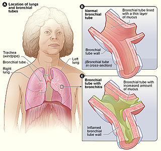
Bronchitis is inflammation of the bronchi in the lungs. Symptoms include coughing up mucus, wheezing, shortness of breath, and chest discomfort. Bronchitis is divided into two types: acute and chronic. Acute bronchitis is also known as a chest cold.

A-T is caused by a defect in the ATM gene, [3] The protein produced by the ATM gene recognizes that there is a break in dsDNA, recruits other proteins to fix the break, and stops the cell from making new DNA until the repair is complete. [4] The prevalence of A-T is estimated to be as high as 1 in 40,000 to as low as 1 in 300,000 people. [5] [6]

ATM serine/threonine kinase protein-coding gene in the species Homo sapiens

ATM serine/threonine kinase, symbol ATM, is a serine/threonine protein kinase that is recruited and activated by DNA double-strand breaks. It phosphorylates several key proteins that initiate activation of the DNA damage checkpoint, leading to cell cycle arrest, DNA repair or apoptosis. Several of these targets, including p53, CHK2, BRCA1, NBS1 and H2AX are tumor suppressors.

Symptoms

There is substantial variability in the severity of features of A-T among affected individuals, and at different ages. The following symptoms or problems are either common or important features of A-T:

Dysarthria is a motor speech disorder resulting from neurological injury of the motor component of the motor-speech system and is characterized by poor articulation of phonemes. In other words, it is a condition in which problems effectively occur with the muscles that help produce speech, often making it very difficult to pronounce words. It is unrelated to problems with understanding language, although a person can have both. Any of the speech subsystems can be affected, leading to impairments in intelligibility, audibility, naturalness, and efficiency of vocal communication. Dysarthria that has progressed to a total loss of speech is referred to as anarthria.

Many children are initially misdiagnosed as having cerebral palsy. The diagnosis of A-T may not be made until the preschool years when the neurologic symptoms of impaired gait, hand coordination, speech and eye movement appear or worsen, and the telangiectasia first appear. Because A-T is so rare, doctors may not be familiar with the symptoms, or methods of making a diagnosis. The late appearance of telangiectasia may be a barrier to the diagnosis. [7] It may also take some time before doctors consider A-T as a possibility because of the early stability of symptoms and signs.[ citation needed ]

Ataxia and other neurologic problems

The first indications of A-T usually occur during the toddler years. [8] [9] Children start walking at a normal age, but may not improve much from their initial wobbly gait. Sometimes they have problems standing or sitting still and tend to sway backward or from side to side. In primary school years, walking becomes more difficult, and children will use doorways and walls for support. Children with A-T often appear better when running or walking quickly in comparison to when they are walking slowly or standing in one place. Around the beginning of their second decade, children with the more severe ("classic") form of A-T start using a wheelchair for long distances. During school years, children may have increasing difficulty with reading because of impaired coordination of eye movement. At the same time, other problems with fine-motor functions (writing, coloring, and using utensils to eat), and with slurring of speech (dysarthria) may arise. Most of these neurologic problems stop progressing after the age of about 12 – 15 years, though involuntary movements may start at any age and may worsen over time. These extra movements can take many forms, including small jerks of the hands and feet that look like fidgeting (chorea), slower twisting movements of the upper body (athetosis), adoption of stiff and twisted postures (dystonia), occasional uncontrolled jerks (myoclonic jerks), and various rhythmic and non-rhythmic movements with attempts at coordinated action (tremors).

Telangiectasia

Ocular telangiectasia in a person with A-T Photo Ocular telangiectasia in a person with A-T.jpg
Ocular telangiectasia in a person with A-T

Prominent blood vessels (telangiectasia) over the white (sclera) of the eyes usually occur by the age of 5–8 years, but sometimes appear later or not at all. [10] The absence of telangiectasia does not exclude the diagnosis of A-T. Potentially a cosmetic problem, the ocular telangiectasia do not bleed or itch, though they are sometimes misdiagnosed as chronic conjunctivitis. It is their constant nature, not changing with time, weather or emotion, that marks them as different from other visible blood vessels. Telangiectasia can also appear on sun-exposed areas of skin, especially the face and ears. They occur in the bladder as a late complication of chemotherapy with cyclophosphamide, [11] have been seen deep inside the brain of older people with A-T, [12] and occasionally arise in the liver and lungs.[ citation needed ]

Immune problems

About two-thirds of people with A-T have abnormalities of the immune system. [13] The most common abnormalities are low levels of one or more classes of immunoglobulins (IgG, IgA or IgM subclasses), not making antibodies in response to vaccines or infections, and having low numbers of lymphocytes (especially T-lymphocytes) in the blood. Some people have frequent infections of the upper (colds, sinus and ear infections) and lower (bronchitis and pneumonia) respiratory tract. All children with A-T should have their immune systems evaluated to detect those with severe problems that require treatment to minimize the number or severity of infections. Some people with A-T need additional immunizations (especially with pneumonia and influenza vaccines), antibiotics to provide protection (prophylaxis) from infections, and/or infusions of immunoglobulins (gamma globulin). The need for these treatments should be determined by an expert in the field of immunodeficiency or infectious diseases.

Cancer

People with A-T have a highly increased incidence (approximately 25% lifetime risk) of cancers, particularly lymphomas and leukemia, but other cancers can occur. [14] When possible, treatment should avoid the use of radiation therapy and chemotherapy drugs that work in a way that is similar to radiation therapy (radiomimetic drugs), as these are particularly toxic for people with A-T. The special problems of managing cancer are sufficiently complicated that treatment should be done only in academic oncology centers and after consultation with physicians who have specific expertise in A-T. Unfortunately, there is no way to predict which individuals will develop cancer. Because leukemia and lymphomas differ from solid tumors in not progressing from solitary to metastatic stages, there is less need to diagnose them early in their appearance. Surveillance for leukemia and lymphoma is thus not helpful, other than considering cancer as a diagnostic possibility whenever possible symptoms of cancer (e.g. persistent swollen lymph glands, unexplained fever) arise.

Women who are A-T carriers (who have one mutated copy of the ATM gene), have approximately a two-fold increased risk for the development of breast cancer compared to the general population. [15] [16] This includes all mothers of A-T children and some female relatives. Current consensus is that special screening tests are not helpful, but all women should have routine cancer surveillance.

Skin

A-T can cause features of early aging such as premature graying of the hair. It can also cause vitiligo (an auto-immune disease causing loss of skin pigment resulting in a blotchy “bleach-splashed” look), and warts which can be extensive and recalcitrant to treatment. A small number of people develop a chronic inflammatory skin disease (granulomas). [17]

Lung disease

Chronic lung disease develops in more than 25% of people with A-T. [18] Three major types of lung disease can develop: (1) recurrent and chronic sinopulmonary infections; (2) lung disease caused by ineffective cough, swallowing dysfunction, and impaired airway clearance; and (3) restrictive interstitial lung disease. It is common for individuals with A-T to have more than one of these lung conditions. Chronic lung disease can occur because of recurrent lung infections due to immunodeficiency. Individuals with this problem are at risk of developing bronchiectasis, a condition in which bronchial tubes are permanently damaged, resulting in recurrent lower airway infections. Gamma globulin for people with antibody deficiency and/or chronic antibiotic treatment may reduce the problems of infection. Other individuals with A-T have difficulty with taking deep breaths and may have an ineffective cough, making it difficult to clear oral and bronchial secretions. This can lead to prolonged respiratory symptoms following common viral respiratory illnesses. Techniques that allow clearance of mucus can be helpful in some individuals during respiratory illnesses. Some people will develop swallowing problems as they age, increasing their risk of aspiration pneumonia. Recurrent injury to the lungs caused by chronic infections or aspiration may cause lung fibrosis and scarring. This process may be enhanced by inadequate tissue repair in ATM-deficient cells. A small number of individuals develop interstitial lung disease. They have decreased pulmonary reserve, trouble breathing, a need for supplemental oxygen and chronic cough in the absence of lung infections. They may respond to systemic steroid treatment or other drugs to reduce inflammation.

Lung function tests (spirometry) should be performed at least annually in children old enough to perform them, influenza and pneumococcal vaccines given to eligible individuals, and sinopulmonary infections treated aggressively to limit the development of chronic lung disease.

Feeding, swallowing, and nutrition

Feeding and swallowing can become difficult for people with A-T as they get older. [19] Feeding refers to all aspects of eating and drinking, including getting food and liquids to the mouth; swallowing refers to ingestion or what happens after food or liquids enter the mouth. Primary goals for feeding and swallowing are safe, adequate, and enjoyable mealtimes.

Involuntary movements may make feeding difficult or messy and may excessively prolong mealtimes. It may be easier to finger feed than use utensils (e.g., spoon or fork). For liquids, it is often easier to drink from a closed container with a straw than from an open cup. Caregivers may need to provide foods or liquids so that self-feeding is possible, or they may need to feed the person with A-T. In general, meals should be completed within approximately 30 minutes. Longer meals may be stressful, interfere with other daily activities, and limit the intake of necessary liquids and nutrients.

If swallowing problems (dysphagia) occur, they typically present during the second decade of life. Dysphagia is common because of the neurological changes that interfere with coordination of mouth and pharynx (throat) movements that are needed for safe and efficient swallowing. Coordination problems involving the mouth may make chewing difficult and increase the duration of meals. Problems involving the pharynx may cause liquid, food, and saliva to be inhaled into the airway (aspiration). People with dysphagia may not cough when they aspirate (silent aspiration). Swallowing problems and especially swallowing problems with silent aspiration may cause lung problems due to inability to cough and clear food and liquids from the airway.

Warning signs of a swallowing problem

Eye and vision

Orthopedics

Many individuals with A-T develop deformities of the feet that compound the difficulty they have with walking due to impaired coordination. Early treatment may slow progression of this deformity. Bracing or surgical correction sometimes improves stability at the ankle sufficient to enable an individual to walk with support, or bear weight during assisted standing transfers from one seat to another. Severe scoliosis is relatively uncommon, but probably does occur more often than in those without A-T. Spinal fusion is only rarely indicated.

Pathophysiology

How does loss of the ATM protein create a multisystem disorder?

Characteristics of the ATM protein Table Characteristics of the ATM protein.jpg
Characteristics of the ATM protein

A-T has been described as a genome instability syndrome, a DNA repair disorder and a DNA damage response (DDR) syndrome. ATM, the gene responsible for this multi-system disorder, encodes a protein of the same name which coordinates the cellular response to DNA double strand breaks (DSBs). [21] Radiation therapy, chemotherapy that acts like radiation (radiomimetic drugs) and certain biochemical processes and metabolites can cause DSBs. When these breaks occur, ATM stops the cell from making new DNA (cell cycle arrest) and recruits and activates other proteins to repair the damage. Thus, ATM allows the cell to repair its DNA before the completion of cell division. If DNA damage is too severe, ATM will mediate the process of programmed cell death (apoptosis) to eliminate the cell and prevent genomic instability. [22]

Cancer and radiosensitivity

In the absence of the ATM protein, cell-cycle check-point regulation and programmed cell death in response to DSBs are defective. The result is genomic instability which can lead to the development of cancers. [29]

Irradiation and radiomimetic compounds induce DSBs which are unable to be repaired appropriately when ATM is absent. Consequently, such agents can prove especially cytotoxic to A-T cells and people with A-T.

Delayed pubertal development (gonadal dysgenesis)

Infertility is often described as a characteristic of A-T. Whereas this is certainly the case for the mouse model of A-T, [30] in humans it may be more accurate to characterize the reproductive abnormality as gonadal atrophy or dysgenesis characterized by delayed pubertal development. Because programmed DSBs are generated to initiate genetic recombinations involved in the production of sperm and eggs in reproductive organs (a process known as meiosis), meiotic defects and arrest can occur when ATM is not present. [30] [31] [32]

ATM and the immune system Table ATM and the immune system.jpg
ATM and the immune system

As lymphocytes develop from stem cells in the bone marrow into mature lymphocytes in the periphery, they rearrange special segments of their DNA [V(D)J recombination process]. This process requires them to make DSBs, which are difficult to repair in the absence of ATM. [37] [38] [39] [40] As a result, most people with A-T have reduced numbers of lymphocytes and some impairment of lymphocyte function (such as an impaired ability to make antibodies in response to vaccines or infections). In addition, broken pieces of DNA in chromosomes involved in the above-mentioned rearrangements have a tendency to recombine with other genes (translocation), making the cells prone to the development of cancer (lymphoma and leukemia).

Progeric changes

Cells from people with A-T demonstrate genomic instability, slow growth and premature senescence in culture, shortened telomeres and an ongoing, low-level stress response. [4] [41] These factors may contribute to the progeric (signs of early aging) changes of skin and hair sometimes observed in people with A-T. For example, DNA damage and genomic instability cause melanocyte stem cell (MSC) differentiation which produces graying. Thus, ATM may be a “stemness checkpoint” protecting against MSC differentiation and premature graying of the hair. [42]

Telangiectasia

The cause of telangiectasia or dilated blood vessels in the absence of the ATM protein is not yet known.

Increased alpha-fetoprotein (AFP) levels

Approximately 95% of people with A-T have elevated serum AFP levels after the age of two, and measured levels of AFP appear to increase slowly over time. [43] AFP levels are very high in the newborn, and normally descend to adult levels over the first year to 18 months. The reason for why individuals with A-T have elevated levels of AFP is not yet known.

Neurodegeneration

A-T is one of several DNA repair disorders which result in neurological abnormalities or degeneration. Arguably some of the most devastating symptoms of A-T are a result of progressive cerebellar degeneration, characterized by the loss of Purkinje cells and, to a lesser extent, granule cells (located exclusively in the cerebellum). [8] The cause of this cell loss is not known, though many hypotheses have been proposed based on experiments performed both in cell culture and in the mouse model of A-T. Current hypotheses explaining the neurodegeneration associated with A-T include the following:

These hypotheses may not be mutually exclusive and more than one of these mechanisms may underlie neuronal cell death when there is an absence or deficiency of ATM. Further, cerebellar damage and loss of Purkinje and granule cells do not explain all of the neurologic abnormalities seen in people with A-T. The effects of ATM deficiency on the other areas of the brain outside of the cerebellum are being actively investigated.

Radiation exposure

People with A-T have an increased sensitivity to ionizing radiation (X-rays and gamma rays). Therefore, X-ray exposure should be limited to times when it is medically necessary, as exposing an A-T patient to ionizing radiation can damage cells in such a way that the body cannot repair them. The cells can cope normally with other forms of radiation, such as ultraviolet light, so there is no need for special precautions from sunlight exposure.

Genetics and information about A-T carriers

A-T is inherited in an autosomal recessive fashion Autorecessive.svg
A-T is inherited in an autosomal recessive fashion

A-T is caused by mutations in the ATM (ATM serine/threonine kinase or ataxia-telangiectasia mutated) gene, which was cloned in 1995. [3] ATM is located on human chromosome 11 (11q22.3) and is made up of 69 exons spread across 150kb of genomic DNA. [62]

The mode of inheritance for A-T is autosomal recessive. Each parent is a carrier, meaning that they have one normal copy of the A-T gene (ATM) and one copy which is mutated. A-T occurs if a child inherits the mutated A-T gene from each parent, so in a family with two carrier parents, there is 1 chance in 4 that a child born to the parents will have the disorder. Prenatal diagnosis (and carrier detection) can be carried out in families if the errors (mutation) in an affected child’s two ATM genes have been identified. The process of getting this done can be complicated and, as it requires time, should be arranged before conception.

Looking for mutations in the ATM gene of an unrelated person (for example, the spouse of a known A-T carrier) presents significant challenges. Genes often have variant spellings (polymorphisms) which do not affect function. In a gene as large as ATM, such variant spellings are likely to occur and doctors cannot always predict whether a specific variant will or will not cause disease. Genetic counseling can help family members of an A-T patient understand what can or cannot be tested, and how the test results should be interpreted.

Carriers of A-T, such as the parents of a person with A-T, have one mutated copy of the ATM gene and one normal copy. They are generally healthy, but there is an increased risk of breast cancer in women. This finding has been confirmed in a variety of different ways, and is the subject of current research. Standard surveillance (including monthly breast self-exams and mammography at the usual schedule for age) is recommended, unless additional tests are indicated because the individual has other risk factors (e.g., family history of breast cancer).

Diagnosis

The diagnosis of A-T is usually suspected by the combination of neurologic clinical features (ataxia, abnormal control of eye movement, and postural instability) with telangiectasia and sometimes increased infections, and confirmed by specific laboratory abnormalities (elevated alpha-fetoprotein levels, increased chromosomal breakage or cell death of white blood cells after exposure to X-rays, absence of ATM protein in white blood cells, or mutations in each of the person’s ATM genes).

A variety of laboratory abnormalities occur in most people with A-T, allowing for a tentative diagnosis to be made in the presence of typical clinical features. Not all abnormalities are seen in all patients. These abnormalities include:

The diagnosis can be confirmed in the laboratory by finding an absence or deficiency of the ATM protein in cultured blood cells, [64] [65] an absence or deficiency of ATM function (kinase assay), or mutations in both copies of the cell’s ATM gene. These more specialized tests are not always needed, but are particularly helpful if a child’s symptoms are atypical.

Differential diagnosis

There are several other disorders with similar symptoms or laboratory features that physicians may consider when diagnosing A-T. [66] The three most common disorders that are sometimes confused with A-T are:

Each of these can be distinguished from A-T by the neurologic exam and clinical history.

Cerebral palsy (CP) describes a non-progressive disorder of motor function stemming from malformation or early damage to the brain. CP can manifest in many ways, given the different manner in which brain can be damaged; in common to all forms is the emergence of signs and symptoms of impairment as the child develops. However, milestones that have been accomplished and neurologic functions that have developed do not deteriorate in CP as they often do in children with A-T in the late pre-school years. Most children with ataxia caused by CP do not begin to walk at a normal age, whereas most children with A-T start to walk at a normal age even though they often “wobble” from the start. Pure ataxia is a rare manifestation of early brain damage or malformation, however, and the possibility of an occult genetic disorder of brain should be considered and sought for those in whom ataxia is the chief manif estation of CP. Children with ataxic CP will not manifest the laboratory abnormalities associated with A-T.

Cogan occulomotor apraxia is a rare disorder of development. Affected children have difficulty moving their eyes only to a new visual target, so they will turn their head past the target to “drag” the eyes to the new object of interest, then turn the head back. This tendency becomes evident in late infancy and toddler years, and mostly improves with time. This contrasts to the oculomotor difficulties evident in children with A-T, which are not evident in early childhood but emerge over time. Cogan’s oculomotor apraxia is generally an isolated problem, or may be associated with broader developmental delay.

Friedreich ataxia (FA) is the most common genetic cause of ataxia in children. Like A-T, FA is a recessive disease, appearing in families without a history of the disorder. FA is caused by mutation in the frataxin gene, most often an expansion of a naturally occurring repetition of the three nucleotide bases GAA from the usual 5-33 repetitions of this trinucleotide sequence to greater than 65 repeats on each chromosome. Most often the ataxia appears between 10 and 15 years of age, and differs from A-T by the absence of telangiectasia and oculomotor apraxia, a normal alpha fetalprotein, and the frequent presence of scoliosis, absent tendon reflexes, and abnormal features on the EKG. Individuals with FA manifest difficulty standing in one place that is much enhanced by closure of the eyes (Romberg sign) that is not so apparent in those with A-T – even though those with A-T may have greater difficulty standing in one place with their eyes open.

There are other rare disorders that can be confused with A-T, either because of similar clinical features, a similarity of some laboratory features, or both. These include:

Comparison of clinical and laboratory features of rare genetic disorders than can be confused with A-T Table Comparison of rare genetic disorders that can be confused with A-T revised.jpg
Comparison of clinical and laboratory features of rare genetic disorders than can be confused with A-T

Ataxia oculomotor apraxia type 1 (AOA1) is an autosomal recessive disorder similar to A-T in manifesting increasing problems with coordination and oculomotor apraxia, often at a similar age to those having A-T. It is caused by mutation in the gene coding for the protein aprataxin. Affected individuals differ from those with A-T by the early appearance of peripheral neuropathy, early in their course manifest difficulty with initiation of gaze shifts, and the absence of ocular telangiectasia, but laboratory features are of key importance in the differentiation of the two. Individuals with AOA1 have a normal AFP, normal measures of immune function, and after 10–15 years have low serum levels of albumin. Genetic testing of the aprataxin gene can confirm the diagnosis. There is no enhanced risk for cancer.

Ataxia oculomotor apraxia type 2 (AOA2) is an autosomal recessive disorder also similar to A-T in manifesting increasing problems with coordination and peripheral neuropathy, but oculomotor apraxia is present in only half of affected individuals. Ocular telangiectasia do not develop. Laboratory abnormalities of AOA2 are like A-T, and unlike AOA1, in having an elevated serum AFP level, but like AOA1 and unlike A-T in having normal markers of immune function. Genetic testing of the senataxin gene (SETX) can confirm the diagnosis. There is no enhanced risk for cancer.

Ataxia-telangiectasia like disorder (ATLD) is an extremely rare condition, caused by mutation in the hMre11 gene, that could be considered in the differential diagnosis of A-T. Patients with ATLD are very similar to those with A-T in showing a progressive cerebellar ataxia, hypersensitivity to ionizing radiation and genomic instability. Those rare individuals with ATLD who are well described differ from those with A-T by the absence of telangiectasia, normal immunoglobulin levels, a later onset, and a slower progression of the symptoms. Because of its rarity, it is not yet known whether or not ATLD carries an increased risk to develop cancer. Because those mutations of Mre11 that severely impair the MRE11 protein are incompatible with life, individuals with ATLD all have some partial function of the Mre11 protein, and hence likely all have their own levels of disease severity.

Nijmegen breakage syndrome (NBS) is a rare genetic disorder that has similar chromosomal instability to that seen in people with A-T, but the problems experienced are quite different. Children with NBS have significant microcephaly, a distinct facial appearance, short stature, and moderate cognitive impairment, but do not experience any neurologic deterioration over time. Like those with A-T, children with NBS have enhanced sensitivity to radiation, disposition to lymphoma and leukemia, and some laboratory measures of impaired immune function, but do not have ocular telangiectasia or an elevated level of AFP.

The proteins expressed by the hMre11 (defective in ATLD) and Nbs1 (defective in NBS) genes exist in the cell as a complex, along with a third protein expressed by the hRad50 gene. This complex, known as the MRN complex, plays an important role in DNA damage repair and signaling and is required to recruit ATM to the sites of DNA double strand breaks. Mre11 and Nbs1 are also targets for phosphorylation by the ATM kinase. Thus, the similarity of the three diseases can be explained in part by the fact that the protein products of the three genes mutated in these disorders interact in common pathways in the cell.

Differentiation of these disorders is often possible with clinical features and selected laboratory tests. In cases where the distinction is unclear, clinical laboratories can identify genetic abnormalities of ATM, aprataxin and senataxin, and specialty centers can identify abnormality of the proteins of potentially responsible genes, such as ATM, MRE11, nibrin, TDP1, aprataxin and senataxin as well as other proteins important to ATM function such as ATR, DNA-PK, and RAD50.

Management

Ataxia and other neurologic problems

There is no treatment known to slow or stop the progression of the neurologic problems. Treatment of A-T is symptomatic and supportive. Physical, occupational and speech therapies and exercise may help maintain function but will not slow the course of neurodegeneration. Therapeutic exercises should not be used to the point of fatigue and should not interfere with activities of daily life. Certain anti-Parkinson and anti-epileptic drugs may be useful in the management of symptoms, but should be prescribed in consultation with a neurologist.

Immune problems

All individuals with A-T should have at least one comprehensive immunologic evaluation that measures the number and type of lymphocytes in the blood (T-lymphocytes and B-lymphocytes), the levels of serum immunoglobulins (IgG, IgA, and IgM) and antibody responses to T-dependent (e.g., tetanus, Hemophilus influenzae b) and T-independent (23-valent pneumococcal polysaccharide) vaccines. For the most part, the pattern of immunodeficiency seen in an A-T patient early in life (by age five) will be the same pattern seen throughout the lifetime of that individual. Therefore, the tests need not be repeated unless that individual develops more problems with infection. Problems with immunity sometimes can be overcome by immunization. Vaccines against common bacterial respiratory pathogens such as Hemophilus influenzae, pneumococci and influenza virus (the “flu”) are commercially available and often help to boost antibody responses, even in individuals with low immunoglobulin levels. If the vaccines do not work and the patient continues to have problems with infections, gamma globulin therapy (IV or subcutaneous infusions of antibodies collected from normal individuals) may be of benefit. A small number of people with A-T develop an abnormality in which one or more types of immunoglobulin are increased far beyond the normal range. In a few cases, the immunoglobulin levels can be increased so much that the blood becomes thick and does not flow properly. Therapy for this problem must be tailored to the specific abnormality found and its severity.

If an individual patient’s susceptibility to infection increases, it is important to reassess immune function in case deterioration has occurred and a new therapy is indicated. If infections are occurring in the lung, it is also important to investigate the possibility of dysfunctional swallow with aspiration into the lungs (see above sections under Symptoms: Lung Disease and Symptoms: Feeding, Swallowing and Nutrition.)

Most people with A-T have low lymphocyte counts in the blood. This problem seems to be relatively stable with age, but a rare number of people do have progressively decreasing lymphocyte counts as they get older. In the general population, very low lymphocyte counts are associated with an increased risk for infection. Such individuals develop complications from live viral vaccines (measles, mumps, rubella and chickenpox), chronic or severe viral infections, yeast infections of the skin and vagina, and opportunistic infections (such as pneumocystis pneumonia). Although lymphocyte counts are often as low in people with A-T, they seldom have problems with opportunistic infections. (The one exception to that rule is that problems with chronic or recurrent warts are common.) The number and function of T-lymphocytes should be re-evaluated if a person with A-T is treated with corticosteroid drugs such as prednisone for longer than a few weeks or is treated with chemotherapy for cancer. If lymphocyte counts are low in people taking those types of drugs, the use of prophylactic antibiotics is recommended to prevent opportunistic infections.

If the tests show significant abnormalities of the immune system, a specialist in immunodeficiency or infectious diseases will be able to discuss various treatment options. Absence of immunoglobulin or antibody responses to vaccine can be treated with replacement gamma globulin infusions, or can be managed with prophylactic antibiotics and minimized exposure to infection. If antibody function is normal, all routine childhood immunizations including live viral vaccines (measles, mumps, rubella and varicella) should be given. In addition, several “special” vaccines (that is, licensed but not routine for otherwise healthy children and young adults) should be given to decrease the risk that an A-T patient will develop lung infections. The patient and all household members should receive the influenza (flu) vaccine every fall. People with A-T who are less than two years old should receive three (3) doses of a pneumococcal conjugate vaccine (Prevnar) given at two month intervals. People older than two years who have not previously been immunized with Prevnar should receive two (2) doses of Prevnar. At least 6 months after the last Prevnar has been given and after the child is at least two years old, the 23-valent pneumococcal vaccine should be administered. Immunization with the 23-valent pneumococcal vaccine should be repeated approximately every five years after the first dose.

In people with A-T who have low levels of IgA, further testing should be performed to determine whether the IgA level is low or completely absent. If absent, there is a slightly increased risk of a transfusion reaction. “Medical Alert” bracelets are not necessary, but the family and primary physician should be aware that if there is elective surgery requiring red cell transfusion, the cells should be washed to decrease the risk of an allergic reaction.

People with A-T also have an increased risk of developing autoimmune or chronic inflammatory diseases. This risk is probably a secondary effect of their immunodeficiency and not a direct effect of the lack of ATM protein. The most common examples of such disorders in A-T include immune thrombocytopenia (ITP), several forms of arthritis, and vitiligo.

Lung disease

Recurrent sinus and lung infections can lead to the development of chronic lung disease. [18] Such infections should be treated with appropriate antibiotics to prevent and limit lung injury. Administration of antibiotics should be considered when children and adults have prolonged respiratory symptoms (greater than 7 days), even following what was presumed to have been a viral infection. To help prevent respiratory illnesses from common respiratory pathogens, annual influenza vaccinations should be given and pneumococcal vaccines should be administered when appropriate. Antibiotic treatment should also be considered in children with chronic coughs that are productive of mucous, those who do not respond to aggressive pulmonary clearance techniques and in children with muco-purulent secretions from the sinuses or chest. A wet cough can also be associated with chronic aspiration which should be ruled out through proper diagnostic studies, however aspiration and respiratory infections are not necessarily exclusive of each other. In children and adults with bronchiectasis, chronic antibiotic therapy should be considered to slow chronic lung disease progression.

Culturing of the sinuses may be needed to direct antibiotic therapy. This can be done by an Ear Nose and Throat (ENT) specialist. In addition, diagnostic bronchoscopy may be necessary in people who have recurrent pneumonias, especially those who do not respond or respond incompletely to a course of antibiotics.

Clearance of bronchial secretions is essential for good pulmonary health and can help limit injury from acute and chronic lung infections. Children and adults with increased bronchial secretions can benefit from routine chest therapy using the manual method, an a cappella device or a chest physiotherapy vest. Chest physiotherapy can help bring up mucous from the lower bronchial tree, however an adequate cough is needed to remove secretions. In people who have decreased lung reserve and a weak cough, use of an insufflator-exsufflator (cough-assist) device may be useful as a maintenance therapy or during acute respiratory illnesses to help remove bronchial secretions from the upper airways. Evaluation by a Pulmonology specialist however, should first be done to properly assess patient suitability.

Children and adults with chronic dry cough, increased work of breathing (fast respiratory rate, shortness of breath at rest or with activities) and absence of an infectious process to explain respiratory symptoms should be evaluated for interstitial lung disease or another intrapulmonary process. Evaluation by a Pulmonologist and a CT scan of the chest should be considered in individuals with symptoms of interstitial lung disease or to rule other non-infectious pulmonary processes. People diagnosed with interstitial lung disease may benefit from systemic steroids.

Feeding, swallowing and nutrition

Oral intake may be aided by teaching persons with A-T how to drink, chew and swallow more safely. The propriety of treatments for swallowing problems should be determined following evaluation by an expert in the field of speech-language pathology. Dieticians may help treat nutrition problems by recommending dietary modifications, including high calorie foods or food supplements.

A feeding (gastrostomy) tube is recommended when any of the following occur: [67]

Feeding tubes can decrease the risk of aspiration by enabling persons to avoid liquids or foods that are difficult to swallow and provide adequate calories without the stress and time commitment of prolonged meals. Gastrostomy tubes do not prevent people from eating by mouth. Once a tube is in place, the general goal should be to maintain weight at the 10-25th percentile.

Education and socialization

Most children with A-T have difficulty in school because of a delay in response time to visual, verbal or other cues, slurred and quiet speech (dysarthria), abnormalities of eye control (oculomotor apraxia), and impaired fine motor control. Despite these problems, children with A-T often enjoy school if proper accommodations to their disability can be made. The decision about the need for special education classes or extra help in regular classes is highly influenced by the local resources available. Decisions about proper educational placement should be revisited as often as circumstances warrant. Despite their many neurologic impairments, most individuals with A-T are very socially aware and socially skilled, and thus benefit from sustained peer relationships developed at school. Some individuals are able to function quite well despite their disabilities and a few have graduated from community colleges.

Many of the problems encountered will benefit from special attention, as problems are often related more to “input and output” issues than to intellectual impairment. Problems with eye movement control make it difficult for people with A-T to read, yet most fully understand the meaning and nuances of text that is read to them. Delays in speech initiation and lack of facial expression make it seem that they do not know the answers to questions. Reduction of the skilled effort needed to answer questions, and an increase of the time available to respond, is often rewarded by real accomplishment. It is important to recognize that intellectual disability is not regularly a part of the clinical picture of A-T although school performance may be suboptimal because of the many difficulties in reading, writing, and speech. Children with A-T are often very conscious of their appearance, and strive to appear normal to their peers and teachers. Life within the ataxic body can be tiring. The enhanced effort needed to maintain appearances and increased energy expended in abnormal tone and extra movements all contribute to physical and mental fatigue. As a consequence, for some a shortened school day yields real benefits.

General recommendations

Clinics and support

The US, UK, Australia, Israel, The Netherlands, Germany, Poland, Norway and Japan have specialized clinics for patients with A-T. These clinics house multidisciplinary medical teams, including neurologists, immunologists, pulmonologists and therapists, capable of dealing with the many facets of this disease.

Epidemiology

Individuals of all races and ethnicities are affected equally. The incidence worldwide is estimated to be between 1 in 40,000 and 1 in 100,000 people. [4] [6]

Prognosis

The life expectancy of people with A-T is highly variable. The average is approximately 25 years, but continues to improve with advances in care. The two most common causes of death are chronic lung disease (about one-third of cases) and cancer (about one-third of cases).

Research directions

An open-label Phase II clinical trial studying the use of red blood cells (erythrocytes) loaded with dexamethasone sodium phosphate found that this treatment improved symptoms and appeared to be well tolerated. [68] This treatment uses a unique delivery system for medication by using the patient's own red blood cells as the delivery vehicle for the drug. [69] Given the other immunologic deficits present in individuals with A-T, there remains a need to evaluate the therapeutic potential of steroids further, particularly with respect to the duration of any benefit and its long-term safety.

Related Research Articles

Fanconi anaemia (FA) is a rare genetic disease resulting in impaired response to DNA damage. Although it is a very rare disorder, study of this and other bone marrow failure syndromes has improved scientific understanding of the mechanisms of normal bone marrow function and development of cancer. Among those affected, the majority develop cancer, most often acute myelogenous leukemia, and 90% develop bone marrow failure by age 40. About 60–75% of people have congenital defects, commonly short stature, abnormalities of the skin, arms, head, eyes, kidneys, and ears, and developmental disabilities. Around 75% of people have some form of endocrine problems, with varying degrees of severity.

Bálint's syndrome is an uncommon and incompletely understood triad of severe neuropsychological impairments: inability to perceive the visual field as a whole (simultanagnosia), difficulty in fixating the eyes, and inability to move the hand to a specific object by using vision. It was named in 1909 for the Austro-Hungarian neurologist and psychiatrist Rezső Bálint who first identified it.

Histone deacetylase

Histone deacetylases (EC 3.5.1.98, HDAC) are a class of enzymes that remove acetyl groups (O=C-CH3) from an ε-N-acetyl lysine amino acid on a histone, allowing the histones to wrap the DNA more tightly. This is important because DNA is wrapped around histones, and DNA expression is regulated by acetylation and de-acetylation. Its action is opposite to that of histone acetyltransferase. HDAC proteins are now also called lysine deacetylases (KDAC), to describe their function rather than their target, which also includes non-histone proteins.

Spinocerebellar ataxia hereditary ataxia that has material basis in autosomal dominant inheritance

Spinocerebellar ataxia (SCA), also known as spinocerebellar atrophy or spinocerebellar degeneration, is a progressive, degenerative, genetic disease with multiple types, each of which could be considered a neurological condition in its own right. An estimated 150,000 people in the United States have a diagnosis of spinocerebellar ataxia at any given time. SCA is hereditary, progressive, degenerative, and often fatal. There is no known effective treatment or cure. SCA can affect anyone of any age. The disease is caused by either a recessive or dominant gene. In many cases people are not aware that they carry a relevant gene until they have children who begin to show signs of having the disorder.

Lymphoproliferative disorders (LPDs) refer to several conditions in which lymphocytes are produced in excessive quantities. They typically occur in people who have a compromised immune system. They are sometimes equated with "immunoproliferative disorders", but technically lymphoproliferative disorders are a subset of immunoproliferative disorders, along with hypergammaglobulinemia and paraproteinemias.

Nijmegen breakage syndrome human disease

Nijmegen breakage syndrome (NBS), also known as Berlin breakage syndrome, ataxia telangiectasia variant 1 (AT-V1) and Seemanova syndrome, is a rare autosomal recessive congenital disorder causing chromosomal instability, probably as a result of a defect in the double Holliday junction DNA repair mechanism and/or the synthesis dependent strand annealing mechanism for repairing double strand breaks in DNA.

X-linked severe combined immunodeficiency (X-SCID) is an immunodeficiency disorder in which the body produces very few T cells and NK cells.

Paraneoplastic cerebellar degeneration (PCD) is a paraneoplastic syndrome associated with a broad variety of tumors including lung cancer, ovarian cancer, breast cancer, Hodgkin’s lymphoma and others. PCD is a rare condition that occurs in less than 1% of cancer patients.

Primary immunodeficiencies are disorders in which part of the body's immune system is missing or does not function normally. To be considered a primary immunodeficiency (PID), the cause of the immune deficiency must not be secondary in nature. Most primary immunodeficiencies are genetic disorders; the majority are diagnosed in children under the age of one, although milder forms may not be recognized until adulthood. While there are over 300 recognized PIDs, most are very rare. About 1 in 500 people in the United States are born with a primary immunodeficiency. Immune deficiencies can result in persistent or recurring infections, autoinflammatory disorders, tumors, and disorders of various organs. There are currently limited treatments available for these conditions; most are specific to a particular type of PID. Research is currently evaluating the use of stem cell transplants (HSCT) and experimental gene therapies as avenues for treatment in limited subsets of PIDs.

Ataxia telangiectasia and Rad3 related protein-coding gene in the species Homo sapiens

Serine/threonine-protein kinase ATR also known as ataxia telangiectasia and Rad3-related protein (ATR) or FRAP-related protein 1 (FRP1) is an enzyme that, in humans, is encoded by the ATR gene. ATR belongs to the phosphatidylinositol 3-kinase-related kinase protein family. ATR is activated in response to single strand breaks.

Nibrin protein-coding gene in the species Homo sapiens

Nibrin, also known as NBN or NBS1, is a protein which in humans is encoded by the NBN gene.

An immune disorder is a dysfunction of the immune system. These disorders can be characterized in several different ways:

Aprataxin protein-coding gene in the species Homo sapiens

Aprataxin is a protein that in humans is encoded by the APTX gene.

SETX protein-coding gene in the species Homo sapiens

Probable helicase senataxin is an enzyme that in humans is encoded by the SETX gene.

Genome instability refers to a high frequency of mutations within the genome of a cellular lineage. These mutations can include changes in nucleic acid sequences, chromosomal rearrangements or aneuploidy. Genome instability does occur in bacteria. In multicellular organisms genome instability is central to carcinogenesis, and in humans it is also a factor in some neurodegenerative diseases such as amyotrophic lateral sclerosis or the neuromuscular disease myotonic dystrophy.

The stages of HIV infection are acute infection, latency and AIDS. Acute infection lasts for several weeks and may include symptoms such as fever, swollen lymph nodes, inflammation of the throat, rash, muscle pain, malaise, and mouth and esophageal sores. The latency stage involves few or no symptoms and can last anywhere from two weeks to twenty years or more, depending on the individual. AIDS, the final stage of HIV infection, is defined by low CD4+ T cell counts, various opportunistic infections, cancers and other conditions.

Autosomal recessive cerebellar ataxia type 1 Hereditary ataxia that has material basis in autosomal recessive inheritance

Autosomal recessive cerebellar ataxia type 1 (ARCA1) is a condition characterized by progressive problems with movement. Signs and symptoms of the disorder first appear in early to mid-adulthood. People with this condition initially experience impaired speech (dysarthria), problems with coordination and balance (ataxia), or both. They may also have difficulty with movements that involve judging distance or scale (dysmetria). Other features of ARCA1 include abnormal eye movements (nystagmus) and problems following the movements of objects with their eyes. The movement problems are slowly progressive, often resulting in the need for a cane, walker, or wheelchair.

GATA2 deficiency is a recently defined grouping of several disorders caused by common defect, viz., familial or sporadic inactivating mutations in one of the two parental GATA2 genes. These autosomal dominant mutations cause a reduction, i.e. a haploinsufficiency, in the cellular levels of the gene's product, GATA2. The GATA2 protein is a transcription factor critical for the embryonic development, maintenance, and functionality of blood-forming, lympathic-forming, and other tissue-forming stem cells. In consequence of these mutations, cellular levels of GATA2 are deficient and individuals develop over time hematological, immunological, lymphatic, or other presentations that may begin as apparently benign abnormalities but commonly progress to severe organ failure, opportunistic infections, virus infection-induced cancers, the myelodysplastic syndrome, and/or leukemia. GATA2 deficiency is a life-threatening and precancerous condition.

References

  1. Louis-Bar D (1941). "Sur un syndrome progressif cormprenant des télangiectasies capillaires cutanées et conjonctivales symétriques, à disposition naevoïde et des troubles cérébelleux". Confinia Neurologica. 4: 32–42.
  2. Boder E (1985). "Ataxia-telangiectasia: an overview". Kroc Foundation Series. 19: 1–63. PMID   2415689.
  3. 1 2 3 Savitsky K, Bar-Shira A, Gilad S, Rotman G, Ziv Y, Vanagaite L, Tagle DA, Smith S, Uziel T, Sfez S, Ashkenazi M, Pecker I, Frydman M, Harnik R, Patanjali SR, Simmons A, Clines GA, Sartiel A, Gatti RA, Chessa L, Sanal O, Lavin MF, Jaspers NG, Taylor AM, Arlett CF, Miki T, Weissman SM, Lovett M, Collins FS, Shiloh Y (June 1995). "A single ataxia telangiectasia gene with a product similar to PI-3 kinase". Science. 268 (5218): 1749–53. doi:10.1126/science.7792600. PMID   7792600.
  4. 1 2 3 Shiloh Y, Kastan MB (2001). "ATM: genome stability, neuronal development, and cancer cross paths". Advances in Cancer Research. 83: 209–54. doi:10.1016/s0065-230x(01)83007-4. ISBN   9780120066834. PMID   11665719.
  5. "Ataxia-telangiectasia". Genetics Home Reference. Retrieved 2018-03-29.
  6. 1 2 Swift M, Morrell D, Cromartie E, Chamberlin AR, Skolnick MH, Bishop DT (November 1986). "The incidence and gene frequency of ataxia-telangiectasia in the United States". American Journal of Human Genetics. 39 (5): 573–83. PMC   1684065 . PMID   3788973.
  7. Cabana MD, Crawford TO, Winkelstein JA, Christensen JR, Lederman HM (July 1998). "Consequences of the delayed diagnosis of ataxia-telangiectasia". Pediatrics. 102 (1 Pt 1): 98–100. PMID   9651420.
  8. 1 2 Crawford TO (December 1998). "Ataxia telangiectasia". Seminars in Pediatric Neurology. 5 (4): 287–94. doi:10.1016/s1071-9091(98)80007-7. PMID   9874856.
  9. Crawford TO, Mandir AS, Lefton-Greif MA, Goodman SN, Goodman BK, Sengul H, Lederman HM (April 2000). "Quantitative neurologic assessment of ataxia-telangiectasia". Neurology. 54 (7): 1505–9. doi:10.1212/wnl.54.7.1505. PMID   10751267.
  10. Cabana MD, Crawford TO, Winkelstein JA, Christensen JR, Lederman HM (July 1998). "Consequences of the delayed diagnosis of ataxia-telangiectasia". Pediatrics. 102 (1 Pt 1): 98–100. doi:10.1542/peds.102.1.98. PMID   9651420.
  11. Lavin MF, Gueven N, Bottle S, Gatti RA (2007). "Current and potential therapeutic strategies for the treatment of ataxia-telangiectasia". British Medical Bulletin. 81-82: 129–47. doi:10.1093/bmb/ldm012. PMID   17586848.
  12. Lin DD, Barker PB, Lederman HM, Crawford TO (January 2014). "Cerebral abnormalities in adults with ataxia-telangiectasia". AJNR. American Journal of Neuroradiology. 35 (1): 119–23. doi:10.3174/ajnr.A3646. PMC   4106125 . PMID   23886747.
  13. Nowak-Wegrzyn A, Crawford TO, Winkelstein JA, Carson KA, Lederman HM (April 2004). "Immunodeficiency and infections in ataxia-telangiectasia". The Journal of Pediatrics. 144 (4): 505–11. doi:10.1016/j.jpeds.2003.12.046. PMID   15069401.
  14. Reiman A, Srinivasan V, Barone G, Last JI, Wootton LL, Davies EG, Verhagen MM, Willemsen MA, Weemaes CM, Byrd PJ, Izatt L, Easton DF, Thompson DJ, Taylor AM (August 2011). "Lymphoid tumours and breast cancer in ataxia telangiectasia; substantial protective effect of residual ATM kinase activity against childhood tumours". British Journal of Cancer. 105 (4): 586–91. doi:10.1038/bjc.2011.266. PMC   3170966 . PMID   21792198.
  15. Thompson D, Duedal S, Kirner J, McGuffog L, Last J, Reiman A, Byrd P, Taylor M, Easton DF (June 2005). "Cancer risks and mortality in heterozygous ATM mutation carriers". Journal of the National Cancer Institute. 97 (11): 813–22. doi:10.1093/jnci/dji141. PMID   15928302.
  16. Renwick A, Thompson D, Seal S, Kelly P, Chagtai T, Ahmed M, North B, Jayatilake H, Barfoot R, Spanova K, McGuffog L, Evans DG, Eccles D, Easton DF, Stratton MR, Rahman N, et al. (Breast Cancer Susceptibility Collaboration, (UK)) (August 2006). "ATM mutations that cause ataxia-telangiectasia are breast cancer susceptibility alleles". Nature Genetics. 38 (8): 873–5. doi:10.1038/ng1837. PMID   16832357.
  17. Paller AS, Massey RB, Curtis MA, Pelachyk JM, Dombrowski HC, Leickly FE, Swift M (December 1991). "Cutaneous granulomatous lesions in patients with ataxia-telangiectasia". The Journal of Pediatrics. 119 (6): 917–22. doi:10.1016/s0022-3476(05)83043-4. PMID   1960607.
  18. 1 2 McGrath-Morrow SA, Gower WA, Rothblum-Oviatt C, Brody AS, Langston C, Fan LL, Lefton-Greif MA, Crawford TO, Troche M, Sandlund JT, Auwaerter PG, Easley B, Loughlin GM, Carroll JL, Lederman HM (September 2010). "Evaluation and management of pulmonary disease in ataxia-telangiectasia". Pediatric Pulmonology. 45 (9): 847–59. doi:10.1002/ppul.21277. PMC   4151879 . PMID   20583220.
  19. Lefton-Greif MA, Crawford TO, Winkelstein JA, Loughlin GM, Koerner CB, Zahurak M, Lederman HM (February 2000). "Oropharyngeal dysphagia and aspiration in patients with ataxia-telangiectasia". The Journal of Pediatrics. 136 (2): 225–31. doi:10.1016/s0022-3476(00)70106-5. PMID   10657830.
  20. Farr AK, Shalev B, Crawford TO, Lederman HM, Winkelstein JA, Repka MX (December 2002). "Ocular manifestations of ataxia-telangiectasia". American Journal of Ophthalmology. 134 (6): 891–6. doi:10.1016/s0002-9394(02)01796-8. PMID   12470759.
  21. 1 2 Derheimer FA, Kastan MB (September 2010). "Multiple roles of ATM in monitoring and maintaining DNA integrity". FEBS Letters. 584 (17): 3675–81. doi:10.1016/j.febslet.2010.05.031. PMC   2950315 . PMID   20580718.
  22. 1 2 Kurz EU, Lees-Miller SP (Aug–Sep 2004). "DNA damage-induced activation of ATM and ATM-dependent signaling pathways". DNA Repair. 3 (8–9): 889–900. doi:10.1016/j.dnarep.2004.03.029. PMID   15279774.
  23. 1 2 Dar I, Biton S, Shiloh Y, Barzilai A (July 2006). "Analysis of the ataxia telangiectasia mutated-mediated DNA damage response in murine cerebellar neurons". The Journal of Neuroscience. 26 (29): 7767–74. doi:10.1523/JNEUROSCI.2055-06.2006. PMID   16855104.
  24. Gorodetsky E, Calkins S, Ahn J, Brooks PJ (November 2007). "ATM, the Mre11/Rad50/Nbs1 complex, and topoisomerase I are concentrated in the nucleus of Purkinje neurons in the juvenile human brain". DNA Repair. 6 (11): 1698–707. doi:10.1016/j.dnarep.2007.06.011. PMC   2797317 . PMID   17706468.
  25. 1 2 Valentin-Vega YA, Maclean KH, Tait-Mulder J, Milasta S, Steeves M, Dorsey FC, Cleveland JL, Green DR, Kastan MB (February 2012). "Mitochondrial dysfunction in ataxia-telangiectasia". Blood. 119 (6): 1490–500. doi:10.1182/blood-2011-08-373639. PMC   3286212 . PMID   22144182.
  26. 1 2 Guo Z, Kozlov S, Lavin MF, Person MD, Paull TT (October 2010). "ATM activation by oxidative stress". Science. 330 (6003): 517–21. doi:10.1126/science.1192912. hdl:2152/9689. PMID   20966255.
  27. Bakkenist CJ, Kastan MB (January 2003). "DNA damage activates ATM through intermolecular autophosphorylation and dimer dissociation". Nature. 421 (6922): 499–506. doi:10.1038/nature01368. PMID   12556884.
  28. Kanu N, Behrens A (November 2008). "ATMINistrating ATM signalling: regulation of ATM by ATMIN". Cell Cycle. 7 (22): 3483–6. doi:10.4161/cc.7.22.7044. PMID   19001856.
  29. Shiloh Y (March 2003). "ATM and related protein kinases: safeguarding genome integrity". Nature Reviews. Cancer. 3 (3): 155–68. doi:10.1038/nrc1011. PMID   12612651.
  30. 1 2 Barlow C, Hirotsune S, Paylor R, Liyanage M, Eckhaus M, Collins F, Shiloh Y, Crawley JN, Ried T, Tagle D, Wynshaw-Boris A (July 1996). "Atm-deficient mice: a paradigm of ataxia telangiectasia". Cell. 86 (1): 159–71. doi:10.1016/S0092-8674(00)80086-0. PMID   8689683.
  31. Plug AW, Peters AH, Xu Y, Keegan KS, Hoekstra MF, Baltimore D, de Boer P, Ashley T (December 1997). "ATM and RPA in meiotic chromosome synapsis and recombination". Nature Genetics. 17 (4): 457–61. doi:10.1038/ng1297-457. PMID   9398850.
  32. Barchi M, Roig I, Di Giacomo M, de Rooij DG, Keeney S, Jasin M (May 2008). "ATM promotes the obligate XY crossover and both crossover control and chromosome axis integrity on autosomes". PLoS Genetics. 4 (5): e1000076. doi:10.1371/journal.pgen.1000076. PMC   2374915 . PMID   18497861. Open Access logo PLoS transparent.svg
  33. Lumsden JM, McCarty T, Petiniot LK, Shen R, Barlow C, Wynn TA, Morse HC, Gearhart PJ, Wynshaw-Boris A, Max EE, Hodes RJ (November 2004). "Immunoglobulin class switch recombination is impaired in Atm-deficient mice". The Journal of Experimental Medicine. 200 (9): 1111–21. doi:10.1084/jem.20041074. PMC   2211853 . PMID   15504820.
  34. Franco S, Alt FW, Manis JP (September 2006). "Pathways that suppress programmed DNA breaks from progressing to chromosomal breaks and translocations". DNA Repair. 5 (9–10): 1030–41. doi:10.1016/j.dnarep.2006.05.024. PMID   16934538.
  35. Callén E, Jankovic M, Wong N, Zha S, Chen HT, Difilippantonio S, Di Virgilio M, Heidkamp G, Alt FW, Nussenzweig A, Nussenzweig M (May 2009). "Essential role for DNA-PKcs in DNA double-strand break repair and apoptosis in ATM-deficient lymphocytes". Molecular Cell. 34 (3): 285–97. doi:10.1016/j.molcel.2009.04.025. PMC   2709792 . PMID   19450527.
  36. Bagley J, Singh G, Iacomini J (April 2007). "Regulation of oxidative stress responses by ataxia-telangiectasia mutated is required for T cell proliferation". Journal of Immunology. 178 (8): 4757–63. doi:10.4049/jimmunol.178.8.4757. PMID   17404255.
  37. Bredemeyer AL, Sharma GG, Huang CY, Helmink BA, Walker LM, Khor KC, Nuskey B, Sullivan KE, Pandita TK, Bassing CH, Sleckman BP (July 2006). "ATM stabilizes DNA double-strand-break complexes during V(D)J recombination". Nature. 442 (7101): 466–70. doi:10.1038/nature04866. PMID   16799570.
  38. Bredemeyer AL, Huang CY, Walker LM, Bassing CH, Sleckman BP (August 2008). "Aberrant V(D)J recombination in ataxia telangiectasia mutated-deficient lymphocytes is dependent on nonhomologous DNA end joining". Journal of Immunology. 181 (4): 2620–5. doi:10.4049/jimmunol.181.4.2620. PMC   3598579 . PMID   18684952.
  39. Bredemeyer AL, Helmink BA, Innes CL, Calderon B, McGinnis LM, Mahowald GK, Gapud EJ, Walker LM, Collins JB, Weaver BK, Mandik-Nayak L, Schreiber RD, Allen PM, May MJ, Paules RS, Bassing CH, Sleckman BP (December 2008). "DNA double-strand breaks activate a multi-functional genetic program in developing lymphocytes". Nature. 456 (7223): 819–23. doi:10.1038/nature07392. PMC   2605662 . PMID   18849970.
  40. Callén E, Jankovic M, Difilippantonio S, Daniel JA, Chen HT, Celeste A, Pellegrini M, McBride K, Wangsa D, Bredemeyer AL, Sleckman BP, Ried T, Nussenzweig M, Nussenzweig A (July 2007). "ATM prevents the persistence and propagation of chromosome breaks in lymphocytes". Cell. 130 (1): 63–75. doi:10.1016/j.cell.2007.06.016. PMID   17599403.
  41. Shiloh Y, Tabor E, Becker Y (July 1982). "Colony-forming ability of ataxia-telangiectasia skin fibroblasts is an indicator of their early senescence and increased demand for growth factors". Experimental Cell Research. 140 (1): 191–9. doi:10.1016/0014-4827(82)90169-0. PMID   6213420.
  42. Inomata K, Aoto T, Binh NT, Okamoto N, Tanimura S, Wakayama T, Iseki S, Hara E, Masunaga T, Shimizu H, Nishimura EK (June 2009). "Genotoxic stress abrogates renewal of melanocyte stem cells by triggering their differentiation". Cell. 137 (6): 1088–99. doi:10.1016/j.cell.2009.03.037. PMID   19524511.
  43. Stray-Pedersen A, Borresen-Dale AL, Paus E, Lindman CR, Burgers T, Abrahamsen TG (November 2007). "Alpha fetoprotein is increasing with age in ataxia-telangiectasia". European Journal of Paediatric Neurology. 11 (6): 375–80. doi:10.1016/j.ejpn.2007.04.001. PMID   17540590.
  44. Biton S, Dar I, Mittelman L, Pereg Y, Barzilai A, Shiloh Y (June 2006). "Nuclear ataxia-telangiectasia mutated (ATM) mediates the cellular response to DNA double strand breaks in human neuron-like cells". The Journal of Biological Chemistry. 281 (25): 17482–91. doi:10.1074/jbc.M601895200. PMID   16627474.
  45. Herzog KH, Chong MJ, Kapsetaki M, Morgan JI, McKinnon PJ (May 1998). "Requirement for Atm in ionizing radiation-induced cell death in the developing central nervous system". Science. 280 (5366): 1089–91. doi:10.1126/science.280.5366.1089. PMID   9582124.
  46. Lee Y, Chong MJ, McKinnon PJ (September 2001). "Ataxia telangiectasia mutated-dependent apoptosis after genotoxic stress in the developing nervous system is determined by cellular differentiation status". The Journal of Neuroscience. 21 (17): 6687–93. PMID   11517258.
  47. Sordet O, Redon CE, Guirouilh-Barbat J, Smith S, Solier S, Douarre C, Conti C, Nakamura AJ, Das BB, Nicolas E, Kohn KW, Bonner WM, Pommier Y (August 2009). "Ataxia telangiectasia mutated activation by transcription- and topoisomerase I-induced DNA double-strand breaks". EMBO Reports. 10 (8): 887–93. doi:10.1038/embor.2009.97. PMC   2726680 . PMID   19557000.
  48. Das BB, Antony S, Gupta S, Dexheimer TS, Redon CE, Garfield S, Shiloh Y, Pommier Y (December 2009). "Optimal function of the DNA repair enzyme TDP1 requires its phosphorylation by ATM and/or DNA-PK". The EMBO Journal. 28 (23): 3667–80. doi:10.1038/emboj.2009.302. PMC   2790489 . PMID   19851285.
  49. Iourov IY, Vorsanova SG, Liehr T, Kolotii AD, Yurov YB (July 2009). "Increased chromosome instability dramatically disrupts neural genome integrity and mediates cerebellar degeneration in the ataxia-telangiectasia brain". Human Molecular Genetics. 18 (14): 2656–69. doi:10.1093/hmg/ddp207. PMID   19414482.
  50. Alexander A, Cai SL, Kim J, Nanez A, Sahin M, MacLean KH, Inoki K, Guan KL, Shen J, Person MD, Kusewitt D, Mills GB, Kastan MB, Walker CL (March 2010). "ATM signals to TSC2 in the cytoplasm to regulate mTORC1 in response to ROS". Proceedings of the National Academy of Sciences of the United States of America. 107 (9): 4153–8. doi:10.1073/pnas.0913860107. PMC   2840158 . PMID   20160076.
  51. Cosentino C, Grieco D, Costanzo V (February 2011). "ATM activates the pentose phosphate pathway promoting anti-oxidant defence and DNA repair". The EMBO Journal. 30 (3): 546–55. doi:10.1038/emboj.2010.330. PMC   3034007 . PMID   21157431.
  52. Biton S, Barzilai A, Shiloh Y (July 2008). "The neurological phenotype of ataxia-telangiectasia: solving a persistent puzzle". DNA Repair. 7 (7): 1028–38. doi:10.1016/j.dnarep.2008.03.006. PMID   18456574.
  53. D'Souza AD, Parish IA, Krause DS, Kaech SM, Shadel GS (January 2013). "Reducing mitochondrial ROS improves disease-related pathology in a mouse model of ataxia-telangiectasia". Molecular Therapy. 21 (1): 42–8. doi:10.1038/mt.2012.203. PMC   3538311 . PMID   23011031.
  54. Sharma NK, Lebedeva M, Thomas T, Kovalenko OA, Stumpf JD, Shadel GS, Santos JH (January 2014). "Intrinsic mitochondrial DNA repair defects in Ataxia Telangiectasia". DNA Repair. 13: 22–31. doi:10.1016/j.dnarep.2013.11.002. PMID   24342190.
  55. Yang Y, Herrup K (March 2005). "Loss of neuronal cell cycle control in ataxia-telangiectasia: a unified disease mechanism". The Journal of Neuroscience. 25 (10): 2522–9. doi:10.1523/JNEUROSCI.4946-04.2005. PMID   15758161.
  56. Li J, Han YR, Plummer MR, Herrup K (December 2009). "Cytoplasmic ATM in neurons modulates synaptic function". Current Biology. 19 (24): 2091–6. doi:10.1016/j.cub.2009.10.039. PMC   2805770 . PMID   19962314.
  57. Li J, Chen J, Ricupero CL, Hart RP, Schwartz MS, Kusnecov A, Herrup K (May 2012). "Nuclear accumulation of HDAC4 in ATM deficiency promotes neurodegeneration in ataxia telangiectasia". Nature Medicine. 18 (5): 783–90. doi:10.1038/nm.2709. PMC   3378917 . PMID   22466704.
  58. Herrup K (October 2013). "ATM and the epigenetics of the neuronal genome". Mechanisms of Ageing and Development. 134 (10): 434–9. doi:10.1016/j.mad.2013.05.005. PMC   3791148 . PMID   23707635.
  59. Li J, Hart RP, Mallimo EM, Swerdel MR, Kusnecov AW, Herrup K (December 2013). "EZH2-mediated H3K27 trimethylation mediates neurodegeneration in ataxia-telangiectasia". Nature Neuroscience. 16 (12): 1745–53. doi:10.1038/nn.3564. PMC   3965909 . PMID   24162653.
  60. Jiang D, Zhang Y, Hart RP, Chen J, Herrup K, Li J (December 2015). "Alteration in 5-hydroxymethylcytosine-mediated epigenetic regulation leads to Purkinje cell vulnerability in ATM deficiency". Brain. 138 (Pt 12): 3520–36. doi:10.1093/brain/awv284. PMC   4668921 . PMID   26510954.
  61. Wood LM, Sankar S, Reed RE, Haas AL, Liu LF, McKinnon P, Desai SD (January 2011). "A novel role for ATM in regulating proteasome-mediated protein degradation through suppression of the ISG15 conjugation pathway". PLOS One. 6 (1): e16422. doi:10.1371/journal.pone.0016422. PMC   3027683 . PMID   21298066.
  62. Gatti RA, Berkel I, Boder E, Braedt G, Charmley P, Concannon P, Ersoy F, Foroud T, Jaspers NG, Lange K (December 1988). "Localization of an ataxia-telangiectasia gene to chromosome 11q22-23". Nature. 336 (6199): 577–80. doi:10.1038/336577a0. PMID   3200306.
  63. Sun X, Becker-Catania SG, Chun HH, Hwang MJ, Huo Y, Wang Z, Mitui M, Sanal O, Chessa L, Crandall B, Gatti RA (June 2002). "Early diagnosis of ataxia-telangiectasia using radiosensitivity testing". The Journal of Pediatrics. 140 (6): 724–31. doi:10.1067/mpd.2002.123879. PMID   12072877.
  64. Chun HH, Sun X, Nahas SA, Teraoka S, Lai CH, Concannon P, Gatti RA (December 2003). "Improved diagnostic testing for ataxia-telangiectasia by immunoblotting of nuclear lysates for ATM protein expression". Molecular Genetics and Metabolism. 80 (4): 437–43. doi:10.1016/j.ymgme.2003.09.008. PMID   14654357.
  65. Taylor AM, Byrd PJ (October 2005). "Molecular pathology of ataxia telangiectasia". Journal of Clinical Pathology. 58 (10): 1009–15. doi:10.1136/jcp.2005.026062. PMC   1770730 . PMID   16189143.
  66. Anheim M, Tranchant C, Koenig M (February 2012). "The autosomal recessive cerebellar ataxias". The New England Journal of Medicine. 366 (7): 636–46. doi:10.1056/NEJMra1006610. PMID   22335741.
  67. Lefton-Greif MA, Crawford TO, McGrath-Morrow S, Carson KA, Lederman HM (May 2011). "Safety and caregiver satisfaction with gastrostomy in patients with Ataxia Telangiectasia". Orphanet Journal of Rare Diseases. 6: 23. doi:10.1186/1750-1172-6-23. PMC   3116459 . PMID   21569628.
  68. Chessa L, Leuzzi V, Plebani A, Soresina A, Micheli R, D'Agnano D, Venturi T, Molinaro A, Fazzi E, Marini M, Ferremi Leali P, Quinti I, Cavaliere FM, Girelli G, Pietrogrande MC, Finocchi A, Tabolli S, Abeni D, Magnani M (January 2014). "Intra-erythrocyte infusion of dexamethasone reduces neurological symptoms in ataxia teleangiectasia patients: results of a phase 2 trial". Orphanet Journal of Rare Diseases. 9 (1): 5. doi:10.1186/1750-1172-9-5. PMC   3904207 . PMID   24405665.
  69. Yousefpour P, Chilkoti A (September 2014). "Co-opting biology to deliver drugs". Biotechnology and Bioengineering. 111 (9): 1699–716. doi:10.1002/bit.25307. PMC   4251460 . PMID   24916780.
Classification
D
External resources