Dyschronometria

Last updated
Cerebellum
Cerebellum NIH.png
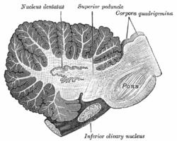
A human brain, with the cerebellum colored in purple
Gray677.png
Drawing of the human brain, showing cerebellum and pons
Details
Part of Metencephalon
Artery SCA, AICA, PICA
Vein superior, inferior
Identifiers
NeuroLex ID birnlex_1489
Anatomical terms of neuroanatomy

Dyschronometria is a condition of cerebellar dysfunction in which an individual cannot accurately estimate the amount of time that has passed (i.e., distorted time perception). It is associated with cerebellar ataxia, [1] [2] when the cerebellum has been damaged and does not function to its fullest ability. Lesions to the cerebellum can cause dyssynergia, dysmetria, dysdiadochokinesia, dysarthria, and ataxia of stance and gait. [3] Dyschronometria can result from autosomal dominant cerebellar ataxia (ADCA). [4]

Contents

Andreas Vesalius Fabrica, published in 1543, showing the base of the human brain, including optic chiasma, cerebellum, olfactory bulbs, etc. 1543, Andreas Vesalius' Fabrica, Base Of The Brain.jpg
Andreas Vesalius Fabrica, published in 1543, showing the base of the human brain, including optic chiasma, cerebellum, olfactory bulbs, etc.

Signs and symptoms

Common signs of dyschronometria are often generic to cerebellar ataxia, including a lack of spatial awareness, poor short term memory, and inability to keep track of time.[ citation needed ] The defining symptoms, while not completely understood, involve time perception. For example, when asked to wait for thirty seconds, or tap every second that has gone by, those affected will be able to perform the task for a short time and then become derailed. This can result from a loss of focus, however, more often than not the individual affected can no longer tell what they are doing and they become disoriented. [5] This often takes form in forgetting basic time keeping unless a timer is set, such as when cooking for example. Dyschronometria does not affect the 24-hour circadian rhythm, which is sustained by a different biological process.[ citation needed ]

Cross-section of human cerebellum, showing the dentate nucleus, as well as the pons and inferior olivary nucleus Gray707.png
Cross-section of human cerebellum, showing the dentate nucleus, as well as the pons and inferior olivary nucleus

Causes

The most common cause of cerebellar ataxia, and by extension dyschronometria, is cerebellar damage. This can be by form of a trauma, or by disease and genetics. Examples of trauma include a car accident, stroke, epilepsy, and head trauma. [6] These traumas are especially detrimental to children and the elderly due to decreased brain matter, increasing the risk that trauma may damage the cerebellum. This also explains why dyschronometria is seen more commonly in the elderly due to the deterioration of physical brain matter with age. Other probable causes for the deterioration of brain matter in the elderly include increased supranational activation, decreased cerebellar activation (which is consistent with fronto-cerebellar dissociation). [7]

Dyslexia

An interesting case of dyschronometria has to do with dyslexia. When dyslexia was studied within children, it was found that dyslexic children were often stressed as well as mentally exhausted. These children would place little to no importance on their present state, a behavior that would continue into adulthood. [8] It remains unclear as to whether dyslexia is a symptom of dyschronometria, a cause, or both.[ citation needed ]

Dementia

Dementia has a huge effect on dyschronometria and was one of the main sources of how dyschronometria was discovered. Through studies, dementia is both a cause and an effect of dyschronometria. This has to do completely with the fact that with dementia the brain is constantly rewiring itself and thus information becomes lost causing the person who has dementia to become confused as well as disoriented, and in most cases completely unaware of the passage of time.[ citation needed ] As a cause, dyschronometria causes the person to become disoriented and completely unaware of time, thus making bits and pieces of their brain involving a memory to become lost, ultimately leading to dementia in the long term. [9]

Errors and Inaccuracies

Diagnosing

Despite dyschronometria's easily recognizable symptoms, the fact that they may also be present in other cerebellar ataxias can make diagnosis difficult. Other ataxias may also have symptoms that affect gait, speech, thought process, spatial awareness, and time orientation used in their diagnoses, covering up the fact that most of these patients also have dyschronometria. The most common ataxias dyschronometria has been seen to be evident in are dyssynergia, dysmetria, dysdiadochokinesia, dysarthria as well as ataxias effecting stance and gait. Dyslexia can be another problem in those individuals affected by dyschronometria, however it is uncertain whether dyslexia is developed or worsened by having it, or if it is the opposite in that having dyslexia increases the chance of developing dyschronometria. Another problem that dyschronometria faces in detection is that it is a relatively new term for this side effect and precursor to dementia, compared with other cerebellar ataxias such as those mentioned above. Even when dyschronometria is detected, it has usually progressed to the point where it cannot be reversed, and there is no benefit in taking the testing medication to either slow down the dyschronometria or the process of dementia setting in, which is what dyschronometria is a signal for.

However the greatest error in diagnosing dyschronometria is that this cerebellar ataxia hides itself in its symptoms and signs. The signs seen in those diagnosed with dyschronometria are not obvious, and are often mistaken for other cerebellar ataxias or dementia by medical professionals. In addition, medical professionals usually expect to see circadian rhythm being disrupted by noting sleeping cycles and patterns that have no logical sense to them, which has nothing to do with dyschronometria. Other errors in diagnosing dyschronometria include the idea that those who have dyschronometria have a speech impediment, experience delusions bordering psychosis, impairment of long term memory, or the complete loss of conscious understanding of time. These misconceptions mostly stem from the fact that this cerebellar ataxia is rarely diagnosed without being seen in dementia or with another ataxia. [1]

Clinical testing

Testing and diagnosing for dyschronometria have also been shown to be ineffective. Dementia is caught so late despite the signals seeming obvious because psychological tests that try to catch signs of dementia, such as dyschronometria, are not very helpful. With these tests, the bell curves formed after statistical analysis and a wide range among normal test takers ensure that only patients that have extremely abnormal tests are given a positive diagnosis, and these cases are often already obvious to diagnose. [10] Diagnosis of dyschronometria is also difficult due to the lack of research and professional concentrating on this cerebellar ataxia. Neuroscientists are only just beginning to conduct more research into this lack of awareness and keeping of time. When the science and the tests are more specialized and dyschronometria has been researched at greater depth, the sensitivity of future tests conducted will likely yield more insights into the phenomenon.[ citation needed ]

Treatments

It has not been determined what role drugs may play in the treatment of cerebellar ataxia. In research done by Trouillas in Lyon, France, the pharmacology of cerebellar ataxia was examined by manipulating key components found at the nerve level within the cerebellum or the inferior Olive. This was done mostly through the modification of the GABA, dopamine, and serotonin receptors which did seem to show positive results in the primary stages of the experimentation. The clinical benefits presented in this study justifying the prescription of d-l-5-HTP or better with the l-5-HTP with benserazide to patients with certain cerebellar ataxias including that of dyschronometria. At the present as stated, this is the best indication for treatment of the cerebellar cortical atrophies. Even still, it is important to stress that the response to this treatment may be slow and irregular. [11]

Neuroplastic rehabilitation

Previously, neuroplasticity used as a rehabilitation method was considered as a potential treatment for dyschronometria. However these studies were not further developed since the 1980s. With current techniques and research from the neuroscience community, this is still a viable option not to eliminate cerebellar ataxia, but to slow its progress of development. [12]

Related Research Articles

Ataxia is a neurological sign consisting of lack of voluntary coordination of muscle movements that can include gait abnormality, speech changes, and abnormalities in eye movements, that indicates dysfunction of parts of the nervous system that coordinate movement, such as the cerebellum.

<span class="mw-page-title-main">Olivopontocerebellar atrophy</span> Medical condition

Olivopontocerebellar atrophy (OPCA) is the degeneration of neurons in specific areas of the brain – the cerebellum, pons, and inferior olivary nucleus. OPCA is present in several neurodegenerative syndromes, including inherited and non-inherited forms of ataxia and multiple system atrophy (MSA), with which it is primarily associated.

<span class="mw-page-title-main">Progressive supranuclear palsy</span> Medical condition

Progressive supranuclear palsy (PSP) is a late-onset neurodegenerative disease involving the gradual deterioration and death of specific volumes of the brain. The condition leads to symptoms including loss of balance, slowing of movement, difficulty moving the eyes, and cognitive impairment. PSP may be mistaken for other types of neurodegeneration such as Parkinson's disease, frontotemporal dementia and Alzheimer's disease. The cause of the condition is uncertain, but involves the accumulation of tau protein within the brain. Medications such as levodopa and amantadine may be useful in some cases.

<span class="mw-page-title-main">Spinocerebellar ataxia</span> Medical condition

Spinocerebellar ataxia (SCA) is a progressive, degenerative, genetic disease with multiple types, each of which could be considered a neurological condition in its own right. An estimated 150,000 people in the United States have a diagnosis of spinocerebellar ataxia at any given time. SCA is hereditary, progressive, degenerative, and often fatal. There is no known effective treatment or cure. SCA can affect anyone of any age. The disease is caused by either a recessive or dominant gene. In many cases people are not aware that they carry a relevant gene until they have children who begin to show signs of having the disorder.

Dysmetria is a lack of coordination of movement typified by the undershoot or overshoot of intended position with the hand, arm, leg, or eye. It is a type of ataxia. It can also include an inability to judge distance or scale.

Dysdiadochokinesia (DDK) is the medical term for an impaired ability to perform rapid, alternating movements. Complete inability is called adiadochokinesia. The term is from Greek δυςdys "bad", διάδοχοςdiadochos "succeeding", κίνησιςkinesis "movement".

Cerebellar ataxia is a form of ataxia originating in the cerebellum. Non-progressive congenital ataxia (NPCA) is a classical presentation of cerebral ataxias.

Intention tremor is a dyskinetic disorder characterized by a broad, coarse, and low-frequency tremor evident during deliberate and visually-guided movement. An intention tremor is usually perpendicular to the direction of movement. When experiencing an intention tremor, one often overshoots or undershoots one's target, a condition known as dysmetria. Intention tremor is the result of dysfunction of the cerebellum, particularly on the same side as the tremor in the lateral zone, which controls visually guided movements. Depending on the location of cerebellar damage, these tremors can be either unilateral or bilateral.

Paraneoplastic cerebellar degeneration (PCD) is a paraneoplastic syndrome associated with a broad variety of tumors including lung cancer, ovarian cancer, breast cancer, Hodgkin’s lymphoma and others. PCD is a rare condition that occurs in less than 1% of cancer patients.

Alcohol-related dementia (ARD) is a form of dementia caused by long-term, excessive consumption of alcoholic beverages, resulting in neurological damage and impaired cognitive function.

Uner Tan syndrome (UTS) is a syndrome that was discovered by the Turkish evolutionary biologist Üner Tan. People affected by UTS walk with a quadrupedal locomotion and often have severe learning disabilities. Tan postulated that this is an example of "reverse evolution" (atavism). The proposed syndrome was featured in the 2006 BBC2 documentary The Family That Walks On All Fours.

Ramsay Hunt syndrome type 1 is a rare, degenerative, neurological disorder characterized by myoclonus epilepsy, intention tremor, progressive ataxia and occasionally cognitive impairment

Bruns apraxia, or frontal ataxia, is a gait apraxia found in patients with bilateral frontal lobe disorders. It is characterised by an inability to initiate the process of walking, despite the power and coordination of the legs being normal when tested in the seated or lying position. The gait is broad-based with short steps with a tendency to fall backwards. It was originally described in patients with frontal lobe tumours, but is now more commonly seen in patients with cerebrovascular disease.

<span class="mw-page-title-main">Ataxic cerebral palsy</span> Medical condition

Ataxic cerebral palsy is clinically in approximately 5–10% of all cases of cerebral palsy, making it the least frequent form of cerebral palsy diagnosed. Ataxic cerebral palsy is caused by damage to cerebellar structures, differentiating it from the other two forms of cerebral palsy, which are spastic cerebral palsy and dyskinetic cerebral palsy.

<span class="mw-page-title-main">Post viral cerebellar ataxia</span> Medical condition

Post-viral cerebellar ataxia also known as acute cerebellitis and acute cerebellar ataxia (ACA) is a disease characterized by the sudden onset of ataxia following a viral infection. The disease affects the function or structure of the cerebellum region in the brain.

Scanning speech is a type of ataxic dysarthria in which spoken words are broken up into separate syllables, often separated by a noticeable pause, and spoken with varying force. The sentence "Walking is good exercise", for example, might be pronounced as "Walk (pause) ing is good ex (pause) er (pause) cise". Additionally, stress may be placed on unusual syllables.

<span class="mw-page-title-main">Autosomal recessive cerebellar ataxia type 1</span> Hereditary ataxia that has material basis in autosomal recessive inheritance

Autosomal recessive cerebellar ataxia type 1 (ARCA1) is a condition characterized by progressive problems with movement. Signs and symptoms of the disorder first appear in early to mid-adulthood. People with this condition initially experience impaired speech (dysarthria), problems with coordination and balance (ataxia), or both. They may also have difficulty with movements that involve judging distance or scale (dysmetria). Other features of ARCA1 include abnormal eye movements (nystagmus) and problems following the movements of objects with their eyes. The movement problems are slowly progressive, often resulting in the need for a cane, walker, or wheelchair.

Dyssynergia is any disturbance of muscular coordination, resulting in uncoordinated and abrupt movements. This is also an aspect of ataxia. It is typical for dyssynergic patients to split a movement into several smaller movements. Types of dyssynergia include Ramsay Hunt syndrome type 1, bladder sphincter dyssynergia, and anal sphincter dyssynergia.

<span class="mw-page-title-main">Spinocerebellar ataxia type 1</span> Rare neurodegenerative disorder

Spinocerebellar ataxia type 1 (SCA1) is a rare autosomal dominant disorder, which, like other spinocerebellar ataxias, is characterized by neurological symptoms including dysarthria, hypermetric saccades, and ataxia of gait and stance. This cerebellar dysfunction is progressive and permanent. First onset of symptoms is normally between 30 and 40 years of age, though juvenile onset can occur. Death typically occurs within 10 to 30 years from onset.

<span class="mw-page-title-main">Cerebellar degeneration</span> Medical condition

Cerebellar degeneration is a condition in which cerebellar cells, otherwise known as neurons, become damaged and progressively weaken in the cerebellum. There are two types of cerebellar degeneration; paraneoplastic cerebellar degeneration, and alcoholic or nutritional cerebellar degeneration. As the cerebellum contributes to the coordination and regulation of motor activities, as well as controlling equilibrium of the human body, any degeneration to this part of the organ can be life-threatening. Cerebellar degeneration can result in disorders in fine movement, posture, and motor learning in humans, due to a disturbance of the vestibular system. This condition may not only cause cerebellar damage on a temporary or permanent basis, but can also affect other tissues of the central nervous system, those including the cerebral cortex, spinal cord and the brainstem.

References

  1. 1 2 Lagarde, J.; Hajjioui, A.; Yelnik, A. (2009). "Neuropsychological disorders induced by cerebellar damage". Annals of Physical and Rehabilitation Medicine. 52 (4): 360–370. doi:10.1016/j.rehab.2009.02.002. PMID   19874739.
  2. Ferrarin, M; Gironi, M; Mendozzi, L; Nemni, R; Mazzoleni, P; Rabuffetti, M (2005). "Procedure for the quantitative evaluation of motor disturbances in cerebellar ataxic patients". Med. Biol. Eng. Comput. 43 (3): 349–356. doi:10.1007/BF02345812. PMID   16035223. S2CID   1307431.
  3. Diener, HC; Dichgans, J (1992). "Pathophysiology of Cerebellar Ataxia". Movement Disorders. 7 (2): 95–109. doi:10.1002/mds.870070202. PMID   1584245. S2CID   19100385.
  4. Whaley, N.R.; Fujioka, S.; Wszolek, Z.K. (2011). "Autosomal dominant cerebellar ataxia type I: A review of the phenotypic and genotypic characteristics". Orphanet Journal of Rare Diseases. 6: 33. doi: 10.1186/1750-1172-6-33 . PMC   3123548 . PMID   21619691.
  5. "Cerebellar ataxia". BBC News. 2004-11-30. Retrieved 2007-07-29.
  6. Tobe, E.H. (2012). "Behavioral Effects of Incomplete Temporal Lobe Necrosis and Cerebellar Damage". Biological Psychiatry. 71 (9): e41–2. doi:10.1016/j.biopsych.2012.01.005. PMID   22305977. S2CID   876527.
  7. Lapresle, J.; Metreau, R.; Annabi, A. (1977-05-13). "Transient achromatopsia in vertebrobasilar insufficiency". Journal of Neurology. 215 (2): 155–158. doi:10.1007/BF02402149. ISSN   0340-5354. PMID   68108. S2CID   19610994.
  8. Bruno, J.E.; Maguire, S.R. (1993). "Perception and Allocation of time by dyslexic children". Perceptual and Motor Skills. 77 (2): 419–32. doi:10.2466/pms.1993.77.2.419. PMID   8247661. S2CID   21301291.
  9. Mochizuki, Hitoshi; Ugawa, Yoshikazu (November 2010). "[Cerebellar ataxic gait]". Brain and Nerve = Shinkei Kenkyu No Shinpo. 62 (11): 1203–1210. ISSN   1881-6096. PMID   21068457.
  10. Shibusawa, Nobuyuki; Hashimoto, Koshi; Yamada, Masanobu (2008). "Thyrotropin-releasing hormone (TRH) in the cerebellum". Cerebellum (London, England). 7 (1): 84–95. doi:10.1007/s12311-008-0033-0. ISSN   1473-4230. PMID   18418668. S2CID   11811519.
  11. Trouillas, P. (September 1984). "Regression of cerebellar syndrome with long-term administration of 5-HTP or the combination 5-HTP-benserazide". Italian Journal of Neurological Sciences. 5 (3): 253–266. doi:10.1007/BF02043955. ISSN   0392-0461. PMID   6334064. S2CID   20237588.
  12. Morgan, M. H. (1980). "Ataxia — its causes, measurement, and management". Disability and Rehabilitation. 2 (3): 126–132. doi:10.3109/09638288009163972. PMID   6110638.