Penicillium marneffei

Last updated

Penicillium marneffei
Scientific classification Red Pencil Icon.png
Kingdom: Fungi
Division: Ascomycota
Class: Eurotiomycetes
Order: Eurotiales
Family: Trichocomaceae
Genus: Penicillium
Species:
P. marneffei
Binomial name
Penicillium marneffei
Segretain
The surface of a Penicillium marneffei colony. Image: James Gathany, CDC Penicillium marneffei colony.jpg
The surface of a Penicillium marneffei colony. Image: James Gathany, CDC

Penicillium species are usually regarded as unimportant in terms of causing human disease. Penicillium marneffei, now called Talaromyces marneffei, [1] discovered in 1956, is an exception. It is now regarded as one of the world's ten most feared fungi [2] . This is the only known thermally dimorphic species of Penicillium, and it can cause a lethal systemic infection (penicilliosis) with fever and anaemia similar to disseminated cryptococcosis.

<i>Penicillium</i> genus of fungi

Penicillium ascomycetous fungi are of major importance in the natural environment as well as food and drug production.

Disease abnormal condition negatively affecting organisms

A disease is a particular abnormal condition that negatively affects the structure or function of part or all of an organism, and that is not due to any external injury. Diseases are often construed as medical conditions that are associated with specific symptoms and signs. A disease may be caused by external factors such as pathogens or by internal dysfunctions. For example, internal dysfunctions of the immune system can produce a variety of different diseases, including various forms of immunodeficiency, hypersensitivity, allergies and autoimmune disorders.

Dimorphic fungi are fungi that can reproduce as either a mycelial or a yeast-like state. Generally the mycelial saprotrophic form grows at 25° C, and the yeast-like pathogenic form at 37° C. This dimorphism is important in the identification of mycoses, as it makes rapid identification of many pathogenic organisms possible.

Contents

Epidemiology

There is a high incidence of penicilliosis in AIDS patients in SE Asia; 10% of patients in Hong Kong get penicillosis as an AIDS-related illness. Cases of P. marneffei human infections (penicillosis) have also been reported in HIV-positive patients in Australia, Europe, Japan, the UK and the U.S.. All the patients, except one, [3] had visited Southeast Asia previously. The disease is considered an AIDS-defining illness.

Hong Kong East Asian city

Hong Kong, officially the Hong Kong Special Administrative Region of the People's Republic of China and commonly abbreviated as HK, is a special administrative region of China on the eastern side of the Pearl River estuary in southern China. With over 7.4 million people of various nationalities in a 1,104-square-kilometre (426 sq mi) territory, Hong Kong is the world's fourth-most densely populated region.

HIV human retrovirus, cause of AIDS

The human immunodeficiency viruses (HIV) are two species of Lentivirus that causes HIV infection and over time acquired immunodeficiency syndrome (AIDS). AIDS is a condition in humans in which progressive failure of the immune system allows life-threatening opportunistic infections and cancers to thrive. Without treatment, average survival time after infection with HIV is estimated to be 9 to 11 years, depending on the HIV subtype. In most cases, HIV is a sexually transmitted infection and occurs by contact with or transfer of blood, pre-ejaculate, semen, and vaginal fluids. Non-sexual transmission can occur from an infected mother to her infant during pregnancy, during childbirth by exposure to her blood or vaginal fluid, and through breast milk. Within these bodily fluids, HIV is present as both free virus particles and virus within infected immune cells.

Australia Country in Oceania

Australia, officially the Commonwealth of Australia, is a sovereign country comprising the mainland of the Australian continent, the island of Tasmania and numerous smaller islands. It is the largest country in Oceania and the world's sixth-largest country by total area. The neighbouring countries are Papua New Guinea, Indonesia and East Timor to the north; the Solomon Islands and Vanuatu to the north-east; and New Zealand to the south-east. The population of 25 million is highly urbanised and heavily concentrated on the eastern seaboard. Australia's capital is Canberra, and its largest city is Sydney. The country's other major metropolitan areas are Melbourne, Brisbane, Perth and Adelaide.

Discovered in bamboo rats ( Rhizomys ) in Vietnam, [4] it is associated with these rats and the tropical Southeast Asia area. Penicillium marneffei is endemic in Myanmar (Burma), Cambodia, Southern China, Indonesia, Laos, Malaysia, Thailand and Vietnam.

Bamboo rat

The bamboo rats are four species of rodents of the subfamily Rhizomyinae. They are the sole living representatives of the tribe Rhizomyini. All are found in the eastern half of Asia.

<i>Rhizomys</i> genus of mammals

Rhizomys is a genus of rodents in the family Spalacidae. They are all stocky burrowers with short, naked tails. Rhizomys contains these species:

Vietnam Country in Southeast Asia

Vietnam, officially the Socialist Republic of Vietnam, is the easternmost country on the Indochina Peninsula. With an estimated 94.6 million inhabitants as of 2016, it is the 15th most populous country in the world. Vietnam is bordered by China to the north, Laos and Cambodia to the west, part of Thailand to the southwest, and the Philippines, Malaysia, and Indonesia across the South China Sea to the east and southeast. Its capital city has been Hanoi since the reunification of North and South Vietnam in 1976, while its most populous city is Ho Chi Minh City.

Although both the immunocompetent and the immunocompromised can be infected, it is extremely rare to find systemic infections in HIV-negative patients. The incidence of P. marneffei is increasing as HIV spreads throughout Asia. An increase in global travel and migration means it will be of increased importance as an infection in AIDS sufferers.

Penicillium marneffei has been found in bamboo rat faeces, liver, lungs and spleen. It has been suggested that these animals serve as a reservoir for the fungus. It is not clear whether the rats are affected by P. marneffei or are merely asymptomatic carriers of the disease.

Liver vital organ in vertebrates and some other animals

The liver, an organ only found in vertebrates, detoxifies various metabolites, synthesizes proteins, and produces biochemicals necessary for digestion. In humans, it is located in the right upper quadrant of the abdomen, below the diaphragm. Its other roles in metabolism include the regulation of glycogen storage, decomposition of red blood cells and the production of hormones.

Spleen internal organ in most vertebrate animals

The spleen is an organ found in virtually all vertebrates. Similar in structure to a large lymph node, it acts primarily as a blood filter. The word spleen comes from Ancient Greek σπλήν (splḗn).

One study of 550 AIDS patients showed that the incidence was higher during the rainy season, which is when the rats breed. But this season also has conditions that are more favorable for production of fungal spores (conidia), which can become airborne and be inhaled by susceptible individuals.

Wet season yearly period of high rainfall, especially in the tropics

The monsoon season is the time of year when most of a region's average annual rainfall occurs. Generally the season lasts at least a month. The term "green season" is also sometimes used as a euphemism by tourist authorities. Areas with wet seasons are dispersed across portions of the tropics and subtropics.

Another study could not establish contact with bamboo rats as a risk factor, but exposure to the soil was the critical risk factor. However, soil samples failed to yield much of the fungus.

It is not known whether people get the disease by eating infected rats, or by inhaling fungi from their faeces.

One HIV-positive physician is known to have been infected while attending a course on tropical microbiology. He did not handle the organism, though students in the same laboratory did. It is presumed he contracted the infection by inhaling aerosol containing P. marneffei conidia. This shows that airborne infections are possible.

Clinical presentation

Patients commonly present with symptoms and signs of infection of the reticuloendothelial system, including generalized lymphadenopathy, hepatomegaly, and splenomegaly. The respiratory system is commonly involved as well; cough, fever, dyspnea, and chest pain may be present, reflecting the probable inhalational route of acquisition. Approximately one-third of patients may also exhibit gastrointestinal symptoms, such as diarrhea. [5] [6] [7]

Laboratory diagnosis

The fact that Penicillium marneffei is thermally dimorphic is a relevant clue when trying to identify it. However, it should be kept in mind that other human-pathogenic fungi are thermally dimorphic as well. Cultures should be done from bone marrow, skin, blood and sputum samples.

Plating samples out onto two Sabouraud agar plates, then incubating one at 30 °C and the other at 37 °C, should result in two different morphologies. A mold-form will grow at 30 °C, and a yeast-form at 37 °C.

Mycelial colonies will be visible on the 30 °C plate after two days. Growth is initially fluffy and white and eventually turns green and granular after sporulation has occurred. A soluble red pigment is produced, which diffuses into the agar, causing the reverse side of the plate to appear red or pink. The periphery of the mold may appear orange-coloured, and radial sulcate folds will develop.

Under the microscope, the mold phase will look like a typical Penicillium , with hyaline, septate and branched hyphae; the conidiophores are located both laterally and terminally. Each conidiophore gives rise to three to five phialides, where chains of lemon-shaped conidia are formed.

On the 37 °C plate, the colonies grow as yeasts. These colonies can be cerebriform, convoluted, or smooth. There is a decreased production in pigment, the colonies appearing cream/light-tan/light-pink in colour. Microscopically, sausage-shaped cells are mixed with hyphae-like structures. As the culture ages, segments begin to form. The cells divide by binary fission, rather than budding. The cells are not yeast cells, but rather arthroconidia. Culturing isn't the only method of diagnosis. A skin scraping can be prepared, and stained with Wright's stain. Many intracellular and extracellular yeast cells with crosswalls are suggestive of P. marneffei infection. Smears from bone marrow aspirates may also be taken; this is regarded as the most sensitive method. These samples can be stained with the Giemsa stain. Histological examination can also be done on skin, bone marrow or lymph nodes.

The patient's history also is a diagnostic help. If they have traveled to Southeast Asia and are HIV-positive, then there is an increased risk of them having penicilliosis.

Antigen testing of urine and serum, and PCR amplification of specific nucleotide sequences have been tried, with high sensitivity and specificity. Rapid identification of penicilliosis is sought, as prompt treatment is critical. Treatment should be provided as soon as penicilliosis is suspected.

Treatment

2 weeks of amphotericin B, then 10 weeks of oral itraconazole.

Research

Genomics

Sexual reproduction

P. marneffei had been assumed to reproduce exclusively by asexual means based on the highly clonal population structure of this species. However, studies by Henk et al. [8] (2012) revealed that the genes required for meiosis are present in P. marneffei. In addition, they obtained evidence for mating and genetic recombination in this species. Henk et al. [8] concluded that P. marneffei is sexually reproducing, but recombination in natural populations is most likely to occur across spatially and genetically limited distances resulting in a highly clonal population structure. It appears that sex can be maintained in this species even though very little genetic variability is produced.

Mycovirology

The study by Lau et al [9] (2018) described the first evidence of a mycovirus in a thermally dimporhic fungus. Talaromyces marneffeipartitivirus-1 (TmPV1), a dsRNA mycovirus, was detected in 12.7% (7 out of 55) of clinical T. marneffei isolates. Phylogenetic analysis showed that TmPV1 occupied a distinct clade among the members of the genus Gammapartitivirus. Two virus-free isolates were successfully infected by purified TmPV1 using protoplast transfection. Mice challenged with TmPV1-infected T. marneffei isolates showed significantly shortened survival time and higher fungal burden in organs than mice challenged with isogenic TmPV1-free isolates. Transcriptomic analysis showed that TmPV1 causes aberrant expression of various genes in T. marneffei, with upregulation of potential virulence factors and suppression of RNA interference (RNAi)-related genes.

MicroRNA-like RNAs

Talaromyces marneffei dicer-dependent microRNA-like RNAs (milRNAs) were identified and theses milRNAs were found to differential expressed in different growth phases of T. marneffei. Furthermore, the phylogeny of RNAi genes of T. marneffei were also described in the same study [10] . Phylogenetic analysis of both ITS, and dcl-1 gene showed that the corresponding sequences in T. marneffei were most closely related to Talaromyces stipitatus (a teleomorph of Penicillium emmonsii ), Penicillium chrysogenum and Aspergillus spp. However, phylogenetic analysis of dcl-2 and qde-2 genes showed a different evolutionary topology. The dcl-2 of T. marneffei and its homologue in T. stipitatus are more closely related to those of the thermal dimorphic pathogenic fungi, Histoplasma capsulatum , Blastomyces dermatitidis , Paracoccidioides brasiliensis and Coccidioides immitis than to P. chrysogenum and Aspergillus spp., suggesting the co-evolution of dcl-2 among the thermal dimorphic fungi. On the other hand, qde-2 of T. marneffei is most closely related to its homologues in other thermal dimorphic fungi than to that in T. stipitatus, P. chrysogenum and Aspergillus spp.

Related Research Articles

Histoplasmosis human disease

Histoplasmosis is a disease caused by the fungus Histoplasma capsulatum. Symptoms of this infection vary greatly, but the disease affects primarily the lungs. Occasionally, other organs are affected; this is called disseminated histoplasmosis, and it can be fatal if left untreated.

Cryptococcus neoformans species of fungus

Cryptococcus neoformans is an encapsulated yeast and an obligate aerobe that can live in both plants and animals. Its teleomorph is Filobasidiella neoformans, a filamentous fungus belonging to the class Tremellomycetes. It is often found in bird excrement. Cryptococcus neoformans is an encapsulated fungal organism and it can cause disease in apparently immunocompetent, as well as immunocompromised, hosts.

<i>Candida</i> (fungus) genus of fungi

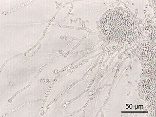
Candida is a genus of yeasts and is the most common cause of fungal infections worldwide. Many species are harmless commensals or endosymbionts of hosts including humans; however, when mucosal barriers are disrupted or the immune system is compromised they can invade and cause disease, known as an opportunistic infection. Candida is located on most of mucosal surfaces and mainly the gastrointestinal tract, along with the skin. Candida albicans is the most commonly isolated species and can cause infections in humans and other animals. In winemaking, some species of Candida can potentially spoil wines.

Mycosis fungal infection of animals, including humans

Mycosis is a fungal infection of animals, including humans. Mycoses are common and a variety of environmental and physiological conditions can contribute to the development of fungal diseases. Inhalation of fungal spores or localized colonization of the skin may initiate persistent infections; therefore, mycoses often start in the lungs or on the skin.

AIDS-defining clinical conditions is the list of diseases published by the Centers for Disease Control and Prevention (CDC) that are associated with AIDS, and used worldwide as a guideline for AIDS diagnosis. CDC exclusively uses the term AIDS-defining clinical conditions, but the other terms remain in common use.

Penicilliosis is an infection caused by Penicillium marneffei.

<i>Pneumocystis jirovecii</i> species of fungus

Pneumocystis jirovecii is a yeast-like fungus of the genus Pneumocystis. The causative organism of Pneumocystis pneumonia, it is an important human pathogen, particularly among immunocompromised hosts. Prior to its discovery as a human-specific pathogen, P. jirovecii was known as P. carinii.

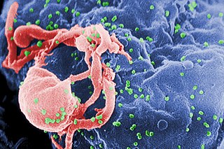
HIV superinfection is a condition in which a person with an established human immunodeficiency virus infection acquires a second strain of HIV, often of a different subtype. These can form a recombinant strain that co-exists with the strain from the initial infection, as well as the strain from the new virus, and may cause more rapid disease progression or carry multiple resistances to certain HIV medications.

Mycoviruses, also known as mycophages, are viruses that infect fungi. The majority of mycoviruses have double-stranded RNA (dsRNA) genomes and isometric particles, but approximately 30% have positive-sense, single-stranded RNA (+ssRNA) genomes.

<i>Sporothrix schenckii</i> species of fungus

Sporothrix schenckii is a fungus that can be found worldwide in the environment. The species is present in soil as well as in and on living and decomposing plant material such as peat moss. It can infect humans as well as animals and is the causative agent of sporotrichosis, commonly known as "rose handler's disease". The most common route of infection is the introduction of spores to the body through a cut or puncture wound in the skin. Infection commonly occurs in otherwise healthy individuals but is rarely life-threatening and can be treated with antifungals. In the environment it is found growing as filamentous hyphae. In host tissue it is found as a yeast. The transition between the hyphal and yeast forms is temperature dependent making S. schenckii a thermally dimorphic fungus.

Chrysovirus is a genus of viruses. It is the only genus in the family Chrysoviridae. They are class III double stranded RNA viruses which infect fungi, in particular Penicillium. Their name is derived from the Greek word chrysos which means yellow-green. There are currently nine species in this genus including the type species Penicillium chrysogenum virus.

Dimorphic fungus

Dimorphic fungi are fungi that can exist in the form of both mold and yeast. An example is Penicillium marneffei, a human pathogen that grows as a mold at room temperature, and as a yeast at human body temperature.

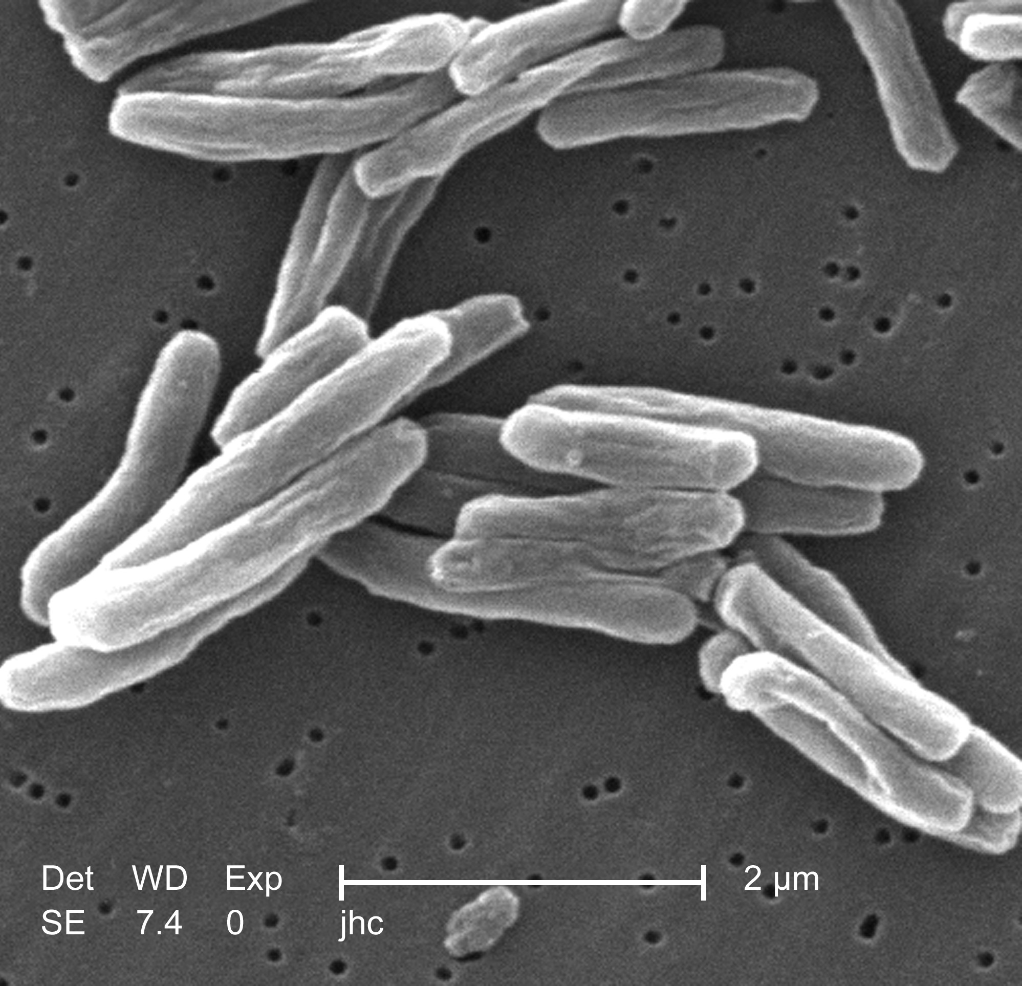
Pathogenic fungi are fungi that cause disease in humans or other organisms. Approximately 300 fungi are known to be pathogenic to humans. The study of fungi pathogenic to humans is called "medical mycology". Although fungi are eukaryotic, many pathogenic fungi are microorganisms. The study of fungi and other organisms pathogenic to plants is called plant pathology.

Tat (HIV)

In molecular biology, Tat is a protein that is encoded for by the tat gene in HIV-1. Tat is a regulatory protein that drastically enhances the efficiency of viral transcription.

The stages of HIV infection are acute infection, latency and AIDS. Acute infection lasts for several weeks and may include symptoms such as fever, swollen lymph nodes, inflammation of the throat, rash, muscle pain, malaise, and mouth and esophageal sores. The latency stage involves few or no symptoms and can last anywhere from two weeks to twenty years or more, depending on the individual. AIDS, the final stage of HIV infection, is defined by low CD4+ T cell counts, various opportunistic infections, cancers and other conditions.

<i>Talaromyces</i> genus of fungi

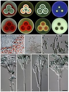
Talaromyces is a genus of fungi in the family Trichocomaceae. Described in 1955 by American mycologist Chester Ray Benjamin, species in the genus form soft, cottony fruit bodies (ascocarps) with cell walls made of tightly interwoven hyphae. The fruit bodies are often yellowish or are surrounded by yellowish granules. A 2008 estimate placed 42 species in the genus, but several new species have since been described.

Histoplasma duboisii is a saprotrophic fungus responsible for the invasive infection known as African histoplasmosis. This species is a close relative of Histoplasma capsulatum, the agent of classical histoplasmosis, and the two occur in similar habitats. Histoplasma duboisii is restricted to continental Africa and Madagascar, although scattered reports have arisen from other places usually in individuals with an African travel history. Like, H. capsulatum, H. duboisii is dimorphic – growing as a filamentous fungus at ambient temperature and a yeast at body temperature. It differs morphologically from H. capsulatum by the typical production of a large-celled yeast form. Both agents cause similar forms of disease, although H. duboisii predominantly causes cutaneous and subcutaneous disease in humans and non-human primates. The agent responds to many antifungal drug therapies used to treat serious fungal diseases.

References

  1. Chan JF, Lau SK, Yuen KY, Woo PC (March 2016). "Talaromyces (Penicillium) marneffei infection in non-HIV-infected patients". Emerging Microbes & Infections. 5 (3): e19. doi:10.1038/emi.2016.18. PMC   4820671 . PMID   26956447.
  2. Hyde, Kevin D.; Al-Hatmi, Abdullah M. S.; Andersen, Birgitte; Boekhout, Teun; Buzina, Walter; Dawson, Thomas L.; Eastwood, Dan C.; Jones, E. B. Gareth; de Hoog, Sybren (2018-11-10). "The world's ten most feared fungi". Fungal Diversity. 93: 161–194. doi:10.1007/s13225-018-0413-9. ISSN   1560-2745.
  3. Lo Y, Tintelnot K, Lippert U, Hoppe T (2000). "Disseminated Penicillium marneffei infection in an African AIDS patient". Transactions of the Royal Society of Tropical Medicine and Hygiene. 94 (2): 187. doi:10.1016/S0035-9203(00)90271-2. PMID   10897365.
  4. Capponi M, Segretain G, Sureau P (1956). "Pénicillose de Rhizomys sinensis". Bull Soc Pathol Exot. 49 (3): 418–21.
  5. Louthrenoo W, Thamprasert K, Sirisanthana T (December 1994). "Osteoarticular penicilliosis marneffei. A report of eight cases and review of the literature". British Journal of Rheumatology. 33 (12): 1145–50. doi:10.1093/rheumatology/33.12.1145. PMID   8000744.
  6. Duong TA (July 1996). "Infection due to Penicillium marneffei, an emerging pathogen: review of 155 reported cases". Clinical Infectious Diseases. 23 (1): 125–30. doi:10.1093/clinids/23.1.125. PMID   8816141.
  7. Supparatpinyo K, Khamwan C, Baosoung V, Nelson KE, Sirisanthana T (July 1994). "Disseminated Penicillium marneffei infection in southeast Asia". Lancet. 344 (8915): 110–3. doi:10.1016/s0140-6736(94)91287-4. PMID   7912350.
  8. 1 2 Henk DA, Shahar-Golan R, Devi KR, Boyce KJ, Zhan N, Fedorova ND, Nierman WC, Hsueh PR, Yuen KY, Sieu TP, Kinh NV, Wertheim H, Baker SG, Day JN, Vanittanakom N, Bignell EM, Andrianopoulos A, Fisher MC (2012). "Clonality despite sex: the evolution of host-associated sexual neighborhoods in the pathogenic fungus Penicillium marneffei". PLoS Pathogens. 8 (10): e1002851. doi:10.1371/journal.ppat.1002851. PMC   3464222 . PMID   23055919.
  9. Lau SK, Lo GC, Chow FW, Fan RY, Cai JJ, Yuen KY, Woo PC (June 2018). "Novel Partitivirus Enhances Virulence of and Causes Aberrant Gene Expression in Talaromyces marneffei". mBio. 9 (3): e00947–18. doi:10.1128/mBio.00947-18. PMC   6016240 . PMID   29895639. CC-BY icon.svg Material was copied from this source, which is available under a Creative Commons Attribution 4.0 International License.
  10. Lau, Susanna K. P.; Chow, Wang-Ngai; Wong, Annette Y. P.; Yeung, Julian M. Y.; Bao, Jessie; Zhang, Na; Lok, Si; Woo, Patrick C. Y.; Yuen, Kwok-Yung (2013-08-22). "Identification of MicroRNA-Like RNAs in Mycelial and Yeast Phases of the Thermal Dimorphic Fungus Penicillium marneffei". PLoS Neglected Tropical Diseases. 7 (8): e2398. doi:10.1371/journal.pntd.0002398. ISSN   1935-2735. PMC   3749987 . PMID   23991243.

Further reading