Peripheral giant-cell granuloma

Last updated
Peripheral giant-cell granuloma
Specialty Dentistry, ENT surgery

Peripheral giant-cell granuloma (PGCG) is an oral pathologic condition that appears in the mouth as an overgrowth of tissue due to irritation or trauma. Because of its overwhelming incidence on the gingiva, the condition is associated with two other diseases, pyogenic granuloma and peripheral ossifying fibroma. These three diseases are associated because they appear frequently on gingiva. Due to its similar microscopic appearance, peripheral giant-cell granuloma is considered to be the soft tissue equivalent of central giant-cell granuloma.

Mouth first portion of the alimentary canal that receives food

In animal anatomy, the mouth, also known as the oral cavity, buccal cavity, or in Latin cavum oris, is the opening through which many animals take in food and issue vocal sounds. It is also the cavity lying at the upper end of the alimentary canal, bounded on the outside by the lips and inside by the pharynx and containing in higher vertebrates the tongue and teeth. This cavity is also known as the buccal cavity, from the Latin bucca ("cheek").

Hyperplasia increase in the amount of organic tissue that results from cell proliferation

Hyperplasia, or hypergenesis, is an increase in the amount of organic tissue that results from cell proliferation. It may lead to the gross enlargement of an organ, and the term is sometimes confused with benign neoplasia or benign tumor.

Irritation, in biology and physiology, is a state of inflammation or painful reaction to allergy or cell-lining damage. A stimulus or agent which induces the state of irritation is an irritant. Irritants are typically thought of as chemical agents but mechanical, thermal (heat), and radiative stimuli can also be irritants. Irritation also has non-clinical usages referring to bothersome physical or psychological pain or discomfort.

Contents

The appearance of peripheral giant-cell granuloma is also similar to pyogenic granuloma. The color ranges from red to bluish-purple, but is usually more blue in comparison to pyogenic granuloma. It can be sessile or pedunculated with the size usually being less than 2 cm.

Color Characteristic of human visual perception

Color, or colour, is the characteristic of human visual perception described through color categories, with names such as red, orange, yellow, green, blue, or purple. This perception of color derives from the stimulation of cone cells in the human eye by electromagnetic radiation in the visible spectrum. Color categories and physical specifications of color are associated with objects through the wavelength of the light that is reflected from them. This reflection is governed by the object's physical properties such as light absorption, emission spectra, etc.

The lesion has a 60% gender predilection to females. The prevalence of the peripheral giant-cell granuloma is highest around 50 - 60 years of age. It appears only on the gingiva or on an edentulous alveolar ridge. It is more often found in the mandible rather than the maxilla, in either anterior or posterior areas. The underlying alveolar bone can be destroyed, leaving a unique appearance referred to as "cupping resorption" or "saucerization".

Female The sex of an organism which produces ova

Female (♀) is the sex of an organism, or a part of an organism, that produces non-mobile ova. Barring rare medical conditions, most female mammals, including female humans, have two X chromosomes. Female characteristics vary between different species with some species containing more well defined female characteristics. Both genetics and environment shape the prenatal development of a female.

Maxilla upper jawbone formed from the fusion of two maxillary bones; includes the frontal portion of the palate of the mouth

The maxilla in animals is the upper fixed bone of the jaw formed from the fusion of two maxillary bones. The upper jaw includes the hard palate in the front of the mouth. The two maxillary bones are fused at the intermaxillary suture, forming the anterior nasal spine. This is similar to the mandible, which is also a fusion of two mandibular bones at the mandibular symphysis. The mandible is the movable part of the jaw.

Diagnosis

Peripheral giant-cell granuloma appears microscopically as a large number of multinucleated giant cells, which can have up to dozens of nuclei. Additionally, there are mesenchymal cells that are ovoid and spindle-shaped. Near the borders of the lesion, deposits of hemosiderin and hemorrhage is often found. In 50% of cases, ulcerations are present.

Microscope instrument used to see objects that are too small for the naked eye

A microscope is an instrument used to see objects that are too small to be seen by the naked eye. Microscopy is the science of investigating small objects and structures using such an instrument. Microscopic means invisible to the eye unless aided by a microscope.

Cell nucleus A membrane-bounded organelle of eukaryotic cells in which chromosomes are housed and replicated.

In cell biology, the nucleus is a membrane-bound organelle found in eukaryotic cells. Eukaryotes usually have a single nucleus, but a few cell types, such as mammalian red blood cells, have no nuclei, and a few others including osteoclasts have many.

Hemosiderin

Hemosiderin or haemosiderin is an iron-storage complex. It is only found within cells and appears to be a complex of ferritin, denatured ferritin and other material. The iron within deposits of hemosiderin is very poorly available to supply iron when needed. Hemosiderin can be identified histologically with Perls' Prussian-blue stain; iron in hemosiderin turns blue to black when exposed to potassium ferrocyanide. In normal animals, hemosiderin deposits are small and commonly inapparent without special stains. Excessive accumulation of hemosiderin is usually detected within cells of the mononuclear phagocyte system (MPS) or occasionally within epithelial cells of liver and kidney.

Treatment

Treatment usually involves surgical removal of the lesion down to the bone. If there are any adjacent teeth, they are cleaned thoroughly by scaling and root planing (SRP) to remove any possible source of irritation. Recurrence is around 10%.

Surgery Medical specialty

Surgery is a medical specialty that uses operative manual and instrumental techniques on a patient to investigate or treat a pathological condition such as a disease or injury, to help improve bodily function or appearance or to repair unwanted ruptured areas.

Scaling and root planing removing or eliminating etiologic agents from the teeth

Scaling and root planing, also known as conventional periodontal therapy, non-surgical periodontal therapy, or deep cleaning, is a procedure involving removal of dental plaque and calculus and then smoothing, or planing, of the (exposed) surfaces of the roots, removing cementum or dentine that is impregnated with calculus, toxins, or microorganisms, the etiologic agents that cause inflammation. This helps to establish a periodontium that is in remission of periodontal disease. Periodontal scalers and periodontal curettes are some of the tools involved.

See also

Related Research Articles

Gumma (pathology) A tertiary syphilis that is characterized by granulomatous lesions, called gummas, which are characterized by a center of necrotic tissue with a rubbery texture. They form in the liver, bones, and testes but may affect any organ.

A gumma is a soft, non-cancerous growth resulting from the tertiary stage of syphilis. It is a form of granuloma. Gummas are most commonly found in the liver, but can also be found in brain, heart, skin, bone, testis, and other tissues, leading to a variety of potential problems including neurological disorders or heart valve disease.

Granuloma inflammation consisting of immune cells known as macrophages

A granuloma is a structure formed during inflammation that is found in many diseases. It is a collection of immune cells known as macrophages. Granulomas form when the immune system attempts to wall off substances it perceives as foreign but is unable to eliminate. Such substances include infectious organisms including bacteria and fungi, as well as other materials such as keratin and suture fragments.

Leukoplakia condition of the mucous membranes

Leukoplakia generally refers to a firmly attached white patch on a mucous membrane which is associated with an increased risk of cancer. The edges of the lesion are typically abrupt and the lesion changes with time. Advanced forms may develop red patches. There are generally no other symptoms. It usually occurs within the mouth, although sometimes mucosa in other parts of the gastrointestinal tract, urinary tract, or genitals may be affected.

Chest radiograph

A chest radiograph, colloquially called a chest X-ray (CXR), or chest film, is a projection radiograph of the chest used to diagnose conditions affecting the chest, its contents, and nearby structures. Chest radiographs are the most common film taken in medicine.

Epulis fissuratum is a benign hyperplasia of fibrous connective tissue which develops as a reactive lesion to chronic mechanical irritation produced by the flange of a poorly fitting denture. More simply, epulis fissuratum is where excess folds of firm tissue form inside the mouth, as a result of rubbing on the edge of dentures that do not fit well. It is a harmless condition and does not represent oral cancer. Treatment is by simple surgical removal of the lesion, and also by adjustment of the denture or provision of a new denture.

Pyogenic granuloma

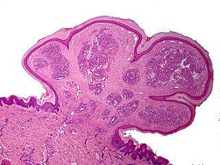
Pyogenic granuloma is a vascular lesion that occurs on both mucosa and skin, and appears as an overgrowth of tissue due to irritation, physical trauma, or hormonal factors. It is often found to involve the gums, the skin and nasal septum, and has also been found far from the head such as in the thigh.

Peripheral ossifying fibroma “a gingival nodule which is composed of a cellular fibroblastic connective tissue stroma which is associated with the formation of randomly dispersed foci of mineralised products, which consists of bone, cementum-like tissue, or a dystrophic calcification. The lesion is considered part of an ossifying fibroma, but that is usually considered to be a jaw tumor. Because of its overwhelming incidence on the gingiva, the condition is associated with two other diseases, though not because they occur together. Instead, the three are associated with each other because they appear frequently on gingiva: pyogenic granuloma and peripheral giant cell granuloma. Some researchers believe peripheral ossifying fibromas to be related to pyogenic fibromas and, in some instances, are the result of a pyogenic granuloma which has undergone fibrosis and calcification.

Oral medicine is a specialty focused on the mouth and nearby structures. It lies at the interface between medicine and dentistry.

Granuloma annulare Human disease

Granuloma annulare is a fairly rare, chronic skin condition which presents as reddish bumps on the skin arranged in a circle or ring. It can initially occur at any age and is four times more common in females.

Osteonecrosis of the jaw area of necrotic bone in the mandible or maxilla

Osteonecrosis of the jaw (ONJ) is a severe bone disease (osteonecrosis) that affects the jaws. Various forms of ONJ have been described over the last 160 years, and a number of causes have been suggested in the literature.

Giant-cell fibroma is a type of fibroma not associated with trauma or irritation. It can occur at any age and on a mucous membrane surface. The most common oral locations are on the gingiva of the mandible, tongue, and palate. It is a localized reactive proliferation of fibrous connective tissue.

Desquamative gingivitis is an erythematous (red), desquamatous (shedding) and ulcerated appearance of the gums. It is a descriptive term and can be caused by several different disorders.

Central giant-cell granuloma

Central giant-cell granuloma (CGCG) is a localised benign condition of the jaws. It is twice as common in females and is more likely to occur before age 30. Central giant-cell granulomas are more common in the anterior mandible, often crossing the midline and causing painless swellings.

Periapical cyst

The periapical cyst is the most common odontogenic cyst. Periapical is defined as "the tissues surrounding the apex of the root of a tooth" and a cyst is "a pathological cavity lined by epithelium, having fluid or gaseous content that is not created by the accumulation of pus." Most frequently located in the maxillary anterior region, it is caused by pulpal necrosis secondary to dental caries or trauma. The cyst has lining that is derived from the epithelial cell rests of Malassez which proliferate to form the cyst. Highly common in the oral cavity, the periapical cyst is asymptomatic, but highly significant because a secondary infection can cause pain and damage. In radiographs, it appears a radiolucency around the apex of a tooth's root.

Gingival enlargement Human disease

Gingival enlargement is an increase in the size of the gingiva (gums). It is a common feature of gingival disease. Gingival enlargement can be caused by a number of factors, including inflammatory conditions and the side effects of certain medications. The treatment is based on the cause. A closely related term is epulis, denoting a localized tumor on the gingiva.

Eosinophilic ulcer of the oral mucosa

Eosinophilic ulcer of the oral mucosa is a condition characterized by an ulcer with an indurated and elevated border. The lesion might be tender, fast-growing and the patient often not be aware of any trauma in the area.

Epulis is any tumor like enlargement situated on the gingival or alveolar mucosa. The word literally means "(growth) on the gingiva", and describes only the location of the mass and has no further implications on the nature of the lesion. There are three types: fibromatous, ossifying and acanthomatous. The related term parulis refers to a mass of inflamed granulation tissue at the opening of a draining sinus on the alveolus over the root of an infected tooth. Another closely related term is gingival enlargement, which tends to be used where the enlargement is more generalized over the whole gingiva rather than a localized mass.

A cyst is a pathological epithelial lined cavity that fills with fluid or soft material and usually grows from internal pressure generated by fluid being drawn into the cavity from osmosis. The bones of the jaws, the mandible and maxilla, are the bones with the highest prevalence of cysts in the human body. This is due to the abundant amount of epithelial remnants that can be left in the bones of the jaws. The enamel of teeth is formed from ectoderm, and so remnants of epithelium can be left in the bone during odontogenesis. The bones of the jaws develop from embryologic processes which fuse together, and ectodermal tissue may be trapped along the lines of this fusion. This "resting" epithelium is usually dormant or undergoes atrophy, but, when stimulated, may form a cyst. The reasons why resting epithelium may proliferate and undergo cystic transformation are generally unknown, but inflammation is thought to be a major factor. The high prevalence of tooth impactions and dental infections that occur in the bones of the jaws is also significant to explain why cysts are more common at these sites.

Amalgam tattoo A common discoloration of tissue in the mouth

Amalgam tattoo is a grey, blue or black area of discoloration on the mucous membranes of the mouth, typically on the gums of the lower jaw. It is a healthcare caused lesion, is due to entry of dental amalgam into the soft tissues. It is common, painless, and benign, but it can be mistaken for melanoma.

References

Classification
D