Retrocuspid papilla (RCP) is a small elevated nodules mostly behind the lower canine teeth in humans(Fig.1,2). [1] It is sometimes associated with reactive arthritis. [2]
Retrocuspid papilla (RCP) is a small elevated nodules mostly behind the lower canine teeth in humans(Fig.1,2). [1] It is sometimes associated with reactive arthritis. [2]
The RCP are first reported in 1947 and 1965. [3] [4] In a Swedish population it was first reported 1994. Among 1150 consecutively examined patients aged 20 –75 years, 10 showed RCP. Among 2000 biopsy cases from 1989 - 1992 in Department of Oral Pathology Lund University, 15 biopsies met the criteria of RCP. [5]
The lesions are bilaterally situated in the attached gingiva or close to the border of the mucosa lingual to the two mandibular canines (Fig.1). [6]

However, they could in a few individuals also be seen simultaneously in the molar region and on the lingual side (Fig.2). [7] They were 2–3 mm wide and high and covered with normal mucosa. Their tips were erected or could be folded down, mimicking the entrance of a periodontal abscess, but no duct was present.

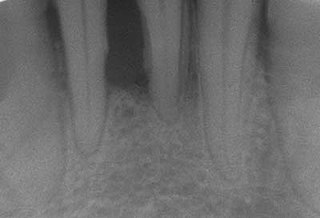
Radiographs showed no bone destruction and the depths of the periodontal pockets could not explain the presence of the lesions. They were nonsymptomatic and were not noticed by the patients. [8]

Immunohistochemical staining with FXIIIa [9] antibody disclosed a population of reactive spindle- or stellate-shaped cells [10] in 11 of 15 cases, located in connective tissue papillae and in a few cases also distributed throughout the lesion. The FXIIIa-stained cells appeared together with the frequently observed stellate, "young" occasionally multinucleated fibroblastic cells observed in more than 50% of patients aged 10–69 years. It is likely that FXIIIA-expressing "mucosal dendrocytes" are pathologically involved in some way. [11] [12] [13]

Human teeth function to mechanically break down items of food by cutting and crushing them in preparation for swallowing and digesting. As such, they are considered part of the human digestive system. Humans have four types of teeth: incisors, canines, premolars, and molars, which each have a specific function. The incisors cut the food, the canines tear the food and the molars and premolars crush the food. The roots of teeth are embedded in the maxilla or the mandible and are covered by gums. Teeth are made of multiple tissues of varying density and hardness.
Periodontal disease, also known as gum disease, is a set of inflammatory conditions affecting the tissues surrounding the teeth. In its early stage, called gingivitis, the gums become swollen and red and may bleed. It is considered the main cause of tooth loss for adults worldwide. In its more serious form, called periodontitis, the gums can pull away from the tooth, bone can be lost, and the teeth may loosen or fall out. Bad breath may also occur.
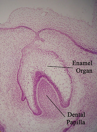
The enamel organ, also known as the dental organ, is a cellular aggregation seen in a developing tooth and it lies above the dental papilla. The enamel organ which is differentiated from the primitive oral epithelium lining the stomodeum.The enamel organ is responsible for the formation of enamel, initiation of dentine formation, establishment of the shape of a tooth's crown, and establishment of the dentoenamel junction.

Tooth development or odontogenesis is the complex process by which teeth form from embryonic cells, grow, and erupt into the mouth. For human teeth to have a healthy oral environment, all parts of the tooth must develop during appropriate stages of fetal development. Primary (baby) teeth start to form between the sixth and eighth week of prenatal development, and permanent teeth begin to form in the twentieth week. If teeth do not start to develop at or near these times, they will not develop at all, resulting in hypodontia or anodontia.
Periodontology or periodontics is the specialty of dentistry that studies supporting structures of teeth, as well as diseases and conditions that affect them. The supporting tissues are known as the periodontium, which includes the gingiva (gums), alveolar bone, cementum, and the periodontal ligament. A periodontist is a dentist that specializes in the prevention, diagnosis and treatment of periodontal disease and in the placement of dental implants.

The alveolar process or alveolar bone is the thickened ridge of bone that contains the tooth sockets on the jaw bones. The structures are covered by gums as part of the oral cavity.

Osteonecrosis of the jaw (ONJ) is a severe bone disease (osteonecrosis) that affects the jaws. Various forms of ONJ have been described since 1861, and a number of causes have been suggested in the literature.

Endometrial intraepithelial neoplasia (EIN) is a premalignant lesion of the uterine lining that predisposes to endometrioid endometrial adenocarcinoma. It is composed of a collection of abnormal endometrial cells, arising from the glands that line the uterus, which have a tendency over time to progress to the most common form of uterine cancer—endometrial adenocarcinoma, endometrioid type.
“Lateral periodontal cysts (LPCs) are defined as non-keratinised and non-inflammatory developmental cysts located adjacent or lateral to the root of a vital tooth.” LPCs are a rare form of jaw cysts, with the same histopathological characteristics as gingival cysts of adults (GCA). Hence LPCs are regarded as the intraosseous form of the extraosseous GCA. They are commonly found along the lateral periodontium or within the bone between the roots of vital teeth, around mandibular canines and premolars. Standish and Shafer reported the first well-documented case of LPCs in 1958, followed by Holder and Kunkel in the same year although it was called a periodontal cyst. Since then, there has been more than 270 well-documented cases of LPCs in literature.
Squamous odontogenic tumors (SOTs) are very rare benign locally infiltrative odontogenic neoplasms of epithelial origin. Only some 50 cases have been documented. They occur mostly from 20-40 and are more common in males. Treatment is by simple enucleation and local curettage, and recurrence is rare.
Lacazia is a genus of fungi containing the single species Lacazia loboi, which is responsible for Lobo's disease. It is a member of the order Onygenales.
Apical periodontitis is typically the body's defense response to the threat of microbial invasion from the root canal. Primary among the members of the host defense mechanism is the polymorphonuclear leukocyte, otherwise known as the neutrophil. The task of the neutrophil is to locate and destroy microbes that intrude into the body – anywhere in the body – and they represent the hallmark of acute inflammation.

Smoker's melanosis is seen with the naked eye as a brown to black pigmentation of the oral tissue i.e. the gums, cheeks or palate as well as in larynx. It is most often seen in the lower labial gingiva of tobacco users. Most easily it is found in Caucasians, due to their lack of a genetically caused melanin pigmentation.
Dental pertains to the teeth, including dentistry. Topics related to the dentistry, the human mouth and teeth include:
Gum depigmentation, also known as gum bleaching, is a procedure used in cosmetic dentistry to lighten or remove black spots or patches on the gums consisting of melanin. Melanin in skin is very common in inhabitants in many parts of the world due to genetic factors. Melanin pigmentation in skin, oral mucosa, inner ear and other organs is a detoxification mechanism. Some toxic agents bind to melanin and will move out of the tissue with the ageing cells and are expelled to the tissue surfaces. Also in the gums and oral mucosa a visible pigmentation is most often caused by genetic factors, but also by tobacco smoking or in a few cases by long-term use of certain medications. If stopping smoking or change of medication do not solve the problem with a disfigurating melanin pigmentation, a surgical operation may be performed. The procedure itself can involve laser ablation techniques.
Verruciform xanthoma is an uncommon benign lesion that has a verruciform (wart-like) appearance, but it may appear polypoid, papillomatous, or sessile. The verruciform was first described by Shafer in 1971 on the oral mucosa. Usually found on the oral mucosa of middle-aged persons, verruciform xanthomas have also been reported on the scrotum and penis of middle-aged to elderly Japanese males. While the most common site is the oral mucosa, lesions that occur elsewhere usually arise on the perineum or on the skin with some predisposing factor, such as lymphedema or an epidermal nevus.

Plasma cell gingivitis is a rare condition, appearing as generalized erythema (redness) and edema (swelling) of the attached gingiva, occasionally accompanied by cheilitis or glossitis. It is called plasma cell gingivitis where the gingiva (gums) are involved, plasma cell cheilitis, where the lips are involved, and other terms such as plasma cell orifacial mucositis, or plasma cell gingivostomatitis where several sites in the mouth are involved. On the lips, the condition appears as sharply outlined, infiltrated, dark red plaque with a lacquer-like glazing of the surface of the involved oral area.
Chronic periodontitis is one of the seven categories of periodontitis as defined by the American Academy of Periodontology 1999 classification system. Chronic periodontitis is a common disease of the oral cavity consisting of chronic inflammation of the periodontal tissues that is caused by the accumulation of profuse amounts of dental plaque. Periodontitis initially begins as gingivitis and can progress onto chronic and subsequent aggressive periodontitis according to the 1999 classification.
Hector L. Sarmiento is an American periodontist involved in dental implant complications research.