Fatty liver

Last updated
Fatty liver
Synonyms Fatty liver disease (FLD), hepatic steatosis, simple steatosis
Non-alcoholic fatty liver disease1.jpg
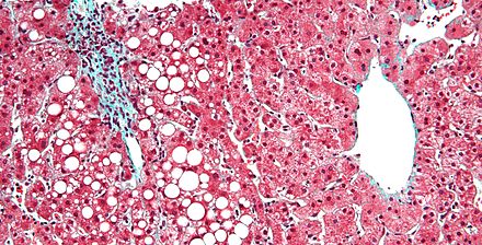
Micrograph showing a fatty liver (macrovesicular steatosis), as seen in non-alcoholic fatty liver disease. Trichrome stain.
Specialty Gastroenterology

Fatty liver , or hepatic steatosis or simple steatosis, is a reversible condition wherein large vacuoles of triglyceride fat accumulate in liver cells via the process of steatosis (i.e., abnormal retention of lipids within a cell). Despite having multiple causes, fatty liver can be considered a single disease that occurs worldwide in those with excessive alcohol intake and the obese (with or without effects of insulin resistance). The condition is also associated with other diseases that influence fat metabolism. [1] When this process of fat metabolism is disrupted, the fat can accumulate in the liver in excessive amounts, thus resulting in a fatty liver. [2] It is difficult to distinguish alcoholic FLD, which is part of alcoholic liver disease, from nonalcoholic FLD (NAFLD), and both show microvesicular and macrovesicular fatty changes at different stages.

Liver vital organ in vertebrates and some other animals

The liver, an organ only found in vertebrates, detoxifies various metabolites, synthesizes proteins, and produces biochemicals necessary for digestion. In humans, it is located in the right upper quadrant of the abdomen, below the diaphragm. Its other roles in metabolism include the regulation of glycogen storage, decomposition of red blood cells and the production of hormones.

Vacuole A closed structure, found only in eukaryotic cells, that is completely surrounded by unit membrane and contains liquid material. Cells contain one or several vacuoles, that may have different functions from each other. Vacuoles have a diverse array o

A vacuole is a membrane-bound organelle which is present in all plant and fungal cells and some protist, animal and bacterial cells. Vacuoles are essentially enclosed compartments which are filled with water containing inorganic and organic molecules including enzymes in solution, though in certain cases they may contain solids which have been engulfed. Vacuoles are formed by the fusion of multiple membrane vesicles and are effectively just larger forms of these. The organelle has no basic shape or size; its structure varies according to the requirements of the cell.

Triglyceride any ester of glycerol having all three hydroxyl groups esterified with fatty acids

A triglyceride is an ester derived from glycerol and three fatty acids. Triglycerides are the main constituents of body fat in humans and other animals, as well as vegetable fat. They are also present in the blood to enable the bidirectional transference of adipose fat and blood glucose from the liver, and are a major component of human skin oils.

Contents

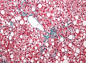
The accumulation of fat in alcoholic or non-alcoholic steatosis may also be accompanied by a progressive inflammation of the liver (hepatitis), called steatohepatitis. This more severe condition may be termed either alcoholic steatohepatitis or non-alcoholic steatohepatitis (NASH).

Hepatitis inflammation of the liver tissue

Hepatitis is inflammation of the liver tissue. Some people have no symptoms whereas others develop yellow discoloration of the skin and whites of the eyes, poor appetite, vomiting, tiredness, abdominal pain, or diarrhea. Hepatitis may be temporary (acute) or long term (chronic) depending on whether it lasts for less than or more than six months. Acute hepatitis can sometimes resolve on its own, progress to chronic hepatitis, or rarely result in acute liver failure. Over time the chronic form may progress to scarring of the liver, liver failure, or liver cancer.

Steatohepatitis steatotis hepatis macrovezacularis

Steatohepatitis is a type of fatty liver disease, characterized by inflammation of the liver with concurrent fat accumulation in liver. Mere deposition of fat in the liver is termed steatosis, and together these constitute fatty liver changes.

Signs and symptoms

Complications

Fatty liver can develop into a fibrosis or a liver cancer. [3] For people affected by NAFLD, the 10-year survival rate was about 80%. The rate of progression of fibrosis in NASH is estimated to one per 7 years and 14 years for NAFLD, with an increasing speed. [4] [5] There is a strong relationship between these pathologies and metabolic illnesses (diabetes type II, metabolic syndrome). These pathologies can also affect non-obese people, who are then at a higher risk. [3]

Less than 10% of people with cirrhotic alcoholic FLD will develop hepatocellular carcinoma, [6] the liver cancer, but up to 45% people with NASH without cirrhosis can develop hepatocellular carcinoma. [7]

Hepatocellular carcinoma liver carcinoma that has material basis in undifferentiated hepatocytes

Hepatocellular carcinoma (HCC) is the most common type of primary liver cancer in adults, and is the most common cause of death in people with cirrhosis.

Causes

Different stages of liver damage Stage of liver damage.JPG
Different stages of liver damage

Fatty liver (FL) is commonly associated with metabolic syndrome (diabetes, hypertension, obesity, and dyslipidemia), but can also be due to any one of many causes: [8] [9]

Metabolic syndrome clustering of medical conditions (giving a total of 16 possible combinations giving the syndrome)

Metabolic syndrome, sometimes known by other names, is a clustering of at least three of the five following medical conditions: central obesity, high blood pressure, high blood sugar, high serum triglycerides, and low serum high-density lipoprotein (HDL).

Hypertension high blood pressure

Hypertension, also known as high blood pressure (HBP), is a long-term medical condition in which the blood pressure in the arteries is persistently elevated. High blood pressure typically does not cause symptoms. Long-term high blood pressure, however, is a major risk factor for coronary artery disease, stroke, heart failure, atrial fibrillation, peripheral vascular disease, vision loss, chronic kidney disease, and dementia.

Obesity medical condition in which excess body fat harms health

Obesity is a medical condition in which excess body fat has accumulated to an extent that it may have a negative effect on health. People are generally considered obese when their body mass index (BMI), a measurement obtained by dividing a person's weight by the square of the person's height, is over 30 kg/m2; the range 25–30 kg/m2 is defined as overweight. Some East Asian countries use lower values. Obesity increases the likelihood of various diseases and conditions, particularly cardiovascular diseases, type 2 diabetes, obstructive sleep apnea, certain types of cancer, osteoarthritis, and depression.

Metabolic
abetalipoproteinemia, glycogen storage diseases, Weber–Christian disease, acute fatty liver of pregnancy, lipodystrophy
Nutritional
obesity, malnutrition, total parenteral nutrition, severe weight loss, refeeding syndrome, jejunoileal bypass, gastric bypass, jejunal diverticulosis with bacterial overgrowth
Drugs and toxins
amiodarone, methotrexate, diltiazem, expired tetracycline, highly active antiretroviral therapy, glucocorticoids, tamoxifen, [10] environmental hepatotoxins (e.g., phosphorus, mushroom poisoning)
Alcohol
Alcoholism is one of the causes of fatty liver due to production of toxic metabolites like aldehydes during metabolism of alcohol in the liver. This phenomenon most commonly occurs with chronic alcoholism.
Other
celiac disease, [11] inflammatory bowel disease, HIV, hepatitis C (especially genotype 3), and alpha 1-antitrypsin deficiency [12]

Pathology

Micrograph of periportal hepatic steatosis, as may be seen due to steroid use, trichrome stain Periportal hepatosteatosis intermed mag.jpg
Micrograph of periportal hepatic steatosis, as may be seen due to steroid use, trichrome stain

Fatty change represents the intracytoplasmatic accumulation of triglycerides (neutral fats). At the beginning, the hepatocytes present small fat vacuoles (liposomes) around the nucleus (microvesicular fatty change). In this stage, liver cells are filled with multiple fat droplets that do not displace the centrally located nucleus. In the late stages, the size of the vacuoles increases, pushing the nucleus to the periphery of the cell, giving characteristic signet ring appearance (macrovesicular fatty change). These vesicles are well-delineated and optically "empty" because fats dissolve during tissue processing. Large vacuoles may coalesce and produce fatty cysts, which are irreversible lesions. Macrovesicular steatosis is the most common form and is typically associated with alcohol, diabetes, obesity, and corticosteroids. Acute fatty liver of pregnancy and Reye's syndrome are examples of severe liver disease caused by microvesicular fatty change. [13] The diagnosis of steatosis is made when fat in the liver exceeds 5–10% by weight. [1] [14] [15]

Cytoplasm all of the contents of a cell excluding the plasma membrane and nucleus, but including other subcellular structures

In cell biology, the cytoplasm is all of the material within a cell, enclosed by the cell membrane, except for the cell nucleus. The material inside the nucleus and contained within the nuclear membrane is termed the nucleoplasm. The main components of the cytoplasm are cytosol – a gel-like substance, the organelles – the cell's internal sub-structures, and various cytoplasmic inclusions. The cytoplasm is about 80% water and usually colorless.

Cell nucleus A membrane-bounded organelle of eukaryotic cells in which chromosomes are housed and replicated.

In cell biology, the nucleus is a membrane-bound organelle found in eukaryotic cells. Eukaryotes usually have a single nucleus, but a few cell types, such as mammalian red blood cells, have no nuclei, and a few others including osteoclasts have many.

Cyst closed sac growth on the body

A cyst is a closed sac, having a distinct membrane and division compared with the nearby tissue. Hence, it is a cluster of cells that has grouped together to form a sac ; however, the distinguishing aspect of a cyst is that the cells forming the "shell" of such a sac are distinctly abnormal when compared with all surrounding cells for that given location. It may contain air, fluids, or semi-solid material. A collection of pus is called an abscess, not a cyst. Once formed, sometimes a cyst may resolve on its own. When a cyst fails to resolve, it may need to be removed surgically, but that would depend upon its type and location.

Mechanism leading to hepatic steatosis Steatosis.jpg
Mechanism leading to hepatic steatosis

Defects in fatty acid metabolism are responsible for pathogenesis of FLD, which may be due to imbalance in energy consumption and its combustion, resulting in lipid storage, or can be a consequence of peripheral resistance to insulin, whereby the transport of fatty acids from adipose tissue to the liver is increased. [1] [16] Impairment or inhibition of receptor molecules (PPAR-α, PPAR-γ and SREBP1) that control the enzymes responsible for the oxidation and synthesis of fatty acids appears to contribute to fat accumulation. In addition, alcoholism is known to damage mitochondria and other cellular structures, further impairing cellular energy mechanism. On the other hand, non-alcoholic FLD may begin as excess of unmetabolised energy in liver cells. Hepatic steatosis is considered reversible and to some extent nonprogressive if the underlying cause is reduced or removed.

Micrograph of inflamed fatty liver (steatohepatitis) Steatohepatitis high mag.jpg
Micrograph of inflamed fatty liver (steatohepatitis)

Severe fatty liver is sometimes accompanied by inflammation, a situation referred to as steatohepatitis. Progression to alcoholic steatohepatitis (ASH) or non-alcoholic steatohepatitis (NASH) depends on the persistence or severity of the inciting cause. Pathological lesions in both conditions are similar. However, the extent of inflammatory response varies widely and does not always correlate with degree of fat accumulation. Steatosis (retention of lipid) and onset of steatohepatitis may represent successive stages in FLD progression. [17]

Liver disease with extensive inflammation and a high degree of steatosis often progresses to more severe forms of the disease. [18] Hepatocyte ballooning and necrosis of varying degrees are often present at this stage. Liver cell death and inflammatory responses lead to the activation of hepatic stellate cells, which play a pivotal role in hepatic fibrosis. The extent of fibrosis varies widely. Perisinusoidal fibrosis is most common, especially in adults, and predominates in zone 3 around the terminal hepatic veins. [19]

The progression to cirrhosis may be influenced by the amount of fat and degree of steatohepatitis and by a variety of other sensitizing factors. In alcoholic FLD, the transition to cirrhosis related to continued alcohol consumption is well-documented, but the process involved in non-alcoholic FLD is less clear.

Diagnosis

Liver steatosis (fatty liver disease) as seen on CT Liversteatosis.png
Liver steatosis (fatty liver disease) as seen on CT
Ultrasound showing diffuse increased echogenicity of the liver. Steatosis hepatis - Sonographie 001.jpg
Ultrasound showing diffuse increased echogenicity of the liver.
Flow chart for diagnosis, modified from [9]


Elevated liver enzyme


Serology to exclude viral hepatitis


Imaging study showing
fatty infiltrate


Alcohol intake


Less than two drinks per day‡
 
More than two drinks per day‡


Nonalcoholic fatty liver disease likely
 
Alcoholic liver disease likely
 
 
Criteria for nonalcoholic fatty liver disease:
consumption of ethanol less than 20 g/day for women and 30 g/day for men [20]

Most individuals are asymptomatic and are usually discovered incidentally because of abnormal liver function tests or hepatomegaly noted in unrelated medical conditions. Elevated liver biochemistry is found in 50% of patients with simple steatosis. [21] The serum alanine transaminase level usually is greater than the aspartate transaminase level in the nonalcoholic variant and the opposite in alcoholic FLD (AST:ALT more than 2:1).

Imaging studies are often obtained during the evaluation process. Ultrasonography reveals a "bright" liver with increased echogenicity. Medical imaging can aid in diagnosis of fatty liver; fatty livers have lower density than spleens on computed tomography (CT), and fat appears bright in T1-weighted magnetic resonance images (MRIs). Magnetic resonance elastography, a variant of magnetic resonance imaging, is investigated as a non-invasive method to diagnose fibrosis progression. [22] Histological diagnosis by liver biopsy is the most accurate measure of fibrosis and liver fat progression as of 2018. [3]

Treatment

The treatment of fatty liver depends on its cause, and, in general, treating the underlying cause will reverse the process of steatosis if implemented at an early stage. Fatty liver can be caused by different factors that are not all identified. However, two known causes of fatty liver disease are an excess consumption of alcohol and a prolonged diet with a high proportion of calories coming from carbohydrates. For people with NAFLD or NASH, weight loss via a combination of diet and exercise was shown to improve or resolve the disease. [3] In more serious cases, medications that decrease insulin resistance, hyperlipidemia, and those that induce weight loss such as bariatric surgery as well as Vitamin E have been shown to improve or resolve liver function. [3] [9]

Bariatric surgery, while not recommended in 2017 as a treatment for fatty liver disease (FLD) alone, has been shown to revert FLD, NAFLD, NASH and advanced steatohepatitis in over 90% of people who have undergone this surgery for the treatment of obesity. [3] [23]

In the case of long-term total parenteral nutrition induced fatty liver disease, choline has been shown to alleviate symptoms. [24] [25] [26] This may be due to a deficiency in the methionine cycle. [27]

Epidemiology

The prevalence of FLD in the general population ranges from 10% to 24% in various countries. [8] However, the NAFLD condition is observed in up to 80% of obese people, 35% of whom progress to NASH, [28] and in up to 20% of normal weight people, [5] despite no evidence of excessive alcohol consumption. FLD is the most common cause of abnormal liver function tests in the United States. [8] Fatty liver is more prevalent in hispanic people than white, with black people having the lowest susceptibility. [5]

See also

Related Research Articles

Parenteral nutrition

Parenteral nutrition (PN) is the feeding of specialist nutritional products to a person intravenously, bypassing the usual process of eating and digestion. The products are made by specialist pharmaceutical compounding companies and is considered to be the highest risk pharmaceutical preparation available as the products cannot undergo any form of terminal sterilization. The person receives highly complex nutritional formulae that contain nutrients such as glucose, salts, amino acids, lipids and added vitamins and dietary minerals. It is called total parenteral nutrition (TPN) or total nutrient admixture (TNA) when no significant nutrition is obtained by other routes, and partial parenteral nutrition (PPN) when nutrition is also partially enteric. It may be called peripheral parenteral nutrition (PPN) when administered through vein access in a limb rather than through a central vein as central venous nutrition (CVN).

Alcoholic liver disease

Alcoholic liver disease is a term that encompasses the liver manifestations of alcohol overconsumption, including fatty liver, alcoholic hepatitis, and chronic hepatitis with liver fibrosis or cirrhosis.

Steatosis

Steatosis, also called fatty change, is the process describing the abnormal retention of lipids within a cell or organ. When not further specified, it is defined as affecting the liver. It reflects an impairment of the normal processes of synthesis and elimination of triglyceride fat. Excess lipid accumulates in vesicles that displace the cytoplasm. When the vesicles are large enough to distort the nucleus, the condition is known as macrovesicular steatosis; otherwise, the condition is known as microvesicular steatosis. While not particularly detrimental to the cell in mild cases, large accumulations can disrupt cell constituents, and in severe cases the cell may even burst.

Alcoholic hepatitis hepatitis (inflammation of the liver) due to excessive intake of alcohol

Alcoholic hepatitis is hepatitis due to excessive intake of alcohol. It is usually found in association with fatty liver, an early stage of alcoholic liver disease, and may contribute to the progression of fibrosis, leading to cirrhosis. Signs and symptoms of alcoholic hepatitis include jaundice, ascites, fatigue and hepatic encephalopathy. Mild cases are self-limiting, but severe cases have a high risk of death. Severe cases may be treated with glucocorticoids.

Liver disease Human disease

Liver disease is a type of damage to or disease of the liver.

Liver biopsy

Liver biopsy is the biopsy from the liver. It is a medical test that is done to aid diagnosis of liver disease, to assess the severity of known liver disease, and to monitor the progress of treatment.

Non-alcoholic fatty liver disease fatty liver disease characterized by the storing of excess fat in liver cells which is is not caused by heavy alcohol use

Non-alcoholic fatty liver disease (NAFLD) is when excess fat builds up in the liver due to causes other than alcohol use. There are two types: non-alcoholic fatty liver (NAFL) and non-alcoholic steatohepatitis (NASH). Non-alcoholic fatty liver usually does not progress to liver damage or NASH. NASH includes both a fatty liver and liver inflammation. It may lead to complications such as cirrhosis, liver cancer, liver failure, or cardiovascular disease.

Acute fatty liver of pregnancy is a rare life-threatening complication of pregnancy that occurs in the third trimester or the immediate period after delivery. It is thought to be caused by a disordered metabolism of fatty acids by mitochondria in the mother, caused by long-chain 3-hydroxyacyl-coenzyme A dehydrogenase deficiency. The condition was previously thought to be universally fatal, but aggressive treatment by stabilizing the mother with intravenous fluids and blood products in anticipation of early delivery has improved prognosis.

Liver failure

Liver failure or hepatic insufficiency is the inability of the liver to perform its normal synthetic and metabolic function as part of normal physiology. Two forms are recognised, acute and chronic. Recently a third form of liver failure known as acute-on-chronic liver failure (ACLF) is increasingly being recognized.

Phosphatidylethanolamine N-methyltransferase protein-coding gene in the species Homo sapiens

Phosphatidylethanolamine N-methyltransferase is a transferase enzyme which converts phosphatidylethanolamine (PE) to phosphatidylcholine (PC) in the liver. In humans it is encoded by the PEMT gene within the Smith-Magenis syndrome region on chromosome 17.

Cirrhosis long-term disease of the liver

Cirrhosis is a condition in which the liver does not function properly due to long-term damage. This damage is characterized by the replacement of normal liver tissue by scar tissue. Typically, the disease develops slowly over months or years. Early on, there are often no symptoms. As the disease worsens, a person may become tired, weak, itchy, have swelling in the lower legs, develop yellow skin, bruise easily, have fluid build up in the abdomen, or develop spider-like blood vessels on the skin. The fluid build-up in the abdomen may become spontaneously infected. Other serious complications include hepatic encephalopathy, bleeding from dilated veins in the esophagus or dilated stomach veins, and liver cancer. Hepatic encephalopathy results in confusion and may lead to unconsciousness.

The AST/ALT ratio is the ratio between the concentrations of the enzymes aspartate transaminase (AST) and alanine transaminase, aka alanine aminotransferase (ALT) in the blood of a human or animal. It is measured with a blood test and is sometimes useful in medical diagnosis to differentiate between causes of liver damage, or hepatotoxicity.

Ballooning degeneration form of liver parenchymal cell (i.e. hepatocyte) death

In histopathology, ballooning degeneration, formally ballooning degeneration of hepatocytes, is a form of liver parenchymal cell death.

Lipotoxicity is a metabolic syndrome that results from the accumulation of lipid intermediates in non-adipose tissue, leading to cellular dysfunction and death. The tissues normally affected include the kidneys, liver, heart and skeletal muscle. Lipotoxicity is believed to have a role in heart failure, obesity, and diabetes, and is estimated to affect approximately 25% of the adult American population.

Elafibranor chemical compound

Elafibranor is an experimental medication that is being studied and developed by Genfit for the treatment of cardiometabolic diseases including diabetes, insulin resistance, dyslipidemia, and non-alcoholic fatty liver disease (NAFLD).

Obeticholic acid chemical compound

Obeticholic acid, is a semi-synthetic bile acid analogue which has the chemical structure 6α-ethyl-chenodeoxycholic acid. It is used as a drug to treat primary biliary cholangitis, and is undergoing development for several other liver diseases and related disorders. Intercept Pharmaceuticals Inc. hold the worldwide rights to develop OCA outside Japan and China, where it is licensed to Dainippon Sumitomo Pharma.

Aramchol is an investigational drug being developed by Galmed Pharmaceuticals as a first-in-class, potentially disease modifying treatment for nonalcoholic steatohepatitis, or NASH, a more advanced condition of non-alcoholic fatty liver disease.

References

  1. 1 2 3 Reddy JK, Rao MS (May 2006). "Lipid metabolism and liver inflammation. II. Fatty liver disease and fatty acid oxidation". American Journal of Physiology. Gastrointestinal and Liver Physiology. 290 (5): G852–8. doi:10.1152/ajpgi.00521.2005. PMID   16603729.
  2. Cutler N. "Symptoms of Fatty Liver" . Retrieved 18 November 2014.
  3. 1 2 3 4 5 6 Chalasani, Naga; Younossi, Zobair; Lavine, Joel E.; Charlton, Michael; Cusi, Kenneth; Rinella, Mary; Harrison, Stephen A.; Brunt, Elizabeth M.; Sanyal, Arun J. (January 2018). "The diagnosis and management of nonalcoholic fatty liver disease: Practice guidance from the American Association for the Study of Liver Diseases". Hepatology. 67 (1): 328–357. doi:10.1002/hep.29367.
  4. Singh, Siddharth; Allen, Alina M.; Wang, Zhen; Prokop, Larry J.; Murad, Mohammad H.; Loomba, Rohit (April 2015). "Fibrosis Progression in Nonalcoholic Fatty Liver vs Nonalcoholic Steatohepatitis: A Systematic Review and Meta-analysis of Paired-Biopsy Studies". Clinical Gastroenterology and Hepatology. 13 (4): 643–654.e9. doi:10.1016/j.cgh.2014.04.014. PMC   4208976 .
  5. 1 2 3 Younossi, Zobair; Anstee, Quentin M.; Marietti, Milena; Hardy, Timothy; Henry, Linda; Eslam, Mohammed; George, Jacob; Bugianesi, Elisabetta (20 September 2017). "Global burden of NAFLD and NASH: trends, predictions, risk factors and prevention". Nature Reviews Gastroenterology & Hepatology. 15 (1): 11–20. doi:10.1038/nrgastro.2017.109.
  6. Qian Y, Fan JG (May 2005). "Obesity, fatty liver and liver cancer". Hepatobiliary & Pancreatic Diseases International. 4 (2): 173–7. PMID   15908310.
  7. Bellentani, Stefano (January 2017). "The epidemiology of non-alcoholic fatty liver disease". Liver International. 37: 81–84. doi:10.1111/liv.13299.
  8. 1 2 3 Angulo P (April 2002). "Nonalcoholic fatty liver disease". The New England Journal of Medicine. 346 (16): 1221–31. doi:10.1056/NEJMra011775. PMID   11961152.
  9. 1 2 3 Bayard M, Holt J, Boroughs E (June 2006). "Nonalcoholic fatty liver disease". American Family Physician. 73 (11): 1961–8. PMID   16770927.
  10. Osman KA, Osman MM, Ahmed MH (January 2007). "Tamoxifen-induced non-alcoholic steatohepatitis: where are we now and where are we going?". Expert Opinion on Drug Safety. 6 (1): 1–4. doi:10.1517/14740338.6.1.1. PMID   17181445.
  11. Marciano F, Savoia M, Vajro P (February 2016). "Celiac disease-related hepatic injury: Insights into associated conditions and underlying pathomechanisms". Digestive and Liver Disease. 48 (2): 112–9. doi:10.1016/j.dld.2015.11.013. PMID   26711682.
  12. Valenti L, Dongiovanni P, Piperno A, Fracanzani AL, Maggioni M, Rametta R, Loria P, Casiraghi MA, Suigo E, Ceriani R, Remondini E, Trombini P, Fargion S (October 2006). "Alpha 1-antitrypsin mutations in NAFLD: high prevalence and association with altered iron metabolism but not with liver damage". Hepatology. 44 (4): 857–64. doi:10.1002/hep.21329. PMID   17006922.
  13. Goldman L (2003). Cecil Textbook of Medicine – 2-Volume Set, Text with Continually Updated Online Reference. Philadelphia: W.B. Saunders Company. ISBN   978-0-7216-4563-6.[ page needed ]
  14. Adams LA, Lymp JF, St Sauver J, Sanderson SO, Lindor KD, Feldstein A, Angulo P (July 2005). "The natural history of nonalcoholic fatty liver disease: a population-based cohort study". Gastroenterology. 129 (1): 113–21. doi:10.1053/j.gastro.2005.04.014. PMID   16012941.
  15. Crabb DW, Galli A, Fischer M, You M (August 2004). "Molecular mechanisms of alcoholic fatty liver: role of peroxisome proliferator-activated receptor alpha". Alcohol. 34 (1): 35–8. doi:10.1016/j.alcohol.2004.07.005. PMID   15670663.
  16. Medina J, Fernández-Salazar LI, García-Buey L, Moreno-Otero R (August 2004). "Approach to the pathogenesis and treatment of nonalcoholic steatohepatitis". Diabetes Care. 27 (8): 2057–66. doi:10.2337/diacare.27.8.2057. PMID   15277442.
  17. Day CP, James OF (April 1998). "Steatohepatitis: a tale of two "hits"?". Gastroenterology. 114 (4): 842–5. doi:10.1016/S0016-5085(98)70599-2. PMID   9547102.
  18. Gramlich T, Kleiner DE, McCullough AJ, Matteoni CA, Boparai N, Younossi ZM (February 2004). "Pathologic features associated with fibrosis in nonalcoholic fatty liver disease". Human Pathology. 35 (2): 196–9. doi:10.1016/j.humpath.2003.09.018. PMID   14991537.
  19. Zafrani ES (January 2004). "Non-alcoholic fatty liver disease: an emerging pathological spectrum". Virchows Archiv. 444 (1): 3–12. doi:10.1007/s00428-003-0943-7. PMID   14685853.
  20. Adams LA, Angulo P, Lindor KD (March 2005). "Nonalcoholic fatty liver disease". CMAJ. 172 (7): 899–905. doi:10.1503/cmaj.045232. PMC   554876 . PMID   15795412.
  21. Sleisenger M (2006). Sleisenger and Fordtran's Gastrointestinal and Liver Disease. Philadelphia: W.B. Saunders Company. ISBN   978-1-4160-0245-1.[ page needed ]
  22. Singh, Siddharth; Venkatesh, Sudhakar K.; Loomba, Rohit; Wang, Zhen; Sirlin, Claude; Chen, Jun; Yin, Meng; Miller, Frank H.; Low, Russell N.; Hassanein, Tarek; Godfrey, Edmund M.; Asbach, Patrick; Murad, Mohammad Hassan; Lomas, David J.; Talwalkar, Jayant A.; Ehman, Richard L. (28 August 2015). "Magnetic resonance elastography for staging liver fibrosis in non-alcoholic fatty liver disease: a diagnostic accuracy systematic review and individual participant data pooled analysis". European Radiology. 26 (5): 1431–1440. doi:10.1007/s00330-015-3949-z.
  23. "Fatty Liver: Overview, Pathophysiology, Etiology". 2017-01-25.
  24. Buchman AL, Dubin MD, Moukarzel AA, Jenden DJ, Roch M, Rice KM, Gornbein J, Ament ME (November 1995). "Choline deficiency: a cause of hepatic steatosis during parenteral nutrition that can be reversed with intravenous choline supplementation". Hepatology. 22 (5): 1399–403. doi:10.1002/hep.1840220510. PMID   7590654.
  25. Buchman AL, Dubin M, Jenden D, Moukarzel A, Roch MH, Rice K, Gornbein J, Ament ME, Eckhert CD (April 1992). "Lecithin increases plasma free choline and decreases hepatic steatosis in long-term total parenteral nutrition patients". Gastroenterology. 102 (4 Pt 1): 1363–70. doi:10.1016/0016-5085(92)70034-9. PMID   1551541.
  26. Buchman AL, Ament ME, Sohel M, Dubin M, Jenden DJ, Roch M, Pownall H, Farley W, Awal M, Ahn C (2016). "Choline deficiency causes reversible hepatic abnormalities in patients receiving parenteral nutrition: proof of a human choline requirement: a placebo-controlled trial". JPEN. Journal of Parenteral and Enteral Nutrition. 25 (5): 260–8. doi:10.1177/0148607101025005260. PMID   11531217.
  27. Hollenbeck CB (August 2010). "The importance of being choline". Journal of the American Dietetic Association. 110 (8): 1162–5. doi:10.1016/j.jada.2010.05.012. PMID   20656090.
  28. Hamaguchi M, Kojima T, Takeda N, Nakagawa T, Taniguchi H, Fujii K, Omatsu T, Nakajima T, Sarui H, Shimazaki M, Kato T, Okuda J, Ida K (November 2005). "The metabolic syndrome as a predictor of nonalcoholic fatty liver disease". Annals of Internal Medicine. 143 (10): 722–8. doi:10.7326/0003-4819-143-10-200511150-00009. PMID   16287793.
Classification
D
External resources