ADP-ribose diphosphatase

Last updated
ADP-ribose diphosphatase
Identifiers
EC no. 3.6.1.13
CAS no. 9024-83-3
Databases
IntEnz IntEnz view
BRENDA BRENDA entry
ExPASy NiceZyme view
KEGG KEGG entry
MetaCyc metabolic pathway
PRIAM profile
PDB structures RCSB PDB PDBe PDBsum
Gene Ontology AmiGO / QuickGO
Search
PMC articles
PubMed articles
NCBI proteins

ADP-ribose diphosphatase (EC 3.6.1.13) is an enzyme that catalyzes a hydrolysis reaction in which water nucleophilically attacks ADP-ribose to produce AMP and D-ribose 5-phosphate. Enzyme hydrolysis occurs by the breakage of a phosphoanhydride bond and is dependent on Mg2+ ions that are held in complex by the enzyme.

Contents

This shows the full enzyme with ADP-Ribose bound in the active site pocket. Blue and red areas on the surface represent polar residues that assist in the creation of the active site pocket. From PDB: 1KHZ . ADP Ribose Diphosphatase (Pocket shown for the substrate).png
This shows the full enzyme with ADP-Ribose bound in the active site pocket. Blue and red areas on the surface represent polar residues that assist in the creation of the active site pocket. From PDB: 1KHZ .

The C-terminal domain of ADP-ribose diphosphatase contains the Nudix sequence, a highly conserved amino acid sequence that is found in over 450 putative proteins in about 90 different species. A part of this sequence known as the Nudix fold is the catalytic part of the sequence. It is a structurally conserved loop-helix-loop motif that creates a scaffold for metal binding and pyrophosphatase chemistry in the enzyme. [1]

ADP-ribose hydrolases in general act as protective agents against excessive intracellular accumulation of ADP-ribose, as high intracellular levels of ADP-ribose can be damaging to the cell. ADP-ribose diphosphatase, in particular, hydrolyzes ADP-ribose into AMP and D-ribose 5-phosphate, both of which are intermediates of central metabolic pathways and therefore are easily reused. [2] [3]

Other common names for ADP-ribose diphosphatase include ADP-ribose pyrophosphatase and ADPRase. ADP-ribose is commonly referred to as ADPR.

Structure

This shows how H-bonds and Mg coordinate and hold the AMPCPR substrate (an analog of ADP-ribose) in complex with the enzyme. Active site amino acids are shown, as well as two important active site water molecules. From PDB: 1KHZ . How ADP-ribose is Held in Complex by H-bonds and Mg2+ coordination.jpg
This shows how H-bonds and Mg coordinate and hold the AMPCPR substrate (an analog of ADP-ribose) in complex with the enzyme. Active site amino acids are shown, as well as two important active site water molecules. From PDB: 1KHZ .

ADPRase is a dimer of two identical monomers, each of which contain 209 amino acids. The two monomers are folded into two distinct structural domains and with two equivalent catalytic sites. The C-terminal domain consists of the Nudix sequence mentioned above and the N-terminal domain is primarily involved in dimer stabilization. As noted earlier, the Nudix fold is the catalytic part of the enzyme, but both domains are involved in the active site and they both help with the attachment and coordination of H2O, Mg2+, and the ADP-ribose substrate. [1]

This shows how two Mg coordinate the attacking water molecule and how the third Mg bridges the two phosphates on ADP-ribose. Distances between the Mg and the substrate are shown in angstroms. H-bonds to the catalytic glutamate are also shown. From PDB: 1KHZ . ADP Ribose Diphosphatase Mechanism (Glutamate 162 as catalyst).png
This shows how two Mg coordinate the attacking water molecule and how the third Mg bridges the two phosphates on ADP-ribose. Distances between the Mg and the substrate are shown in angstroms. H-bonds to the catalytic glutamate are also shown. From PDB: 1KHZ .

The adjacent picture shows the active site of ADPRase in complex with AMPCPR and Mg2+. AMPCPR is a nonhydrolyzable analog of ADP-ribose, and thus functions as an inhibitor of ADPRase. The picture below reveals the first and second Mg2+ ions, which serve to coordinate the attacking water molecule so that it is perfectly in line with the scissile bond (O-P-O bond is 177 degrees) and poised for attack. A glutamate side chain (E162) will deprotonate the water molecule, and then the hydroxide ion will attack nucleophilically. This occurs when the water molecule is 3.0 angstroms away from the phosphorus molecule it is attacking. By using H2O18, it has been found that the water molecule attacks the adenosyl phosphate. This evidence is a refutation of a previous study that suggested that hydrolysis of ADP-ribose could proceed by nucleophilic attack at either alpha- or beta- phosphate (Gabelli, et al. 2001). After the water molecule attacks, an intermediate is formed, and then ribose 5-phosphate is kicked off as a leaving group. The third active site Mg2+ ion is used to stabilize the negative charge of the phosphate group as it leaves, and is thus situated perfectly between the two phosphates. [4]

When the substrate is bound, the structure of the enzyme differs in shape. With the substrate bound, Loop L9 moves 10 angstroms from its original position in the free enzyme, which forms a tighter turn and brings E162 to its catalytic position, meaning this enzyme cycles between an open (free enzyme) and a closed (substrate-metal complex) conformation. This is important because it prevents nonspecific hydrolysis of other nucleotides. [4] ADPRase is highly specific for ADPR, as it has been shown that its Km for ADPR is lower by at least two orders of magnitude than for other sugar nucleotides.

Mechanism

The hydrolysis of ADPR is catalyzed by E162, which improves the nucleophilicity of the water molecule in the active site by deprotonating it. This water is held perfectly in line with the scissile bond by the first and second magnesium ions. The hydroxide ion then attacks the phosphorus atom on the adenosyl phosphate, creating a trigonal bypyramidal intermediate with a negatively charged oxygen attached to the adenosyl phosphate. The double bond is then reformed, effectively discharging ribose 5-phosphate as a leaving group. The third Mg2+ is used to stabilize the negative charge on the leaving group. [4]

Mechanism(2).gif

Enzyme functions

ADP-ribose is an intermediate that is produced during the metabolism of NAD+, mono- or poly-unsaturated proteins, and cyclic-ADP ribose. ADP-ribose is a protein-glycating agent, and excess levels of ADP-ribose in the cell can cause non-enzymatic ADP-ribosylation. Non-enzymatic ADP-ribosylation can inactivate protein targets that contain nucleotide-binding sites when the adenylate moiety of ADP-ribose binds to them, and it can also interfere with metabolic regulation that occurs via enzymatic ADP-ribosylation. [3] For example, actin polymerization is inhibited by non-enzymatic ADP-ribosylation at a Cys residue. Thus, it is believed that ADPRase functions in general as a house-cleaning enzyme to eliminate potentially deleterious ADP-ribose from the cell. [2] [5]

In the literature, the detoxifying role of ADPRase is directly supported in E. coli cells. But in mammalian cells, there is only an indirect evidence linking ADPRase to a detoxifying role, and this comes from studies of the very specific rat liver ADPRibase-I by cytotoxic agents. [3] [6]

Related Research Articles

Hydrolysis is any chemical reaction in which a molecule of water breaks one or more chemical bonds. The term is used broadly for substitution, elimination, and solvation reactions in which water is the nucleophile.

A protein phosphatase is a phosphatase enzyme that removes a phosphate group from the phosphorylated amino acid residue of its substrate protein. Protein phosphorylation is one of the most common forms of reversible protein posttranslational modification (PTM), with up to 30% of all proteins being phosphorylated at any given time. Protein kinases (PKs) are the effectors of phosphorylation and catalyse the transfer of a γ-phosphate from ATP to specific amino acids on proteins. Several hundred PKs exist in mammals and are classified into distinct super-families. Proteins are phosphorylated predominantly on Ser, Thr and Tyr residues, which account for 79.3, 16.9 and 3.8% respectively of the phosphoproteome, at least in mammals. In contrast, protein phosphatases (PPs) are the primary effectors of dephosphorylation and can be grouped into three main classes based on sequence, structure and catalytic function. The largest class of PPs is the phosphoprotein phosphatase (PPP) family comprising PP1, PP2A, PP2B, PP4, PP5, PP6 and PP7, and the protein phosphatase Mg2+- or Mn2+-dependent (PPM) family, composed primarily of PP2C. The protein Tyr phosphatase (PTP) super-family forms the second group, and the aspartate-based protein phosphatases the third. The protein pseudophosphatases form part of the larger phosphatase family, and in most cases are thought to be catalytically inert, instead functioning as phosphate-binding proteins, integrators of signalling or subcellular traps. Examples of membrane-spanning protein phosphatases containing both active (phosphatase) and inactive (pseudophosphatase) domains linked in tandem are known, conceptually similar to the kinase and pseudokinase domain polypeptide structure of the JAK pseudokinases. A complete comparative analysis of human phosphatases and pseudophosphatases has been completed by Manning and colleagues, forming a companion piece to the ground-breaking analysis of the human kinome, which encodes the complete set of ~536 human protein kinases.

<span class="mw-page-title-main">Enzyme catalysis</span> Catalysis of chemical reactions by specialized proteins known as enzymes

Enzyme catalysis is the increase in the rate of a process by a biological molecule, an "enzyme". Most enzymes are proteins, and most such processes are chemical reactions. Within the enzyme, generally catalysis occurs at a localized site, called the active site.

<i>Hin</i>dIII Enzyme

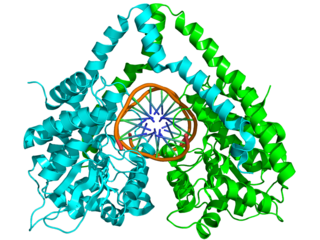
HindIII (pronounced "Hin D Three") is a type II site-specific deoxyribonuclease restriction enzyme isolated from Haemophilus influenzae that cleaves the DNA palindromic sequence AAGCTT in the presence of the cofactor Mg2+ via hydrolysis.

<span class="mw-page-title-main">Cyclic ADP-ribose</span> Chemical compound

Cyclic ADP Ribose, frequently abbreviated as cADPR, is a cyclic adenine nucleotide (like cAMP) with two phosphate groups present on 5' OH of the adenosine (like ADP), further connected to another ribose at the 5' position, which, in turn, closes the cycle by glycosidic bonding to the nitrogen 1 (N1) of the same adenine base (whose position N9 has the glycosidic bond to the other ribose). The N1-glycosidic bond to adenine is what distinguishes cADPR from ADP-ribose (ADPR), the non-cyclic analog. cADPR is produced from nicotinamide adenine dinucleotide (NAD+) by ADP-ribosyl cyclases (EC 3.2.2.5) as part of a second messenger system.

Transition state analogs, are chemical compounds with a chemical structure that resembles the transition state of a substrate molecule in an enzyme-catalyzed chemical reaction. Enzymes interact with a substrate by means of strain or distortions, moving the substrate towards the transition state. Transition state analogs can be used as inhibitors in enzyme-catalyzed reactions by blocking the active site of the enzyme. Theory suggests that enzyme inhibitors which resembled the transition state structure would bind more tightly to the enzyme than the actual substrate. Examples of drugs that are transition state analog inhibitors include flu medications such as the neuraminidase inhibitor oseltamivir and the HIV protease inhibitors saquinavir in the treatment of AIDS.

<span class="mw-page-title-main">Inorganic pyrophosphatase</span> Group of proteins having inorganic pyrophosphatase activity

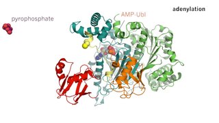
Inorganic pyrophosphatase is an enzyme that catalyzes the conversion of one ion of pyrophosphate to two phosphate ions. This is a highly exergonic reaction, and therefore can be coupled to unfavorable biochemical transformations in order to drive these transformations to completion. The functionality of this enzyme plays a critical role in lipid metabolism, calcium absorption and bone formation, and DNA synthesis, as well as other biochemical transformations.

<span class="mw-page-title-main">6-phosphogluconolactonase</span> Cytosolic enzyme

6-Phosphogluconolactonase (EC 3.1.1.31, 6PGL, PGLS, systematic name 6-phospho-D-glucono-1,5-lactone lactonohydrolase) is a cytosolic enzyme found in all organisms that catalyzes the hydrolysis of 6-phosphogluconolactone to 6-phosphogluconic acid in the oxidative phase of the pentose phosphate pathway:

<span class="mw-page-title-main">Sucrose phosphorylase</span>

Sucrose phosphorylase is an important enzyme in the metabolism of sucrose and regulation of other metabolic intermediates. Sucrose phosphorylase is in the class of hexosyltransferases. More specifically it has been placed in the retaining glycoside hydrolases family although it catalyzes a transglycosidation rather than hydrolysis. Sucrose phosphorylase catalyzes the conversion of sucrose to D-fructose and α-D-glucose-1-phosphate. It has been shown in multiple experiments that the enzyme catalyzes this conversion by a double displacement mechanism.

<span class="mw-page-title-main">Ribose-phosphate diphosphokinase</span> Class of enzymes

Ribose-phosphate diphosphokinase is an enzyme that converts ribose 5-phosphate into phosphoribosyl pyrophosphate (PRPP). It is classified under EC 2.7.6.1.

<span class="mw-page-title-main">ADP-ribosylation</span> Addition of one or more ADP-ribose moieties to a protein.

ADP-ribosylation is the addition of one or more ADP-ribose moieties to a protein. It is a reversible post-translational modification that is involved in many cellular processes, including cell signaling, DNA repair, gene regulation and apoptosis. Improper ADP-ribosylation has been implicated in some forms of cancer. It is also the basis for the toxicity of bacterial compounds such as cholera toxin, diphtheria toxin, and others.

<span class="mw-page-title-main">Biotin carboxylase</span> Class of enzymes

In enzymology, a biotin carboxylase (EC 6.3.4.14) is an enzyme that catalyzes the chemical reaction

In enzymology, an ADP-sugar diphosphatase (EC 3.6.1.21) is an enzyme that catalyzes the chemical reaction

In enzymology, an ATP diphosphatase (EC 3.6.1.8) is an enzyme that catalyzes the chemical reaction

<span class="mw-page-title-main">Nucleoside-diphosphatase</span> Group of proteins having nucleoside-diphosphatase activity

In enzymology, a nucleoside-diphosphatase (EC 3.6.1.6) is an enzyme that catalyzes the chemical reaction

<span class="mw-page-title-main">NUDT9</span> Protein-coding gene in the species Homo sapiens

ADP-ribose pyrophosphatase, mitochondrial is an enzyme that in humans is encoded by the NUDT9 gene.

<span class="mw-page-title-main">Nudix hydrolase</span> Superfamily of hydrolytic enzymes

NUDIX hydrolases are a superfamily of hydrolytic enzymes capable of cleaving nucleoside diphosphates linked to x, hence their name. The reaction yields nucleoside monophosphate (NMP) plus X-P. Substrates hydrolysed by nudix enzymes comprise a wide range of organic pyrophosphates, including nucleoside di- and triphosphates, dinucleoside and diphosphoinositol polyphosphates, nucleotide sugars and RNA caps, with varying degrees of substrate specificity. Enzymes of the NUDIX superfamily are found in all types of organisms, including eukaryotes, bacteria and archaea.

<span class="mw-page-title-main">ADP-ribosylhydrolase</span>

In molecular biology, the (ADP-ribosyl)hydrolase (ARH) family contains enzymes which catalyses the hydrolysis of ADP-ribosyl modifications from proteins, nucleic acids and small molecules.

<span class="mw-page-title-main">Nucleotide pyrophosphatase/phosphodiesterase</span> Class of enzymes

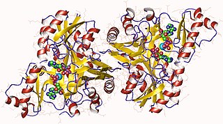
Nucleotide pyrophosphatase/phosphodiesterase (NPP) is a class of dimeric enzymes that catalyze the hydrolysis of phosphate diester bonds. NPP belongs to the alkaline phosphatase (AP) superfamily of enzymes. Humans express seven known NPP isoforms, some of which prefer nucleotide substrates, some of which prefer phospholipid substrates, and others of which prefer substrates that have not yet been determined. In eukaryotes, most NPPs are located in the cell membrane and hydrolyze extracellular phosphate diesters to affect a wide variety of biological processes. Bacterial NPP is thought to localize to the periplasm.

Mn2+-dependent ADP-ribose/CDP-alcohol diphosphatase (EC 3.6.1.53, Mn2+-dependent ADP-ribose/CDP-alcohol pyrophosphatase, ADPRibase-Mn) is an enzyme with systematic name CDP-choline phosphohydrolase. This enzyme catalyses the following chemical reaction

References

  1. 1 2 Gabelli SB, Bianchet MA, Bessman MJ, Amzel LM (May 2001). "The structure of ADP-ribose pyrophosphatase reveals the structural basis for the versatility of the Nudix family". Nature Structural Biology. 8 (5): 467–72. doi:10.1038/87647. PMID   11323725. S2CID   10896410.
  2. 1 2 Galperin MY, Moroz OV, Wilson KS, Murzin AG (January 2006). "House cleaning, a part of good housekeeping". Molecular Microbiology. 59 (1): 5–19. doi: 10.1111/j.1365-2958.2005.04950.x . PMID   16359314. S2CID   10313196.
  3. 1 2 3 Ribeiro JM, Carloto A, Costas MJ, Cameselle JC (April 2001). "Human placenta hydrolases active on free ADP-ribose: an ADP-sugar pyrophosphatase and a specific ADP-ribose pyrophosphatase". Biochimica et Biophysica Acta (BBA) - General Subjects. 1526 (1): 86–94. doi:10.1016/S0304-4165(01)00113-1. PMID   11287126.
  4. 1 2 3 Gabelli SB, Bianchet MA, Ohnishi Y, Ichikawa Y, Bessman MJ, Amzel LM (July 2002). "Mechanism of the Escherichia coli ADP-ribose pyrophosphatase, a Nudix hydrolase". Biochemistry. 41 (30): 9279–85. doi:10.1021/bi0259296. PMID   12135348.
  5. Okuda K, Hayashi H, Nishiyama Y (July 2005). "Systematic characterization of the ADP-ribose pyrophosphatase family in the Cyanobacterium Synechocystis sp. strain PCC 6803". Journal of Bacteriology. 187 (14): 4984–91. doi:10.1128/JB.187.14.4984-4991.2005. PMC   1169527 . PMID   15995214.
  6. Dunn CA, O'Handley SF, Frick DN, Bessman MJ (November 1999). "Studies on the ADP-ribose pyrophosphatase subfamily of the nudix hydrolases and tentative identification of trgB, a gene associated with tellurite resistance". The Journal of Biological Chemistry. 274 (45): 32318–24. doi: 10.1074/jbc.274.45.32318 . PMID   10542272.