Pituitary apoplexy

Last updated

Pituitary apoplexy
Other namesPituitary tumor apoplexy
Specialty Endocrinology   OOjs UI icon edit-ltr-progressive.svg

Pituitary apoplexy is bleeding into or impaired blood supply of the pituitary gland. This usually occurs in the presence of a tumor of the pituitary, although in 80% of cases this has not been diagnosed previously. The most common initial symptom is a sudden headache, often associated with a rapidly worsening visual field defect or double vision caused by compression of nerves surrounding the gland. This is often followed by acute symptoms caused by lack of secretion of essential hormones, predominantly adrenal insufficiency. [1]

Contents

The diagnosis is achieved with magnetic resonance imaging and blood tests. Treatment is by the timely correction of hormone deficiencies. In many cases, surgical decompression is required. Many people who have had a pituitary apoplexy develop pituitary hormone deficiencies and require long-term hormone supplementation. The first case of the disease was recorded in 1898. [1]

Signs and symptoms

Acute symptoms

The initial symptoms of pituitary apoplexy are related to the increased pressure in and around the pituitary gland. The most common symptom, in over 95% of cases, is a sudden-onset headache located behind the eyes or around the temples. It is often associated with nausea and vomiting. [1] [2] [3] Occasionally, the presence of blood leads to irritation of the lining of the brain, which may cause neck rigidity and intolerance to bright light, as well as a decreased level of consciousness. [1] [2] [3] This occurs in 24% of cases. [4]

Visual field loss in bitemporal hemianopsia: compression of the optic chiasm leads to a characteristic pattern of vision loss affecting the outer halves of each eye. Bitempvf labelled.png
Visual field loss in bitemporal hemianopsia : compression of the optic chiasm leads to a characteristic pattern of vision loss affecting the outer halves of each eye.

Pressure on the part of the optic nerve known as the chiasm, which is located above the gland, leads to loss of vision on the outer side of the visual field on both sides, as this corresponds to areas on the retinas supplied by these parts of the optic nerve; it is encountered in 75% of cases. [1] Visual acuity is reduced in half, and over 60% have a visual field defect. [2] [4] The visual loss depends on which part of the nerve is affected. If the part of the nerve between the eye and the chiasm is compressed, the result is vision loss in one eye. If the part after the chiasm is affected, visual loss on one side of the visual field occurs. [2]

Adjacent to the pituitary lies a part of the skull base known as the cavernous sinus. This contains a number of nerves that control the eye muscles. 70% of people with pituitary apoplexy experience double vision due to compression of one of the nerves. In half of these cases, the oculomotor nerve (the third cranial nerve), which controls a number of eye muscles, is affected. This leads to diagonal double vision and a dilated pupil. The fourth (trochlear) and sixth (abducens) cranial nerves are located in the same compartment and can cause diagonal or horizontal double vision, respectively. [1] The oculomotor nerve is predominantly affected as it lies closest to the pituitary. [2] [5] The cavernous sinus also contains the carotid artery, which supplies blood to the brain; occasionally, compression of the artery can lead to one-sided weakness and other symptoms of stroke. [1] [2] [4]

Endocrine dysfunction

The adrenal glands, located above the kidneys, are typically the first to be affected in pituitary apoplexy. When the stimulating hormone ACTH is not secreted, the adrenals cease to produce cortisol. Illu adrenal gland.jpg
The adrenal glands, located above the kidneys, are typically the first to be affected in pituitary apoplexy. When the stimulating hormone ACTH is not secreted, the adrenals cease to produce cortisol.

The pituitary gland consists of two parts, the anterior (front) and posterior (back) pituitary. Both parts release hormones that control numerous other organs. In pituitary apoplexy, the main initial problem is a lack of secretion of adrenocorticotropic hormone (ACTH, corticotropin), which stimulates the secretion of cortisol by the adrenal gland. This occurs in 70% of those with pituitary apoplexy. A sudden lack of cortisol in the body leads to a constellation of symptoms called "adrenal crisis" or "Addisonian crisis" (after a complication of Addison's disease, the main cause of adrenal dysfunction and low cortisol levels). [1] The main problems are low blood pressure (particularly on standing), low blood sugars (which can lead to coma) and abdominal pain; the low blood pressure can be life-threatening and requires immediate medical attention. [6]

Hyponatremia, an unusually low level of sodium in the blood that may cause confusion and seizures, is found in 40% of cases. This may be caused by low cortisol levels or by inappropriate release of antidiuretic hormone (ADH) from the posterior pituitary. [1] Several other hormonal deficiencies may develop in the subacute phase. 50% have a deficiency in thyroid-stimulating hormone (TSH), leading to hyposecretion of thyroid hormone by the thyroid gland and characteristic symptoms such as fatigue, weight gain, and cold intolerance. 75% develop a deficiency to gonadotropins (LH and FSH), which control the reproductive hormone glands. This leads to a disrupted menstrual cycle, infertility, and decreased libido. [1]

Causes

Almost all cases of pituitary apoplexy arise from a pituitary adenoma, a benign tumor of the pituitary gland. In 80%, the patient has been previously unaware of this (although some will retrospectively report associated symptoms). [1] It was previously thought that particular types of pituitary tumors were more prone to apoplexy than others, but this has not been confirmed. [2] In absolute terms, only a very small proportion of pituitary tumors eventually undergoes apoplexy. In an analysis of incidentally found pituitary tumors, apoplexy occurred in 0.2% annually, but the risk was higher in tumors larger than 10 mm ("macroadenomas") and tumors that were growing more rapidly; in a meta-analysis, not all these associations achieved statistical significance. [7]

The majority of cases (60–80%) are not precipitated by a particular cause. [2] A quarter has a history of high blood pressure, [1] but this is a common problem in the general population, and it is not clear whether it significantly increase the risk of apoplexy. [8] A number of cases has been reported in association with particular conditions and situations; it is uncertain whether these were in fact causative. [8] Amongst reported associations are surgery (especially coronary artery bypass graft, where there are significant fluctuations in the blood pressure), disturbances in blood coagulation or medication that inhibits coagulation, radiation therapy to the pituitary, traumatic brain injury, pregnancy (during which the pituitary enlarges) and treatment with estrogens. Hormonal stimulation tests of the pituitary have been reported to provoke episodes. Treatment of prolactinomas (pituitary adenomas that secrete prolactin) with dopamine agonist drugs, as well as withdrawal of such treatment, has been reported to precipitate apoplexy. [1] [2] [4]

Hemorrhage from a Rathke's cleft cyst, a remnant of Rathke's pouch that normally regresses after embryological development, may cause symptoms that are indistinguishable from pituitary apoplexy. [4] Pituitary apoplexy is regarded by some as distinct from Sheehan's syndrome, where the pituitary undergoes infarction as a result of prolonged very low blood pressure, particularly when caused by bleeding after childbirth. This condition usually occurs in the absence of a tumor. [4] Others regard Sheehan's syndrome as a form of pituitary apoplexy. [3] [9]

Mechanism

The pituitary gland is located in a recess in the skull base known as the sella turcica ("Turkish saddle", after its shape). It is attached to the hypothalamus, a part of the brain, by a stalk that also contains the blood vessels that supply the gland. It is unclear why pituitary tumors are five times more likely to bleed than other tumors in the brain. There are various proposed mechanisms by which a tumor can increase the risk of either infarction (insufficient blood supply leading to tissue dysfunction) or hemorrhage. [2] The pituitary gland normally derives its blood supply from vessels that pass through the hypothalamus, but tumors develop a blood supply from the nearby inferior hypophyseal artery that generates a higher blood pressure, possibly accounting for the risk of bleeding. Tumors may also be more sensitive to fluctuations in blood pressure, and the blood vessels may show structural abnormalities that make them vulnerable to damage. It has been suggested that infarction alone causes milder symptoms than either hemorrhage or hemorrhagic infarction (infarction followed by hemorrhage into the damaged tissue). [4] Larger tumors are more prone to bleeding, and more rapidly growing lesions (as evidenced by detection of increased levels of the protein PCNA) may also be at a higher risk of apoplexy. [2]

After an apoplexy, the pressure inside the sella turcica rises, and surrounding structures such as the optic nerve and the contents of the cavernous sinus are compressed. The raised pressure further impairs the blood supply to the pituitary hormone-producing tissue, leading to tissue death due to insufficient blood supply. [2]

Diagnosis

Normal pituitary gland on MRI (T1 sagittal without contrast enhancement). The arrow points at the posterior pituitary (intense signal), and the arrowhead at the anterior pituitary. Hypophyse MRT sag.png
Normal pituitary gland on MRI (T1 sagittal without contrast enhancement). The arrow points at the posterior pituitary (intense signal), and the arrowhead at the anterior pituitary.

It is recommended that magnetic resonance imaging (MRI) scan of the pituitary gland is performed if the diagnosis is suspected; this has a sensitivity of over 90% for detecting pituitary apoplexy; it may demonstrate infarction (tissue damage due to a decreased blood supply) or hemorrhage. [1] Different MRI sequences can be used to establish when the apoplexy occurred, and the predominant form of damage (hemorrhage or infarction). [2] If MRI is not suitable (e.g. due to claustrophobia or the presence of metal-containing implants), a computed tomography (CT) scan may demonstrate abnormalities in the pituitary gland, although it is less reliable. [1] Many pituitary tumors (25%) are found to have areas of hemorrhagic infarction on MRI scans, but apoplexy is not said to exist unless it is accompanied by symptoms. [1] [4]

In some instances, lumbar puncture may be required if there is a suspicion that the symptoms might be caused by other problems (meningitis or subarachnoid hemorrhage). This is the examination of the cerebrospinal fluid that envelops the brain and the spinal cord; the sample is obtained with a needle that is passed under local anesthetic into the spine. In pituitary apoplexy the results are typically normal, although abnormalities may be detected if blood from the pituitary has entered the subarachnoid space. [2] [4] If there is remaining doubt about the possibility of subarachnoid hemorrhage (SAH), a magnetic resonance angiogram (MRI with a contrast agent) may be required to identify aneurysms of the brain blood vessels, the most common cause of SAH. [10]

Professional guidelines recommend that if pituitary apoplexy is suspected or confirmed, the minimal blood tests performed should include a complete blood count, urea (a measure of renal function, usually performed together with creatinine), electrolytes (sodium and potassium), liver function tests, routine coagulation testing, and a hormonal panel including IGF-1, growth hormone, prolactin, luteinizing hormone, follicle-stimulating hormone, thyroid-stimulating hormone, thyroid hormone, and either testosterone in men or estradiol in women. [1]

Visual field testing is recommended as soon as possible after diagnosis, [1] [4] as it quantifies the severity of any optic nerve involvement, and may be required to decide on surgical treatment. [1]

Treatment

The first priority in suspected or confirmed pituitary apoplexy is stabilization of the circulatory system. Cortisol deficiency can cause severe low blood pressure. [1] [6] Depending on the severity of the illness, admission to a high dependency unit (HDU) may be required. [1]

Treatment for acute adrenal insufficiency requires the administration of intravenous saline or dextrose solution; volumes of over two liters may be required in an adult. [6] This is followed by the administration of hydrocortisone, which is pharmaceutical grade cortisol, intravenously or into a muscle. [4] [6] The drug dexamethasone has similar properties, [6] but its use is not recommended unless it is required to reduce swelling in the brain around the area of hemorrhage. [1] Some are well enough not to require immediate cortisol replacement; in this case, blood levels of cortisol are determined at 9:00 AM (as cortisol levels vary over the day). A level below 550  nmol/L indicates a need for replacement. [1]

The decision on whether to surgically decompress the pituitary gland is complex and mainly dependent on the severity of visual loss and visual field defects. If visual acuity is severely reduced, there are large or worsening visual field defects, or the level of consciousness falls consistently, professional guidelines recommend that surgery is performed. [1] Most commonly, operations on the pituitary gland are performed through transsphenoidal surgery. In this procedure, surgical instruments are passed through the nose towards the sphenoid bone, which is opened to give access to the cavity that contains the pituitary gland. [1] [4] Surgery is most likely to improve vision if there was some remaining vision before surgery, [4] and if surgery is undertaken within a week of the onset of symptoms. [4] [11]

Those with relatively mild visual field loss or double vision only may be managed conservatively, with close observation of the level of consciousness, visual fields, and results of routine blood tests. If there is any deterioration, or expected spontaneous improvement does not occur, surgical intervention may still be indicated. [1] [4] If the apoplexy occurred in a prolactin-secreting tumor, this may respond to dopamine agonist treatment. [4] [11]

After recovery, people who have had pituitary apoplexy require follow-up by an endocrinologist to monitor for long-term consequences. MRI scans are performed 3–6 months after the initial episode and subsequently on an annual basis. [1] If after surgery some tumor tissue remains, this may respond to medication, further surgery, or radiation therapy with a "gamma knife". [4]

Prognosis

In larger case series, the mortality was 1.6% overall. In the group of patients who were unwell enough to require surgery, the mortality was 1.9%, with no deaths in those who could be treated conservatively. [8]

After an episode of pituitary apoplexy, 80% of people develop hypopituitarism and require some form of hormone replacement therapy. [1] [2] The most common problem is growth hormone deficiency, which is often left untreated [1] [4] but may cause decreased muscle mass and strength, obesity and fatigue. [12] 60–80% require hydrocortisone replacement (either permanently or when unwell), 50–60% need thyroid hormone replacement, and 60–80% of men require testosterone supplements. Finally, 10–25% develop diabetes insipidus, the inability to retain fluid in the kidneys due to a lack of the pituitary antidiuretic hormone. This may be treated with the drug desmopressin, [1] which can be applied as a nose spray or taken by mouth. [13]

Epidemiology

Pituitary apoplexy is rare. [1] [2] Even in people with a known pituitary tumor, only 0.6–10% experience apoplexy; the risk is higher in larger tumors. [2] Based on extrapolations from existing data, one would expect 18 cases of pituitary apoplexy per one million people every year; the actual figure is probably lower. [14]

The average age at onset is 50; cases have reported in people between 15 and 90 years old. [14] Men are affected more commonly than women, [2] with a male-to-female ratio of 1.6. [4] The majority of the underlying tumors are "null cell" or nonsecretory tumors, which do not produce excessive amounts of hormones; this might explain why the tumor has often gone undetected prior to an episode of apoplexy. [4]

History

The first case description of pituitary apoplexy has been attributed to the American neurologist Pearce Bailey in 1898. [15] [16] This was followed in 1905 by a further report from the German physician Bleibtreu. [4] [17] Surgery for pituitary apoplexy was described in 1925. [16] [18] Before the introduction of steroid replacement, the mortality from pituitary apoplexy approximated 50%. [4]

The name of the condition was coined in 1950 in a case series by physicians from Boston City Hospital and Harvard Medical School. [1] [19] The term "apoplexy" was applied as it referred to both necrosis and bleeding into pituitary tumors. [19]

Related Research Articles

<span class="mw-page-title-main">Adrenal gland</span> Endocrine gland

The adrenal glands are endocrine glands that produce a variety of hormones including adrenaline and the steroids aldosterone and cortisol. They are found above the kidneys. Each gland has an outer cortex which produces steroid hormones and an inner medulla. The adrenal cortex itself is divided into three main zones: the zona glomerulosa, the zona fasciculata and the zona reticularis.

<span class="mw-page-title-main">Cushing's syndrome</span> Symptoms from excessive exposure to glucocorticoids such as cortisol

Cushing's syndrome is a collection of signs and symptoms due to prolonged exposure to glucocorticoids such as cortisol. Signs and symptoms may include high blood pressure, abdominal obesity but with thin arms and legs, reddish stretch marks, a round red face due to facial plethora, a fat lump between the shoulders, weak muscles, weak bones, acne, and fragile skin that heals poorly. Women may have more hair and irregular menstruation. Occasionally there may be changes in mood, headaches, and a chronic feeling of tiredness.

<span class="mw-page-title-main">Addison's disease</span> Endocrine disorder

Addison's disease, also known as primary adrenal insufficiency, is a rare long-term endocrine disorder characterized by inadequate production of the steroid hormones cortisol and aldosterone by the two outer layers of the cells of the adrenal glands, causing adrenal insufficiency. Symptoms generally come on slowly and insidiously and may include abdominal pain and gastrointestinal abnormalities, weakness, and weight loss. Darkening of the skin in certain areas may also occur. Under certain circumstances, an adrenal crisis may occur with low blood pressure, vomiting, lower back pain, and loss of consciousness. Mood changes may also occur. Rapid onset of symptoms indicates acute adrenal failure, which is a clinical emergency. An adrenal crisis can be triggered by stress, such as from an injury, surgery, or infection.

Cushing's disease is one cause of Cushing's syndrome characterised by increased secretion of adrenocorticotropic hormone (ACTH) from the anterior pituitary. This is most often as a result of a pituitary adenoma or due to excess production of hypothalamus CRH that stimulates the synthesis of cortisol by the adrenal glands. Pituitary adenomas are responsible for 80% of endogenous Cushing's syndrome, when excluding Cushing's syndrome from exogenously administered corticosteroids. The equine version of this disease is Pituitary pars intermedia dysfunction.

<span class="mw-page-title-main">Adrenal insufficiency</span> Medical condition

Adrenal insufficiency is a condition in which the adrenal glands do not produce adequate amounts of steroid hormones. The adrenal glands—also referred to as the adrenal cortex—normally secrete glucocorticoids, mineralocorticoids, and androgens. These hormones are important in regulating blood pressure, electrolytes, and metabolism as a whole. Deficiency of these hormones leads to symptoms ranging from abdominal pain, vomiting, muscle weakness and fatigue, low blood pressure, depression, mood and personality changes to organ failure and shock. Adrenal crisis may occur if a person having adrenal insufficiency experiences stresses, such as an accident, injury, surgery, or severe infection; this is a life-threatening medical condition resulting from severe deficiency of cortisol in the body. Death may quickly follow.

<span class="mw-page-title-main">Hypopituitarism</span> Medical condition

Hypopituitarism is the decreased (hypo) secretion of one or more of the eight hormones normally produced by the pituitary gland at the base of the brain. If there is decreased secretion of one specific pituitary hormone, the condition is known as selective hypopituitarism. If there is decreased secretion of most or all pituitary hormones, the term panhypopituitarism is used.

<span class="mw-page-title-main">Sheehan's syndrome</span> Medical condition

Sheehan's syndrome, also known as postpartum pituitary gland necrosis, occurs when the pituitary gland is damaged due to significant blood loss and hypovolemic shock usually during or after childbirth leading to decreased functioning of the pituitary gland (hypopituitarism). The pituitary gland is an endocrine organ, meaning it produces certain hormones and is involved in the regulation of various other hormones. This gland is located in the brain and sits in a pocket of the sphenoid bone known as the sella turcica. The pituitary gland works in conjunction with the hypothalamus, and other endocrine organs to modulate numerous bodily functions including growth, metabolism, menstruation, lactation, and even the "fight-or-flight" response. These endocrine organs release hormones in very specific pathways, known as hormonal axes. For example, the release of a hormone in the hypothalamus will target the pituitary to trigger the release of a subsequent hormone, and the pituitary's released hormone will target the next organ in the pathway. Hence, damage to the pituitary gland can have downstream effects on any of the aforementioned bodily functions.

Optic nerve hypoplasia (ONH) is a medical condition arising from the underdevelopment of the optic nerve(s). This condition is the most common congenital optic nerve anomaly. The optic disc appears abnormally small, because not all the optic nerve axons have developed properly. It is often associated with endocrinopathies, developmental delay, and brain malformations. The optic nerve, which is responsible for transmitting visual signals from the retina to the brain, has approximately 1.2 million nerve fibers in the average person. In those diagnosed with ONH, however, there are noticeably fewer nerves.

<span class="mw-page-title-main">Pituitary adenoma</span> Human disease

Pituitary adenomas are tumors that occur in the pituitary gland. Most pituitary tumors are benign, approximately 35% are invasive and just 0.1% to 0.2% are carcinomas. Pituitary adenomas represent from 10% to 25% of all intracranial neoplasms and the estimated prevalence rate in the general population is approximately 17%.

<span class="mw-page-title-main">Prolactinoma</span> Medical condition

A prolactinoma is a tumor (adenoma) of the pituitary gland that produces the hormone prolactin. It is the most common type of functioning pituitary tumor. Symptoms of prolactinoma are due to abnormally high levels of prolactin in the blood (hyperprolactinemia), or due to pressure of the tumor on surrounding tissues. Based on size, a prolactinoma can be classified as a microprolactinoma or a macroprolactinoma.

<span class="mw-page-title-main">Endocrine gland</span> Glands of the endocrine system that secrete hormones to blood

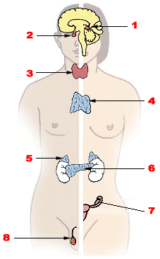
Endocrine glands are ductless glands of the endocrine system that secrete their products, hormones, directly into the blood. The major glands of the endocrine system include the pineal gland, pituitary gland, pancreas, ovaries, testicles, thyroid gland, parathyroid gland, hypothalamus and adrenal glands. The hypothalamus and pituitary glands are neuroendocrine organs.

Nelson's syndrome is a disorder that occurs in about one in four patients who have had both adrenal glands removed to treat Cushing's disease. In patients with pre-existing adrenocorticotropic hormone (ACTH)-secreting pituitary adenomas, loss of adrenal feedback following bilateral adrenalectomy can trigger the rapid growth of the tumor, leading to visual symptoms and hyperpigmentation. The severity of the disease is dependent upon the effect of ACTH release on the skin, pituitary hormone loss from mass compression, as well as invasion into surrounding structures around the pituitary gland.

<span class="mw-page-title-main">Adrenalectomy</span> Surgical removal of adrenal glands

Adrenalectomy is the surgical removal of one or both adrenal glands. It is usually done to remove tumors of the adrenal glands that are producing excess hormones or is large in size. Adrenalectomy can also be done to remove a cancerous tumor of the adrenal glands, or cancer that has spread from another location, such as the kidney or lung. Adrenalectomy is not performed on those who have severe coagulopathy or whose heart and lungs are too weak to undergo surgery. The procedure can be performed using an open incision (laparotomy) or minimally invasive laparoscopic or robot-assisted techniques. Minimally invasive techniques are increasingly the gold standard of care due to shorter length of stay in the hospital, lower blood loss, and similar complication rates.

<span class="mw-page-title-main">Adrenocorticotropic hormone deficiency</span> Medical condition

Adrenocorticotropic hormone deficiency is a rare disorder characterized by secondary adrenal insufficiency with minimal or no cortisol production and normal pituitary hormone secretion apart from ACTH. ACTH deficiency may be congenital or acquired, and its symptoms are clinically similar to those of glucocorticoid deficiency. Symptoms consist of weight loss, diminished appetite, muscle weakness, nausea, vomiting, and hypotension. Low blood sugar and hyponatremia are possible; however, blood potassium levels typically remain normal because affected patients are deficient in glucocorticoids rather than mineralocorticoids because of their intact renin-angiotensin-aldosterone system. ACTH may be undetectable in blood tests, and cortisol is abnormally low. Glucocorticoid replacement therapy is required. With the exception of stressful situations, some patients with mild or nearly asymptomatic disease may not require glucocorticoid replacement therapy. As of 2008 about two hundred cases have been described in the literature.

In humans and other animals, the adrenocortical hormones are hormones produced by the adrenal cortex, the outer region of the adrenal gland. These polycyclic steroid hormones have a variety of roles that are crucial for the body’s response to stress, and they also regulate other functions in the body. Threats to homeostasis, such as injury, chemical imbalances, infection, or psychological stress, can initiate a stress response. Examples of adrenocortical hormones that are involved in the stress response are aldosterone and cortisol. These hormones also function in regulating the conservation of water by the kidneys and glucose metabolism, respectively.

Autoimmune hypophysitis is defined as inflammation of the pituitary gland due to autoimmunity.

<span class="mw-page-title-main">Pituitary disease</span> Medical condition

A pituitary disease is a disorder primarily affecting the pituitary gland.

<span class="mw-page-title-main">Acromegaly</span> Human disease that results in excess growth of certain parts of the body

Acromegaly is a disorder that results in excess growth of certain parts of the human body. It is caused by excess growth hormone (GH) after the growth plates have closed. The initial symptom is typically enlargement of the hands and feet. There may also be an enlargement of the forehead, jaw, and nose. Other symptoms may include joint pain, thicker skin, deepening of the voice, headaches, and problems with vision. Complications of the disease may include type 2 diabetes, sleep apnea, and high blood pressure.

<span class="mw-page-title-main">Myelolipoma</span> Medical condition

Myelolipoma is a benign tumor-like lesion composed of mature adipose (fat) tissue and haematopoietic (blood-forming) elements in various proportions.

Galactorrhea hyperprolactinemia is increased blood prolactin levels associated with galactorrhea. It may be caused by such things as certain medications, pituitary disorders and thyroid disorders. The condition can occur in males as well as females. Relatively common etiologies include prolactinoma, medication effect, kidney failure, granulomatous diseases of the pituitary gland, and disorders which interfere with the hypothalamic inhibition of prolactin release. Ectopic (non-pituitary) production of prolactin may also occur. Galactorrhea hyperprolactinemia is listed as a “rare disease” by the Office of Rare Diseases of the National Institutes of Health. This means that it affects less than 200,000 people in the United States population.

References

  1. 1 2 3 4 5 6 7 8 9 10 11 12 13 14 15 16 17 18 19 20 21 22 23 24 25 26 27 28 29 30 31 32 Rajasekaran S, Vanderpump M, Baldeweg S, et al. (Jan 2011). "UK guidelines for the management of pituitary apoplexy". Clin Endocrinol. 74 (1): 9–20. doi: 10.1111/j.1365-2265.2010.03913.x . PMID   21044119. S2CID   52867017.
  2. 1 2 3 4 5 6 7 8 9 10 11 12 13 14 15 16 17 18 Murad-Kejbou S, Eggenberger E (Nov 2009). "Pituitary apoplexy: evaluation, management, and prognosis". Current Opinion in Ophthalmology. 20 (6): 456–61. doi:10.1097/ICU.0b013e3283319061. PMID   19809320. S2CID   5209711.
  3. 1 2 3 Melmed S, Jameson JL (2005). "Disorders of the anterior pituitary and hypothalamus". In Kasper DL, Braunwald E, Fauci AS, et al. (eds.). Harrison's Principles of Internal Medicine (16th ed.). New York, NY: McGraw-Hill. pp.  2076–97. ISBN   978-0-07-139140-5.
  4. 1 2 3 4 5 6 7 8 9 10 11 12 13 14 15 16 17 18 19 20 21 22 Nawar RN, AbdelMannan D, Selman WR, Arafah BM (Mar–Apr 2008). "Pituitary tumor apoplexy: a review". J. Intens. Care Med. 23 (2): 75–90. doi:10.1177/0885066607312992. PMID   18372348. S2CID   34782106.
  5. Bruce BB, Biousse V, Newman NJ (Jul 2007). "Third nerve palsies". Semin Neurol. 27 (3): 257–68. doi:10.1055/s-2007-979681. PMID   17577867. S2CID   27798175.
  6. 1 2 3 4 5 de Herder WW, van der Lely AJ (May 2003). "Addisonian crisis and relative adrenal failure". Rev Endocr Metab Disord. 4 (2): 143–7. doi:10.1023/A:1022938019091. PMID   12766542. S2CID   38382183.
  7. Fernández-Balsells MM, Murad MH, Barwise A, et al. (April 2011). "Natural history of nonfunctioning pituitary adenomas and incidentalomas: a systematic review and metaanalysis". J. Clin. Endocrinol. Metab. 96 (4): 905–12. doi: 10.1210/jc.2010-1054 . PMID   21474687.
  8. 1 2 3 Russell SJ, Miller K (2008). "Pituitary apoplexy". In Swearingen B, Biller BM (eds.). Diagnosis and management of pituitary disorders. Totowa, NJ: Humana Press. p. 368. ISBN   978-1-58829-922-2.
  9. Thorner MO, Vance ML, Horvath E, Kovacs K (1992). "The anterior pituitary". In Wilson JD, Foster DW (eds.). Williams Textbook of Endocrinology, 8th edition. Philadelphia: W.B. Saunders. pp. 294–5. ISBN   978-0-7216-9514-3.
  10. Post KD, Shiau JS, Walsh J (2008). "Pituitary apoplexy". In Loftus CM (ed.). Neurosurgical Emergencies (2nd ed.). New York, NY: Thieme Publishing Group. pp. 78–83. ISBN   978-3-13-135052-7.
  11. 1 2 Adams CB (2003). "The surgery of pituitary tumours". In Wass JA, Shalet SM (eds.). Oxford Textbook of Endocrinology and Diabetes. Oxford: Oxford University Press. pp. 161–2. ISBN   978-0-19-263045-2.
  12. van Aken MO, Lamberts SW (2005). "Diagnosis and treatment of hypopituitarism: an update". Pituitary. 8 (3–4): 183–91. doi:10.1007/s11102-006-6039-z. PMID   16508719. S2CID   581096.
  13. Jane JA Jr, Vance ML, Laws ER (2006). "Neurogenic diabetes insipidus". Pituitary. 9 (4): 327–9. doi:10.1007/s11102-006-0414-7. PMID   17080264. S2CID   2692261.
  14. 1 2 Russell SJ, Miller K (2008). "Pituitary apoplexy". In Swearingen B, Biller BM (eds.). Diagnosis and management of pituitary disorders. Totowa, NJ: Humana Press. p. 356. ISBN   978-1-58829-922-2.
  15. Bailey P (1898). "Pathological report of a case of akromegaly, with special reference to the lesions in the hypophysis cerebri and in the thyroid gland; and a case of haemorrhage into the pituitary". Phila Med J. 1: 789–92.
  16. 1 2 Russell SJ, Klahr Miller K (2008). "Pituitary apoplexy". In Swearingen B, Biller BM (eds.). Diagnosis and management of pituitary disorders. Totowa, NJ: Humana Press. p. 354. ISBN   978-1-58829-922-2.
  17. Bleibtreu L (1905). "Ein Fall von Akromegalia (Zerstorung der Hypophysis durch Blutung)". Munch Med Wochenschr (in German). 41: 2079–80.
  18. Dott NM, Bailey P, Cushing H (1925). "A consideration of the hypophyseal adenomata". Br J Surg. 13 (50): 314–66. doi:10.1002/bjs.1800135009. S2CID   70559984.
  19. 1 2 Brougham M, Heusner AP, Adams RD (Sep 1950). "Acute degenerative changes in adenomas of the pituitary body--with special reference to pituitary apoplexy". J Neurosurg. 7 (5): 421–39. doi:10.3171/jns.1950.7.5.0421. PMID   14774761.