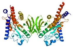
Protein tyrosine phosphatase non-receptor type 22 (PTPN22) is a cytoplasmatic protein encoded by gene PTPN22 and a member of PEST family of protein tyrosine phosphatases. This protein is also called "PEST-domain Enriched Phosphatase" ("PEP") or "Lymphoid phosphatase" ("LYP"). The name LYP is used strictly for the human protein encoded by PTPN22, but the name PEP is used only for its mouse homolog. However, both proteins have similar biological functions and show 70% identity in amino acid sequence. PTPN22 functions as a negative regulator of T cell receptor (TCR) signaling, which maintains homeostasis of T cell compartment. [5] [6]
Gene PTPN22 is located on the p arm of the human chromosome 1. It is nearly 58 000 base pairs long and contains 21 exons. [7] In the case of mouse genome, it is located on the q arm of the chromosome 3. It is nearly 55 700 base pairs long and contains 23 exons. [8]
PTPN22 is composed from 807 amino acids, and it weighs 91,705 kDa. On its N terminus it possesses catalytic domain, which shows the highest level of conservation between human and mouse proteins. Other parts of PTPN22 are less conserved. After catalytic domain PTPN22 has approximately 300 amino acids long domain called "Interdomain". On the C terminus of PTPN22 there are 4 proline-rich motifs (P1 - P4), which can mediate interactions with other proteins. P1 motif is the most important among them, because it is crucial for binding of CSK kinase, and allele encoding PTPN22 with mutated P1 motif is associated with increased risk of numerous autoimmune diseases. [5]
A T cell receptor activation by a cognate peptide triggers a signaling pathway activating a T cell. The first event of this pathway is activation of the SRC family kinase LCK by a dephosphorylation of its C terminal inhibition tyrosine (Y505) and by a phosphorylation of its activation tyrosine (Y394). [9] LCK then phosphorylate tyrosines in the CD3 complex creating a docking site for the SH2 domain of the SYK family kinase ZAP70, which is there phosphorylated too. The Phosphorylated ZAP70 then propagate a signal from a TCR, phosphorylating other proteins and creating a multi-protein complex, which activates downstream signaling pathways. [10] PTPN22 possess the ability to dephosphorylate proteins included in proximal events of the TCR signaling and serves as an important negative regulator of a T cell activation. PTPN22 is able to bind the LCK with phosphorylated Y394, the phosphorylated ZAP70 and the phosphorylated ζ chain of CD3 complex. Thus, it binds molecules of a proximal TCR signaling only after their activation. PTPN22 can dephosphorylate those proteins and decrease the activating signal obtained by a T cell. Dephosphorylation of kinases LCK and ZAP70 by a PTPN22 is specific concerning the phosphorylated tyrosine in those proteins – only the Y394 of LCK and the Y493 of ZAP70 are dephosphorylated. [11] In the absence of PTPN22, an activated T cell receive a stronger activation signal, which is reflected by a greater influx of Ca2+ cations into the cytosol, bigger phosphorylation of an LCK, ZAP70 and ERK and larger expansion of those cells. [5] [6] [12] [13] [14] [15] The inhibitory effect on a TCR signaling was also verified with the usage of PTPN22 inhibitor on a Jurkat T cell line and on human primary T cells, [14] and also with the experiments of PTPN22 overexpression in vitro. [16] [17] The expression of PTPN22 is upregulated after an activation of T cells and an antigen-experienced T cell have higher expression of PTPN22 than a naive T cell. [17] [18]
The regulatory function of PTPN22 is particularly important during an activation by low affinity peptides. In the absence of PTPN22, T cell cannot discriminate between strong and weak antigens sufficiently and those T cells become more responsive, which can be detected like increased upregulation of transcription factors and CD69, increased ERK phosphorylation, increased ability to expand in vivo and to produce cytokines. Increased responsiveness can also break the tolerance against low affinity self-antigens and is well visible, when PTPN22-deficient T cells get into a lymphopenic environment. [15] [19]
One particular population of T cells, which is influenced by a PTPN22 deficiency is the population of regulatory T cells (Treg cells). PTPN22-deficient mice contain higher amount of Treg cells in lymph nodes and spleens and this difference is more visible with increasing age of mice. There is also a change of the effector Treg cells : central Treg cells ratio in favor of the effector Treg cells. PTPN22 deficiency increases abilities of Treg cells to survive, differentiation of Treg cells from naive T cells, but not the ability to proliferate in vivo, and it also supports transition of central Treg cells to effector Treg cells. [13] [20] [21] [22] One of the reasons, of the increased survival of PTPN22-deficient Treg cells, is that those cells have upregulated expression of GITR, which increases their expansion in vivo. Treatment of PTPN22-deficient mice with an anti-GITR-L blocking antibody suppresses the expansion of Treg cells. [20] PTPN22 deficiency does not impair the suppressive function of Treg cells. Actually there are some articles suggesting that PTPN22-deficient Treg cells possess an enhanced suppressive function or have a bigger ability to obtain an effector phenotype. [13] [19] [21]
Next to a TCR signaling PTPN22 regulates an adhesiveness and a motility of T cells. PTPN22-deficient T cells have a prolonged interval of contact with an antigen presenting cell, which present a low affinity peptide. With a high affinity peptide the difference is not detectable. Part of the reason of the increased adhesiveness of those T cells is that enhanced TCR signaling results in a higher activation of the RAP1 and a boosted inside-out signaling to activate the adhesive molecule LFA-1. [15] In migrating T cells we can see the polarized localization of the PTPN22 at the leading edge of a migrating T cell, where it colocalizes with its substrates LCK and ZAP70. A downregulation of the PTPN22 increases motility, adhesivity and levels of phosphorylated LCK and phosphorylated ZAP70 in those cells. On the contrary, an overexpression of the PTPN22, but not the catalytically inactive PTPN22, increases motility of migrating T cells. An association of the PTPN22, but not its disease associated mutant form, with the LFA-1 results in a decreased LFA-1 clustering and a decreased adhesion. [23] The role of the PTPN22 in the regulation of LFA-1-mediated adhesion and motility is also supported by the observation of increased LFA-1 expression in PTPN22-/- Treg cells. [13]
The C-terminal part of the PTPN22 bare proline-rich motifs providing binding sites for putative interaction partners. One of those interaction partners is the cytoplasmatic tyrosine kinase CSK, which is a negative regulator of SRC family kinases and a TCR signaling as well as the PTPN22. CSK binds two prolin-rich motifs (P1 and P2) in the structure of PTPN22 through its SH3 domain and the P1 motif is more important in this interaction. A deletion of the P1 motif greatly diminish the inhibitory effect of the PTPN22 on a TCR signaling. The Interaction of those enzymes is needed for their optimal function and the inhibition of TCR signaling. [16] [24] It was also proposed that the interaction of PTPN22 and CSK regulate a localization of the PTPN22 and a dissociation of this complex enables translocation of the PTPN22 to lipid rafts of a plasma membrane, where it can inhibit a TCR signaling. The mutant PTPN22, which is unable to bind CSK, is effectively recruited to a plasma membrane. [14]
Another interaction partner of the PTPN22 is TRAF3. This protein bind the PTPN22 and regulate its translocalization to a plasma membrane, in the absence of TRAF3 there is a bigger amount of the PTPN22 localized at a plasma membrane. [25]
It was revealed that PTPN22 is regulated by a phosphorylation. PTPN22 is phosphorylated on the serine in the position 751 by the protein PKC (most probably isoform PKCα) after activation of a T cell. This phosphorylation negatively regulates the TCR-suppressing function of the PTPN22. It also suppresses the polyubiquitination of PTPN22, which targets this protein for degradation, and by this mean, it prolongs half-life of the PTPN22. Phosphorylared PTPN22 interacts better with the CSK which hold PTPN22 away from a plasma membrane, where it can dephosphorylate proteins of a TCR signaling pathway. PTPN22 with the mutated serine 751 has shorter half-life, enhanced recruitment to plasma membrane and reduced interaction with CSK. [26]
Young PTPN22-deficient mice do not display any abnormality in peripheral lymphoid organs, but older PTPN22-deficient mice (older than 6 months) develop a splenomegaly and a lymphadenopathy. In these older mice we can see an increased number of the T cells with phenotype of the effector/memory T cells (CD44 hi, CD62L lo), which have higher expression of the PTPN22 than naive T cells in Wild Type mice. The expansion of those T cells is supported by the PTPN22 deficiency. A compartment of Treg cell is also influenced by the PTPN22 deficiency in vivo. [12] Same as with the effector/memory T cells, PTPN22-deficient mice contain a bigger amount of Treg cells in lymph nodes and spleens and this difference is more visible with increasing age of mice. There is also a change of the effector Treg cells : central Treg cells ratio in favor of the effector Treg cells. [13] [20] [21] [22] Influence of the PTPN22 deficiency on Treg cells number is consistent with the higher expression of PTPN22 in Treg cells than in naive T cells. [17] Another abnormality of PTPN22-deficient mice is a spontaneous formation of large germinal centers in spleens and peyer's patches. This formation of germinal centers is dependent on the costimulation molecule CD40L and it is another consequence of the T cell dysregulation. PTPN22-deficient mice have increased levels of antibodies. However, there is no increase in levels of autoantibodies. Despite those effects of the PTPN22 deficiency on a T cell compartment and an antibody production, PTPN22-deficient mice do not show signs of any autoimmune disease. [12] [13]
In 2004, Bottini et al. discovered the single-nucleotide polymorphism in the PTPN22 gene at nucleotide 1858. In this variant of the gene, normally occurring cytosine is substituted by thymine (C1858T). This cytosine encodes the codon for an amino acid arginine in the position 620 of the linear protein structure, but the mutation to thymine cause change of an arginine to a tryptophan (R620W). The amino acid 620 is placed in the P1 motif, which is involved in the association with CSK and the mutation to tryptophan diminish the ability of the PTPN22 to bind CSK. The article reporting the existence of this variant also discovered that it is more frequent in Diabetes mellitus type 1 patients. [27] The association of C1858T allele with type 1 diabetes was then confirmed by other studies. [28] [29] [30] In addition, C1858T allele of PTPN22 is associated with other autoimmune diseases including Rheumatoid arthritis, [31] systemic lupus erythematosus, juvenile idiopathic arthritis, [32] anti-neutrophil cytoplasmic antibody (ANCA)-associated vasculitis, Graves’ disease, myasthenia gravis, Addison's disease. The contribution of the C1858T PTPN22 allele to those diseases was confirmed by more robust meta-analysis. On the other hand, this allele is not linked to autoimmune diseases like multiple sclerosis, Ulcerative colitis, pephigus vulgaris and others. [33] [34] [6] The exact way how the function of the PTPN22 is influenced by this mutation is still unknown. Throughout past years there were appearing evidences supporting that C1858T mutation is a loss-of-function mutation, but also evidences supporting that it is a gain-of-function mutation. [6] [5]
The JAK-STAT signaling pathway is a chain of interactions between proteins in a cell, and is involved in processes such as immunity, cell division, cell death, and tumor formation. The pathway communicates information from chemical signals outside of a cell to the cell nucleus, resulting in the activation of genes through the process of transcription. There are three key parts of JAK-STAT signalling: Janus kinases (JAKs), signal transducer and activator of transcription proteins (STATs), and receptors. Disrupted JAK-STAT signalling may lead to a variety of diseases, such as skin conditions, cancers, and disorders affecting the immune system.
In molecular biology, CD4 is a glycoprotein that serves as a co-receptor for the T-cell receptor (TCR). CD4 is found on the surface of immune cells such as helper T cells, monocytes, macrophages, and dendritic cells. It was discovered in the late 1970s and was originally known as leu-3 and T4 before being named CD4 in 1984. In humans, the CD4 protein is encoded by the CD4 gene.
The T-cell receptor (TCR) is a protein complex found on the surface of T cells, or T lymphocytes, that is responsible for recognizing fragments of antigen as peptides bound to major histocompatibility complex (MHC) molecules. The binding between TCR and antigen peptides is of relatively low affinity and is degenerate: that is, many TCRs recognize the same antigen peptide and many antigen peptides are recognized by the same TCR.
Cytotoxic T-lymphocyte associated protein 4, (CTLA-4) also known as CD152, is a protein receptor that functions as an immune checkpoint and downregulates immune responses. CTLA-4 is constitutively expressed in regulatory T cells but only upregulated in conventional T cells after activation – a phenomenon which is particularly notable in cancers. It acts as an "off" switch when bound to CD80 or CD86 on the surface of antigen-presenting cells. It is encoded by the gene CTLA4 in humans.
Tyrosin-protein kinase Lck is a 56 kDa protein that is found inside lymphocytes and encoded in the human by the LCK gene. The Lck is a member of Src kinase family (SFK) and is important for the activation of T-cell receptor (TCR) signaling in both naive T cells and effector T cells. The role of Lck is less prominent in the activation or in the maintenance of memory CD8 T cells in comparison to CD4 T cells. In addition, the constitutive activity of the mouse Lck homolog varies among memory T cell subsets. It seems that in mice, in the effector memory T cell (TEM) population, more than 50% of Lck is present in a constitutively active conformation, whereas less than 20% of Lck is present as active form in central memory T cells. These differences are due to differential regulation by SH2 domain–containing phosphatase-1 (Shp-1) and C-terminal Src kinase.
Nuclear factor of activated T-cells (NFAT) is a family of transcription factors shown to be important in immune response. One or more members of the NFAT family is expressed in most cells of the immune system. NFAT is also involved in the development of cardiac, skeletal muscle, and nervous systems. NFAT was first discovered as an activator for the transcription of IL-2 in T cells but has since been found to play an important role in regulating many more body systems. NFAT transcription factors are involved in many normal body processes as well as in development of several diseases, such as inflammatory bowel diseases and several types of cancer. NFAT is also being investigated as a drug target for several different disorders.
In immunology, a naive T cell (Th0 cell) is a T cell that has differentiated in the thymus, and successfully undergone the positive and negative processes of central selection in the thymus. Among these are the naive forms of helper T cells (CD4+) and cytotoxic T cells (CD8+). Any naive T cell is considered immature and, unlike activated or memory T cells, has not encountered its cognate antigen within the periphery. After this encounter, the naive T cell is considered a mature T cell.
ZAP-70 is a protein normally expressed near the surface membrane of lymphocytes. It is most prominently known to be recruited upon antigen binding to the T cell receptor (TCR), and it plays a critical role in T cell signaling.
Tyrosine-protein kinase SYK, also known as spleen tyrosine kinase, is an enzyme which in humans is encoded by the SYK gene.
CD22, or cluster of differentiation-22, is a molecule belonging to the SIGLEC family of lectins. It is found on the surface of mature B cells and to a lesser extent on some immature B cells. Generally speaking, CD22 is a regulatory molecule that prevents the overactivation of the immune system and the development of autoimmune diseases.
The Linker for activation of T cells, also known as linker of activated T cells or LAT, is a protein involved in the T-cell antigen receptor signal transduction pathway which in humans is encoded by the LAT gene. Alternative splicing results in multiple transcript variants encoding different isoforms.
Tyrosine-protein kinase JAK3 is a tyrosine kinase enzyme that in humans is encoded by the JAK3 gene.
Signal transducer and activator of transcription 4 (STAT4) is a transcription factor belonging to the STAT protein family, composed of STAT1, STAT2, STAT3, STAT4, STAT5A, STAT5B, STAT6. STAT proteins are key activators of gene transcription which bind to DNA in response to cytokine gradient. STAT proteins are a common part of Janus kinase (JAK)- signalling pathways, activated by cytokines.STAT4 is required for the development of Th1 cells from naive CD4+ T cells and IFN-γ production in response to IL-12. There are two known STAT4 transcripts, STAT4α and STAT4β, differing in the levels of interferon-gamma production downstream.
Tyrosine-protein kinase Lyn is a protein that in humans is encoded by the LYN gene.
Tyrosine-protein kinase Tec is a tyrosine kinase that in humans is encoded by the TEC gene. Tec kinase is expressed in hematopoietic, liver, and kidney cells and plays an important role in T-helper cell processes. Tec kinase is the name-giving member of the Tec kinase family, a family of non-receptor protein-tyrosine kinases.
Lymphocyte-activation gene 3, also known as LAG-3, is a protein which in humans is encoded by the LAG3 gene. LAG3, which was discovered in 1990 and was designated CD223 after the Seventh Human Leucocyte Differentiation Antigen Workshop in 2000, is a cell surface molecule with diverse biological effects on T cell function but overall has an immune inhibitory effect. It is an immune checkpoint receptor and as such is the target of various drug development programs by pharmaceutical companies seeking to develop new treatments for cancer and autoimmune disorders. In soluble form it is also being developed as a cancer drug in its own right.
A non-receptor tyrosine kinase (nRTK) is a cytosolic enzyme that is responsible for catalysing the transfer of a phosphate group from a nucleoside triphosphate donor, such as ATP, to tyrosine residues in proteins. Non-receptor tyrosine kinases are a subgroup of protein family tyrosine kinases, enzymes that can transfer the phosphate group from ATP to a tyrosine residue of a protein (phosphorylation). These enzymes regulate many cellular functions by switching on or switching off other enzymes in a cell.
Kinetic-segregation is a model proposed for the mechanism of T-cell receptor (TCR) triggering. It offers an explanation for how TCR binding to its ligand triggers T-cell activation, based on size-sensitivity for the molecules involved. Simon J. Davis and Anton van der Merwe, University of Oxford, proposed this model in 1996. According to the model, TCR signalling is initiated by segregation of phosphatases with large extracellular domains from the TCR complex when binding to its ligand, allowing small kinases to phosphorylate intracellular domains of the TCR without inhibition. Its might also be applicable to other receptors of the Non-catalytic tyrosine-phosphorylated receptors family such as CD28.
Tyrosine-protein kinase CSK also known as C-terminal Src kinase is an enzyme that, in humans, is encoded by the CSK gene. This enzyme phosphorylates tyrosine residues located in the C-terminal end of Src-family kinases (SFKs) including SRC, HCK, FYN, LCK, LYN and YES1.
Non-catalytic tyrosine-phosphorylated receptors (NTRs), also called immunoreceptors or Src-family kinase-dependent receptors, are a group of cell surface receptors expressed by leukocytes that are important for cell migration and the recognition of abnormal cells or structures and the initiation of an immune response. These transmembrane receptors are not grouped into the NTR family based on sequence homology, but because they share a conserved signalling pathway utilizing the same signalling motifs. A signaling cascade is initiated when the receptors bind their respective ligand resulting in cell activation. For that tyrosine residues in the cytoplasmic tail of the receptors have to be phosphorylated, hence the receptors are referred to as tyrosine-phosphorylated receptors. They are called non-catalytic receptors, as the receptors have no intrinsic tyrosine kinase activity and cannot phosphorylate their own tyrosine residues. Phosphorylation is mediated by additionally recruited kinases. A prominent member of this receptor family is the T-cell receptor.
Overview of all the structural information available in the PDB for UniProt : Q9Y2R2 (Tyrosine-protein phosphatase non-receptor type 22) at the PDBe-KB .