Campylobacter

Last updated

Campylobacter
ARS Campylobacter jejuni.jpg
Campylobacter jejuni
Scientific classification OOjs UI icon edit-ltr.svg
Domain: Bacteria
Phylum: Campylobacterota
Class: "Campylobacteria"
Order: Campylobacterales
Family: Campylobacteraceae
Genus: Campylobacter
Sebald & Véron 1963
Type species
Campylobacter fetus
(Smith & Taylor 1919) Sebald & Veron 1963
Species

See text

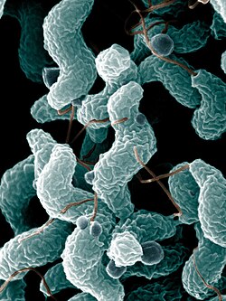
Campylobacter is a type of bacteria that can cause a diarrheal disease in people. [1] Its name means "curved bacteria", as the germ typically appears in a comma or "s" shape. According to its scientific classification, it is a genus of gram-negative bacteria that is motile. [lower-alpha 1] [2]

Contents

The germ is common in nature and in domestic animals. It is frequently found in raw food of vegetable and animal origin. Its numbers can be very high in some foods, like raw poultry. [3] Due to their diverse natural reservoir, some Campylobacter can also be detected in the air, although not at an epidemiologically significant level. [4] The disease that some of the species of the bacteria can cause is called campylobacteriosis. [lower-alpha 2]

At least a dozen species of Campylobacter have been implicated in human disease, with C. jejuni (80–90%) and C. coli (5–10%) being the most common. [6] [1] C. jejuni is recognized as one of the main causes of bacterial foodborne disease in many developed countries. [6] [7] It is the number one cause of bacterial gastroenteritis in Europe, with over 246,000 cases confirmed annually. [8] C. jejuni infection can also cause bacteremia in immunocompromised people, while C. lari is a known cause of recurrent diarrhea in children. [9] C. fetus can cause spontaneous abortions in cattle and sheep, and is an opportunistic pathogen in humans. [10]

Morphology and phenotype

Campylobacter spp. generally appear as curved or comma-shaped rods, and are able to move via unipolar or bipolar flagella. [2] They grow best between 37–42 °C in a microaerophilic environment. [11] When exposed to atmospheric oxygen, C. jejuni is able to change into a coccus form. [12] Most species of Campylobacter are positive by the oxidase test and catalase test and are able to reduce nitrate. The number of known quinolone-resistant Campylobacter strains is growing. It is suggested that this is caused by the overuse of quinolone antibiotics in animal agriculture. [12]

History

Theodor Escherich was the first to describe in 1886 what are known today as Campylobacters in the stool samples of infants, who perished from a disease he named "cholera infantum". [13] In the following years until the end of the century, a number of publications appeared, describing the occurrence of such "spirilla" in cases of "cholera-like" and "dysenteric" disease. These organisms were mainly found in the colon or associated with mucus in diarrhoeal stool specimens. Vibrio-like bacteria were also described by Sir John McFadyean and Stockman in 1913 in fetal tissues of aborted sheep. [14] For several years Campylobacters were continuously referred to as ‘‘Vibrio-like organisms’’, until 1963 when Sebald and Veron gave the name "Campylobacter" to the genus based on their shape and microaerophilic growth requirement and after showing significant biological differences with Vibrio species. [13]

Genomics

The genomes of several Campylobacter species have been sequenced, beginning with C. jejuni in 2000. [15] [16] These genome studies have identified molecular markers specific to members of Campylobacter.[ citation needed ]Campylobacter ssp. genomes are rather small compared to those of other gastrointestinal pathogens, with sizes ranging between 1.60 and 1.90 Mbp. [16] A characteristic of most Campylobacter genomes is the presence of hypervariable regions, which can differ greatly between different strains. [16]

Studies have investigated the genes responsible for motility in Campylobacter species. Some Campylobacter species contain two flagellin genes in tandem for motility, flaA and flaB. These genes undergo intergenic recombination, further contributing to their virulence. [17] A single Type VI secretion system (T6SS) cluster was also predicted in approximately one-third of Campylobacter species, grouping into three distinct organisations and harbouring up to five vgrG genes. [18]

Phylogeny

The currently accepted taxonomy is based on the List of Prokaryotic names with Standing in Nomenclature (LPSN) [19] and National Center for Biotechnology Information (NCBI) [20]

16S rRNA based LTP_08_2023 [21] [22] [23] 120 single copy marker proteins based GTDB 08-RS214 [24] [25] [26]
Campylobacter

C. portucalensis

C. corcagiensis

C. ureolyticus

C. hominis

C. blaseri

C. geochelonis

C. pinnipediorum

C. gracilis

C. rectus

C. massiliensis

C. showae

C. concisus

C. curvus

C. sputorum

C. anatolicusAydin et al. 2022

C. majalisLynch et al. 2022

C. mucosalis

C. suisLynch et al. 2022

C. fetus

Campylobacter fetus testudinum

C. hyointestinalis

C. iguaniorum

C. lanienae

Campylobacter hyointestinalis lawsoniiOn et al. 1995

C. canadensis

C. coli

C. jejuni

Campylobacter jejuni doyleiSteele & Owen 1988

C. estrildidarum

C. aviculae

C. taeniopygiae

C. insulaenigrae

C. bilis

C. hepaticus

Campylobacter lari concheus

C. ornithocola

C. subantarcticus

C. lari

C. volucris

C. armoricus

C. peloridis

C. novaezeelandiae

C. cuniculorum

C. helveticus

C. upsaliensis

C. vulpis

C. avium

C. troglodytis

Campylobacter

C. canadensisInglis et al. 2007

C. insulaenigraeFoster et al. 2004

C. volucrisDebruyne et al. 2010

C. peloridisDebruyne et al. 2009

C. armoricusBoukerb et al. 2019

C. subantarcticusDebruyne et al. 2010

C. ornithocolaCaceres et al. 2017

Campylobacter lari concheusDebruyne et al. 2009

C. lari corrig. Benjamin et al. 1984

C. aviumRossi et al. 2009

C. troglodytisKaur et al. 2023

C. cuniculorumZanoni et al. 2009

C. helveticusStanley et al. 1993

C. upsaliensis Sandstedt & Ursing 1991

C. vulpisParisi et al. 2021

C. novaezeelandiaeBloomfield et al. 2020

C. jejuni (Jones, Orcutt & Little 1931) Veron & Chatelain 1973

C. bilisPhung et al. 2022

C. hepaticusVan et al. 2016

C. coli (Doyle 1948) Veron & Chatelain 1973

C. taeniopygiaeBryant et al. 2021

C. aviculaeBryant et al. 2021

C. estrildidarumBryant et al. 2021

C. rectus (Tanner et al. 1981) Vandamme et al. 1991

C. massiliensisAntezack et al. 2021

C. showae Etoh et al. 1993

C. concisus Tanner et al. 1981

C. curvus(Tanner, Listgarten & Ebersole 1984) Vandamme et al. 1991

C. mucosalis (Lawson et al. 1981) Roop et al. 1985

C. pinnipediorumGilbert et al. 2017

C. sputorum(Prévot 1940) Veron & Chatelain 1973

C. gracilis(Tanner et al. 1981) Vandamme et al. 1995

C. hominisLawson et al. 2001

C. blaseriGilbert et al. 2018

C. geochelonisPiccirillo et al. 2016

C. portucalensisSilva et al. 2021

C. corcagiensisKoziel et al. 2014

C. ureolyticus(Jackson & Goodman 1978) Vandamme et al. 2010

"Ca. C. infans" Bian et al. 2020

C. lanienae Logan et al. 2000

C. hyointestinalis Gebhart et al. 1985

C. iguaniorumGilbert et al. 2015

Campylobacter fetus testudinumFitzgerald et al. 2014

C. fetus (Smith & Taylor 1919) Sebald & Veron 1963

Species incertae sedis:

Campylobacter-specific bacteriophages

Campylobacter-specific bacteriophages are natural viral predators of the organism. [27] Bacteriophages specific to the species now known as C. coli and C. fetus (previously Vibrio coli and V. fetus), were first isolated from cattle and pigs during the 1960s, and Campylobacter bacteriophage therapy is an ongoing area of research in the age of bacterial antibiotic resistance. [27] [28] [29]

Pathogenesis

Campylobacter can cause a gastrointestinal infection, campylobacteriosis. The incubation period is 24–72 hours after infection. [30] This is characterized by an inflammatory, sometimes bloody diarrhea or dysentery syndrome, mostly including cramps, fever, and pain. [3] [31] The most common routes of transmission are fecal-oral, ingestion of contaminated food or water, and the eating of raw meat. Foods implicated in campylobacteriosis include raw or under-cooked poultry, raw dairy products, and contaminated produce. [31] Campylobacter is sensitive to the stomach's normal production of hydrochloric acid: as a result, the infectious dose is relatively high, and the bacteria rarely cause illness when a person is exposed to less than 10,000 organisms. [9] Nevertheless, people taking antacid medication (e. g. people with gastritis or stomach ulcers) are at higher risk of contracting disease from a smaller number of organisms, since this type of medication neutralizes normal gastric acid.[ citation needed ]

In humans, the sites of tissue injury include the jejunum, the ileum, and the colon.[ citation needed ] Most strains of C. jejuni produce cytolethal distending toxin, which inhibits cell division and impedes activation of the immune system. This helps the bacteria to evade the immune system and survive for a limited time inside intestinal cells.[ citation needed ]Campylobacter has, on rare occasions, been suggested to cause hemolytic uremic syndrome [32] and thrombotic thrombocytopenic purpura, [33] though no unequivocal case reports exist. Campylobacter infection is the most common trigger of Guillain–Barré syndrome. [34] Gastrointestinal perforation is a rare complication of ileal infection. [35]

Campylobacter has also been associated with periodontitis. [3]

Detection

Campylobacter testing needs to be done to manage the risk of foodborne Campylobacter and reducing the level of foodborne Campoboteriosis, to protect people and to determine if a person is infected with Campylobacter.[ citation needed ]

In humans

Usually, detection of Campylobacter in humans is done by laboratory culturing a stool sample or swab of the rectum collected by a healthcare provider. Results take about 48–72 hours for preliminary results. Confirmation test and testing to determine the species of Campylobacter or drug sensitivities of the organism require additional time. [36]

In livestock

Usually, detection of Campylobacter in livestock is done by laboratory culturing a faecal sample. Results take about 48–72 hours. [37]

In meat

Usually, detection of Campylobacter in meat is done by laboratory culturing a homogenised sample. Results takes about 48–72 hours. [37]

Treatment

The infection is usually self-limiting and, in most cases, symptomatic treatment by liquid and electrolyte replacement is sufficient to treat human infections. Symptoms typically last 5–7 days. [31] Treatment with antibiotics has only a minor effect on the typical duration of the infection in non-complex cases, and is discouraged except in high-risk patients. [38] Diagnosis of campylobacteriosis is made by testing a fecal specimen. Standard treatment in high-risk cases is azithromycin, a macrolide antibiotic, especially for Campylobacter infections in children, [39] although other antibiotics, such as quinolones, tetracycline and other macrolides are sometimes used to treat gastrointestinal Campylobacter infections in adults. [40] In case of systemic infection, other bactericidal antibiotics are used, such as ampicillin, amoxicillin/clavulanic acid, or aminoglycosides. Fluoroquinolone antibiotics, such as ciprofloxacin or levofloxacin, may no longer be effective in some cases, due to resistance. [41] In addition to antibiotics, dehydrated patients may require intravenous fluid treatment in a hospital. [42]

Epidemiology

United Kingdom

In January 2013, the UK's Food Standards Agency (FSA) warned that two-thirds of all raw chicken bought from UK shops was contaminated with Campylobacter, affecting an estimated half a million people annually and killing about 100 of them. [43] In June 2014, the FSA started a campaign against washing raw chicken, as washing can spread germs onto clean surfaces by splashing. [44] In May 2015, cumulative results for samples taken from fresh chickens between February 2014 and February 2015 were published by the FSA and showed 73% of chickens tested positive for the presence of Campylobacter. [45]

United States

Campylobacter infections increased 14% in the United States in 2012 compared to the rate from 2006 to 2008. This represents the highest reported number of infections since calendar year 2000. [31]

High prevalence of Campylobacter (40% or more) has been reported in raw chicken meat in regional retail stores in the US, which remained steady from 2005 through 2011. [46] The last USDA quarterly progress report on Salmonella and Campylobacter testing of meat and poultry, for July–September 2014, showed a low prevalence of Campylobacter spp. in ground chicken meat, but a larger prevalence (20%) in mechanically separated chicken meat (which is sold only for further processing). [47]

Canada

FoodNet Canada has reported that Campylobacter was the most common pathogen found on packaged chicken breast, with nearly half of all samples testing positive. Additionally, Campylobacter and Salmonella were the most common causes of gastrointestinal illness in Canada. [48]

New Zealand

In August 2016, an estimated 8,000+ residents of Havelock North, a town with around 13,000 residents, had gastric illness after the water supply was thought to be contaminated by Campylobacter. [49] [50] [51]

Italy

In Italy, the annual prevalence of Campylobacter infections appears to be relatively stable based on findings from a national survey conducted on more than 5000 isolates. The survey revealed that the most common species of Campylobacter were C. jejuni, accounting for 83.7% of isolates, followed by C. coli (13.5%) and C. fetus (0.6%). The mean age of affected patients was 34.61 years, with males constituting 57.1% of cases. Outpatients represented the majority of cases, comprising 54% of the total. Campylobacter infections were predominantly isolated from feces, accounting for 97.3% of cases, while a smaller proportion (2.7%) was isolated from blood. Notably, C. fetus was primarily isolated from blood samples, constituting 88.2% of cases. Regarding antibiotic resistance patterns, the survey found that resistance to ciprofloxacin and tetracyclines was relatively high, with rates of 75.5% and 54.8%, respectively. In contrast, resistance to macrolides, including erythromycin, clarithromycin, and azithromycin, was lower, with rates ranging from 2% to 4.8%. Additionally, approximately 50% of C. jejuni and C. coli isolates exhibited resistance to two or more antibiotics. There was a significant decrease in resistance to ciprofloxacin and tetracyclines over time, while resistance to macrolides remained stable. [52]

Norway

In June 2019, an estimated 2,000 residents of Askøy municipality got sick due to the presence of C. jejuni in the water supply. Two deaths were connected to the outbreak, and it was the largest outbreak of Campylobacter in Norway. [53] The suspected source of the contamination was thought to be horse faeces, which leaked into a drinking water pool. [54] A C. jejuni water isolate thought to be the cause of the outbreak was examined with human isolates, and showed the highest pathogenic potential in vitro, transcriptomic and genomic investigations. This could suggest why the isolate was able to cause an outbreak. [55]

Sweden

During the period of August 2016 to June 2017 there was a large outbreak of C. jejuni in Sweden. It was the largest outbreak that has been reported so far. 5000 more cases than would be expected during this period were reported to the authorities. The source of the outbreak was contaminated chicken meat that came from the same producer. The reason for the increased incidence and elevated levels of Campylobacter was reported to be an improperly installed washing plant, where dirty water was accidentally used to wash transport cages. [56]

See also

Related Research Articles

<i>Escherichia coli</i> O157:H7 Serotype of the bacteria Escherichia coli

Escherichia coli O157:H7 is a serotype of the bacterial species Escherichia coli and is one of the Shiga-like toxin–producing types of E. coli. It is a cause of disease, typically foodborne illness, through consumption of contaminated and raw food, including raw milk and undercooked ground beef. Infection with this type of pathogenic bacteria may lead to hemorrhagic diarrhea, and to kidney failure; these have been reported to cause the deaths of children younger than five years of age, of elderly patients, and of patients whose immune systems are otherwise compromised.

<i>Vibrio</i> Genus of bacteria and the disease it can cause

Vibrio is a genus of Gram-negative bacteria, possessing a curved-rod (comma) shape, several species of which can cause foodborne infection, usually associated with eating undercooked seafood. Being highly salt tolerant and unable to survive in fresh water, Vibrio spp. are commonly found in various salt water environments. Vibrio spp. are facultative anaerobes that test positive for oxidase and do not form spores. All members of the genus are motile. They are able to have polar or lateral flagellum with or without sheaths. Vibrio species typically possess two chromosomes, which is unusual for bacteria. Each chromosome has a distinct and independent origin of replication, and are conserved together over time in the genus. Recent phylogenies have been constructed based on a suite of genes.

<span class="mw-page-title-main">Enteritis</span> Intestinal inflammatory disease

Enteritis is inflammation of the small intestine. It is most commonly caused by food or drink contaminated with pathogenic microbes, such as Serratia, but may have other causes such as NSAIDs, radiation therapy as well as autoimmune conditions like Crohn's disease and coeliac disease. Symptoms include abdominal pain, cramping, diarrhoea, dehydration, and fever. Related diseases of the gastrointestinal system involve inflammation of the stomach and large intestine.

<span class="mw-page-title-main">Foodborne illness</span> Illness from eating spoiled food

Foodborne illness is any illness resulting from the contamination of food by pathogenic bacteria, viruses, or parasites, as well as prions, and toxins such as aflatoxins in peanuts, poisonous mushrooms, and various species of beans that have not been boiled for at least 10 minutes.

<i>Campylobacter jejuni</i> Species of bacterium

Campylobacter jejuni is a species of pathogenic bacteria that is commonly associated with poultry, and is also often found in animal feces. This species of microbe is one of the most common causes of food poisoning in Europe and in the US, with the vast majority of cases occurring as isolated events rather than mass outbreaks. Active surveillance through the Foodborne Diseases Active Surveillance Network (FoodNet) indicates that about 20 cases are diagnosed each year for each 100,000 people in the US, while many more cases are undiagnosed or unreported; the CDC estimates a total of 1.5 million infections every year. The European Food Safety Authority reported 246,571 cases in 2018, and estimated approximately nine million cases of human campylobacteriosis per year in the European Union. Campylobacter jejuni infections are increasing at an alarming rate in Europe, North America, and Australia. In Africa, Asia, and the Middle East, data indicates that C. jejuni infections are endemic.

<span class="mw-page-title-main">Phage therapy</span> Therapeutic use of bacteriophages to treat bacterial infections

Phage therapy, viral phage therapy, or phagotherapy is the therapeutic use of bacteriophages for the treatment of pathogenic bacterial infections. This therapeutic approach emerged at the beginning of the 20th century but was progressively replaced by the use of antibiotics in most parts of the world after the Second World War. Bacteriophages, known as phages, are a form of virus that attach to bacterial cells and inject their genome into the cell. The bacteria's production of the viral genome interferes with its ability to function, halting the bacterial infection. The bacterial cell causing the infection is unable to reproduce and instead produces additional phages. Phages are very selective in the strains of bacteria they are effective against.

<span class="mw-page-title-main">Campylobacteriosis</span> Medical condition

Campylobacteriosis is among the most common infections caused by a bacterium in humans, often as a foodborne illness. It is caused by the Campylobacter bacterium, most commonly C. jejuni. It produces an inflammatory, sometimes bloody, diarrhea or dysentery syndrome, and usually cramps, fever and pain.

<span class="mw-page-title-main">Salmonellosis</span> Infection caused by Salmonella bacteria

Salmonellosis is a symptomatic infection caused by bacteria of the Salmonella type. It is the most common disease to be known as food poisoning, these are defined as diseases, usually either infectious or toxic in nature, caused by agents that enter the body through the ingestion of food. In humans, the most common symptoms are diarrhea, fever, abdominal cramps, and vomiting. Symptoms typically occur between 12 hours and 36 hours after exposure, and last from two to seven days. Occasionally more significant disease can result in dehydration. The old, young, and others with a weakened immune system are more likely to develop severe disease. Specific types of Salmonella can result in typhoid fever or paratyphoid fever. Typhoid fever and paratyphoid fever are specific types of salmonellosis, known collectively as enteric fever, and are, respectively, caused by salmonella typhi & paratyphi bacteria, which are only found in humans. Most commonly, salmonellosis cases arise from salmonella bacteria from animals, and chicken is a major source for these infections.

<i>Campylobacter fetus</i> Species of bacterium

Campylobacter fetus is a rod-shaped, gram-negative species of bacteria within the genus Campylobacter of phylum Pseudomonadota. Identification of C. fetus species in infected animals or people is routinely performed by culture on blood or cefoperazone deoxycholate agar. Subspecies of C. fetus commonly causes reproductive disease in ruminants and gastrointestinal disease in humans. Transmission of C. fetus subspecies venerealis occurs mainly through venereal contact while transmission of C. fetus subspecies fetus occurs mainly through ingestion of bacteria in a contaminated environment. Infertility in cattle and abortion in sheep are common outcomes of infection associated with C. fetus subspecies venerealis and C. fetus subspecies fetus, respectively. Disease in humans occurs through zoonotic transmission of C. fetus mainly via ingestion of contaminated food or water sources. C. fetus can be diagnosed with polymerase chain reaction assays, enzyme linked immunosorbent assays and vaginal mucus agglutination testing. As vaccines are typically not efficient in preventing future spread, infected bulls are often culled. Human infections may be treated with erythromycin as antimicrobial resistance has been emerging for the fluoroquinolones.

Intralytix, Inc. is a privately owned biotech venture incorporated in the State of Maryland on July 28, 1998. Intralytix specializes in bacteriophage based products used to control bacterial pathogens in environmental, agricultural, food processing, and medical settings.

Campylobacter upsaliensis is a gram-negative bacteria in the Campylobacter genus. C. upsaliensis is found worldwide, and is a common cause of campylobacteriosis in humans, as well as gastroenteritis in dogs and cats. Human infections are primarily associated with raw or undercooked meat and contaminated water sources, however there is some zoonotic risk associated with the spread from dogs and cats. C. upsaliensis primarily affects the gastrointestinal tract as it damages gastrointestinal epithelial cells. There are many methods for detecting C.upsaliensis including PCR and ELISA, however there is no current gold standard in detection techniques. Infection is typically self limiting, however there is antimicrobial therapy available.

<span class="mw-page-title-main">Pathogenic bacteria</span> Disease-causing bacteria

Pathogenic bacteria are bacteria that can cause disease. This article focuses on the bacteria that are pathogenic to humans. Most species of bacteria are harmless and are often beneficial but others can cause infectious diseases. The number of these pathogenic species in humans is estimated to be fewer than a hundred. By contrast, several thousand species are part of the gut flora present in the digestive tract.

Raw meat generally refers to any type of uncooked muscle tissue of an animal used for food. In the meat production industry, the term ‘meat’ refers specifically to mammalian flesh, while the words ‘poultry’ and ‘seafood’ are used to differentiate between the tissue of birds and aquatic creatures.

Pathogenic <i>Escherichia coli</i> Strains of E. coli that can cause disease

Escherichia coli is a gram-negative, rod-shaped bacterium that is commonly found in the lower intestine of warm-blooded organisms (endotherms). Most E. coli strains are harmless, but pathogenic varieties cause serious food poisoning, septic shock, meningitis, or urinary tract infections in humans. Unlike normal flora E. coli, the pathogenic varieties produce toxins and other virulence factors that enable them to reside in parts of the body normally not inhabited by E. coli, and to damage host cells. These pathogenic traits are encoded by virulence genes carried only by the pathogens.

Gastrointestinal campylobacteriosis is caused by Campylobacter jejuni or Campylobacter coli. Although it is a commensal in the gastrointestinal tract of many species, it can cause diarrhea - mainly in young animals. It is most commonly seen in cattle, but may also infect many other species, including humans. Campylobacter is spread horizontally via the fecal-oral route.

Campylobacter coli is a Gram-negative, microaerophilic, non-endospore-forming, S-shaped bacterial species within genus Campylobacter. In humans, C. coli can cause campylobacteriosis, a diarrhoeal disease which is the most frequently reported foodborne illness in the European Union. C. coli grows slowly with an optimum temperature of 42 °C. When exposed to air for long periods, they become spherical or coccoid shaped.

Enteroaggregative Escherichia coli are a pathotype of Escherichia coli which cause acute and chronic diarrhea in both the developed and developing world. They may also cause urinary tract infections. EAEC are defined by their "stacked-brick" pattern of adhesion to the human laryngeal epithelial cell line HEp-2. The pathogenesis of EAEC involves the aggregation of and adherence of the bacteria to the intestinal mucosa, where they elaborate enterotoxins and cytotoxins that damage host cells and induce inflammation that results in diarrhea.

<span class="mw-page-title-main">Elizabeth O. King</span> American bacteriologist (1912–1966)

Elizabeth Osborne King was an American microbiologist who discovered and described bacteria of medical importance at the United States Centers for Disease Control and Prevention from the late 1940s through the early 1960s. A 1984 CDC manual dedication referred to King as "internationally known as an authority on a variety of unusual bacteria." The genera Kingella and Elizabethkingia and several species of bacteria are named to honor her for her pioneering work. King died of cancer on April 8, 1966, in Atlanta, where she is interred in Oakland Cemetery.

<span class="mw-page-title-main">Multidrug-resistant bacteria</span>

Multidrug-resistant bacteria are bacteria that are resistant to three or more classes of antimicrobial drugs. MDR bacteria have seen an increase in prevalence in recent years and pose serious risks to public health. MDR bacteria can be broken into 3 main categories: Gram-positive, Gram-negative, and other (acid-stain). These bacteria employ various adaptations to avoid or mitigate the damage done by antimicrobials. With increased access to modern medicine there has been a sharp increase in the amount of antibiotics consumed. Given the abundant use of antibiotics there has been a considerable increase in the evolution of antimicrobial resistance factors, now outpacing the development of new antibiotics.

<span class="mw-page-title-main">Milk borne diseases</span>

Milk borne diseases are any diseases caused by consumption of milk or dairy products infected or contaminated by pathogens. Milk borne diseases are one of the recurrent foodborne illnesses—between 1993 and 2012 over 120 outbreaks related to raw milk were recorded in the US with approximately 1,900 illnesses and 140 hospitalisations. With rich nutrients essential for growth and development such as proteins, lipids, carbohydrates, and vitamins in milk, pathogenic microorganisms are well nourished and are capable of rapid cell division and extensive population growth in this favourable environment. Common pathogens include bacteria, viruses, fungi, and parasites and among them, bacterial infection is the leading cause of milk borne diseases.

References

  1. Motile means that it can move of its own.
  2. It is usually self-limiting and antimicrobial treatment is often not required, except in severe cases or immunocompromised patients. [5]
  1. 1 2 Blaser, Martin J. (December 1997). "Epidemiologic and Clinical Features of Campylobacter jejuni Infections". The Journal of Infectious Diseases. 176 (s2): S103–S105. doi: 10.1086/513780 . PMID   9396691.
  2. 1 2 Garrity, George M.; Bell, Julia A.; Lilburn, Timothy (2005). "Class V. Epsilonproteobacteria class. Nov.". Bergey's Manual® of Systematic Bacteriology. pp. 1145–1194. doi:10.1007/0-387-29298-5_4. ISBN   978-0-387-24145-6.
  3. 1 2 3 Humphrey, Tom; O'Brien, Sarah; Madsen, Mogens (July 2007). "Campylobacters as zoonotic pathogens: a food production perspective". International Journal of Food Microbiology. 117 (3): 237–57. doi:10.1016/j.ijfoodmicro.2007.01.006. PMID   17368847.
  4. Olsen, Katja N.; Lund, Marianne; Skov, Julia; Christensen, Laurids S.; Hoorfar, Jeffrey (2009). "Detection of Campylobacter Bacteria in Air Samples for Continuous Real-Time Monitoring of Campylobacter Colonization in Broiler Flocks". Applied and Environmental Microbiology. 75 (7): 2074–2078. doi:10.1128/AEM.02182-08. ISSN   0099-2240. PMC   2663182 . PMID   19201953.
  5. Skarp CP, Hänninen ML, Rautelin HI (February 2016). "Campylobacteriosis: the role of poultry meat". Clinical Microbiology and Infection. 22 (2): 103–109. doi: 10.1016/j.cmi.2015.11.019 . PMID   26686808.
  6. 1 2 Ryan, Kenneth J.; Ray, C. George, eds. (2004). Sherris Medical Microbiology: An Introduction to Infectious Diseases (4th ed.). McGraw Hill. pp. 378–380. ISBN   978-0-8385-8529-0.
  7. Moore, John E.; Corcoran, Deborah; Dooley, James S.G.; Fanning, Séamus; Lucey, Brigid; Matsuda, Motoo; McDowell, David A.; Mégraud, Francis; Cherie Millar, B.; O'Mahony, Rebecca; O?Riordan, Lisa; O'Rourke, Michele; Rao, Juluri R.; Rooney, Paul J.; Sails, Andrew; Whyte, Paul (May 2005). "Campylobacter". Veterinary Research. 36 (3): 351–382. doi: 10.1051/vetres:2005012 . PMID   15845230.
  8. "Campylobacter". European Food Safety Authority. Retrieved 2020-11-02.
  9. 1 2 Campylobacter Infections at eMedicine
  10. Sauerwein, RW; Bisseling, J; Horrevorts, AM (1993). "Septic abortion associated with Campylobacter fetus subspecies fetus infection: case report and review of the literature". Infection. 21 (5): 331–3. doi:10.1007/BF01712458. PMID   8300253. S2CID   28539930.
  11. "Information for Health Professionals | Campylobacter". Centers for Disease Control and Prevention. 2019-12-23. Retrieved 2020-11-02.
  12. 1 2 Crushell, Ellen; Harty, Sinead; Sharif, Farhana; Bourke, Billy (January 2004). "Enteric Campylobacter: Purging Its Secrets?". Pediatric Research. 55 (1): 3–12. doi: 10.1203/01.PDR.0000099794.06260.71 . ISSN   1530-0447. PMID   14605259.
  13. 1 2 Samie, A.; Obi, C.L.; Barrett, L.J.; Powell, S.M.; Guerrant, R.L. (June 2007). "Prevalence of Campylobacter species, Helicobacter pylori and Arcobacter species in stool samples from the Venda region, Limpopo, South Africa: Studies using molecular diagnostic methods". Journal of Infection. 54 (6): 558–566. doi:10.1016/j.jinf.2006.10.047. ISSN   0163-4453. PMID   17145081.
  14. Altekruse, SF; Stern, NJ; Fields, PI; Swerdlow, DL (1999). "Campylobacter jejuni--an emerging foodborne pathogen". Emerging Infectious Diseases. 5 (1): 28–35. doi:10.3201/eid0501.990104. OCLC   677425436. PMC   2627687 . PMID   10081669.
  15. Fouts, Derrick E.; Mongodin, Emmanuel F.; Mandrell, Robert E.; Miller, William G.; Rasko, David A.; Ravel, Jacques; Brinkac, Lauren M.; DeBoy, Robert T.; Parker, Craig T.; Daugherty, Sean C.; Dodson, Robert J.; Durkin, A. Scott; Madupu, Ramana; Sullivan, Steven A.; Shetty, Jyoti U. (2005-01-04). "Major Structural Differences and Novel Potential Virulence Mechanisms from the Genomes of Multiple Campylobacter Species". PLOS Biology. 3 (1): e15. doi: 10.1371/journal.pbio.0030015 . ISSN   1545-7885. PMC   539331 . PMID   15660156.
  16. 1 2 3 Parkhill, J.; Wren, B. W.; Mungall, K.; Ketley, J. M.; Churcher, C.; Basham, D.; Chillingworth, T.; Davies, R. M.; Feltwell, T.; Holroyd, S.; Jagels, K.; Karlyshev, A. V.; Moule, S.; Pallen, M. J.; Penn, C. W. (February 2000). "The genome sequence of the food-borne pathogen Campylobacter jejuni reveals hypervariable sequences". Nature. 403 (6770): 665–668. doi:10.1038/35001088. ISSN   1476-4687.
  17. Grant, CC; Konkel, ME; Cieplak, W; Tompkins, LS (May 1993). "Role of flagella in adherence, internalization, and translocation of Campylobacter jejuni in nonpolarized and polarized epithelial cell cultures". Infection and Immunity. 61 (5): 1764–71. doi:10.1128/IAI.61.5.1764-1771.1993. PMC   280763 . PMID   8478066.
  18. Robinson, L; Liaw, J; Omole, Z; Corcionivoschi, N; Hachani, A; Gundogdu, O (October 2022). "In silico investigation of the genus Campylobacter type VI secretion system reveals genetic diversity in organization and putative effectors". Microbial Genomics. 8 (10): 000898. doi: 10.1099/mgen.0.000898 . ISSN   2057-5858. PMC   9676060 . PMID   36314601.
  19. A.C. Parte; et al. "Campylobacter". List of Prokaryotic names with Standing in Nomenclature (LPSN). Retrieved 2023-09-09.
  20. Sayers; et al. "Campylobacter". National Center for Biotechnology Information (NCBI) taxonomy database. Retrieved 2023-09-09.
  21. "The LTP" . Retrieved 20 November 2023.
  22. "LTP_all tree in newick format" . Retrieved 20 November 2023.
  23. "LTP_08_2023 Release Notes" (PDF). Retrieved 20 November 2023.
  24. "GTDB release 08-RS214". Genome Taxonomy Database . Retrieved 10 May 2023.
  25. "bac120_r214.sp_label". Genome Taxonomy Database . Retrieved 10 May 2023.
  26. "Taxon History". Genome Taxonomy Database . Retrieved 10 May 2023.
  27. 1 2 Connerton, P.L.; Timms, A.R.; Connerton, I.F. (2011). "Campylobacter bacteriophages and bacteriophage therapy: Campylobacter bacteriophages". Journal of Applied Microbiology. 111 (2): 255–265. doi:10.1111/j.1365-2672.2011.05012.x. PMID   21447013. S2CID   46270047.
  28. Firehammer, BD; Border, M (November 1968). "Isolation of temperate bacteriophages from Vibrio fetus". American Journal of Veterinary Research. 29 (11): 2229–35. PMID   5693467.
  29. Fletcher, RD (1965). "Activity and morphology of Vibrio coli phage". American Journal of Veterinary Research. 26 (111): 361–4.
  30. Zilbauer, Matthias; Dorrell, Nick; Wren, Brendan W.; Bajaj-Elliott, Mona (February 2008). "Campylobacter jejuni-mediated disease pathogenesis: an update". Transactions of the Royal Society of Tropical Medicine and Hygiene. 102 (2): 123–129. doi:10.1016/j.trstmh.2007.09.019. ISSN   0035-9203. PMID   18023831.
  31. 1 2 3 4 "Infections from some foodborne germs increased, while others remained unchanged in 2012" (Press release). CDC. April 18, 2013. Retrieved October 22, 2015.
  32. Keithlin, Jessica; Sargeant, Jan; Thomas, M. Kate; Fazil, Aamir (2014-11-22). "Systematic review and meta-analysis of the proportion of Campylobacter cases that develop chronic sequelae". BMC Public Health. 14: 1203. doi: 10.1186/1471-2458-14-1203 . ISSN   1471-2458. PMC   4391665 . PMID   25416162.
  33. Morton, A. R.; Yu, R.; Waldek, S.; Holmes, A. M.; Craig, A.; Mundy, K. (1985-11-16). "Campylobacter induced thrombotic thrombocytopenic purpura". Lancet. 2 (8464): 1133–1134. doi:10.1016/s0140-6736(85)90729-9. ISSN   0140-6736. PMID   2865609. S2CID   44823836.
  34. Finsterer, Josef (2022-11-17). "Triggers of Guillain-Barré Syndrome: Campylobacter jejuni Predominates". International Journal of Molecular Sciences. 23 (22): 14222. doi: 10.3390/ijms232214222 . ISSN   1422-0067. PMC   9696744 . PMID   36430700.
  35. Jassim, SS; Malik, A; Aldridge, A (2011). "Small bowel perforation: an unusual cause". Grand Rounds. 11 (1): 17–19. doi:10.1102/1470-5206.2011.0006.
  36. Buss, Janice E.; Cresse, Michelle; Doyle, Susan; Buchan, Blake W.; Craft, David W.; Young, Steve (2019). "Campylobacter culture fails to correctly detect Campylobacter in 30% of positive patient stool specimens compared to non-cultural methods". European Journal of Clinical Microbiology & Infectious Diseases. 38 (6): 1087–1093. doi:10.1007/s10096-019-03499-x. ISSN   1435-4373. PMC   6520473 . PMID   30783889.
  37. 1 2 Hong, Yang; Berrang, Mark E.; Liu, Tongrui; Hofacre, Charles L.; Sanchez, Susan; Wang, Lihua; Maurer, John J. (2003). "Rapid Detection of Campylobacter coli , C. jejuni , and Salmonella enterica on Poultry Carcasses by Using PCR-Enzyme-Linked Immunosorbent Assay". Applied and Environmental Microbiology. 69 (6): 3492–3499. Bibcode:2003ApEnM..69.3492H. doi:10.1128/AEM.69.6.3492-3499.2003. ISSN   0099-2240. PMC   161512 . PMID   12788755.
  38. Ternhag, Anders; Asikainen, Tommi; Giesecke, Johan; Ekdahl, Karl (2007-03-01). "A Meta-Analysis on the Effects of Antibiotic Treatment on Duration of Symptoms Caused by Infection with Campylobacter Species". Clinical Infectious Diseases. 44 (5): 696–700. doi:10.1086/509924. ISSN   1058-4838. PMID   17278062.
  39. Vukelic, Dalibor; Trkulja, Vladimir; Salkovic-Petrisic, Melita (April 2010). "Single oral dose of azithromycin versus 5 days of oral erythromycin or no antibiotic in treatment of Campylobacter enterocolitis in children: a prospective randomized assessor-blind study". Journal of Pediatric Gastroenterology and Nutrition. 50 (4): 404–10. doi: 10.1097/MPG.0b013e3181a87104 . PMID   19881393. S2CID   22460970.
  40. Gendrel, D.; Cohen, R.; European Society for Pediatric Infectious Diseases; European Society for Gastroenterology, Hepatology and Nutrition (October 2008). "Diarrhées bactériennes et antibiotiques : les recommandations européennes" [Bacterial diarrheas and antibiotics: European recommendations]. Archives de Pédiatrie (in French). 15: S93–S96. doi:10.1016/S0929-693X(08)74223-4. PMID   19000862.
  41. Lehtopolku, Mirva; Nakari, Ulla-Maija; Kotilainen, Pirkko; Huovinen, Pentti; Siitonen, Anja; Hakanen, Antti J. (March 2010). "Antimicrobial Susceptibilities of Multidrug-Resistant Campylobacter jejuni and C. coli Strains: In Vitro Activities of 20 Antimicrobial Agents". Antimicrobial Agents and Chemotherapy. 54 (3): 1232–1236. doi:10.1128/AAC.00898-09. ISSN   0066-4804. PMC   2825995 . PMID   20038624.
  42. Mortensen, Nicolay; Jonasson, Solveig Aalstad; Lavesson, Ingrid Viola; Emberland, Knut Erik; Litleskare, Sverre; Wensaas, Knut-Arne; Rortveit, Guri; Langeland, Nina; Hanevik, Kurt (2021-03-23). "Characteristics of hospitalized patients during a large waterborne outbreak of Campylobacter jejuni in Norway". PLOS ONE. 16 (3): e0248464. doi: 10.1371/journal.pone.0248464 . ISSN   1932-6203. PMC   7987138 . PMID   33755697.
  43. Ward, Victoria (January 23, 2013). "FSA warns that chicken bacteria could be next meat scandal". The Telegraph.
  44. "Food Safety Week 2014: 'Don't wash raw chicken'". Food Standards Agency. Archived from the original on November 8, 2014.
  45. "Campylobacter survey: cumulative results from the full 12 months (Q1 – Q4)" (Press release). Food Standards Agency. May 28, 2015. Retrieved October 23, 2015.
  46. Williams, Aretha; Oyarzabal, Omar A (August 2012). "Prevalence of Campylobacter spp. in skinless, boneless retail broiler meat from 2005 through 2011 in Alabama, USA". BMC Microbiology. 12: 184. doi: 10.1186/1471-2180-12-184 . PMC   3490988 . PMID   22920043.
  47. "Quarterly Progress Report on Salmonella and Campylobacter". Testing of Selected Raw Meat and Poultry Products: Preliminary Results, July 2014 to September 2014. Food Safety and Inspection Service . United States Department of Agriculture. 2015-04-24.
  48. "FoodNet Canada 2014 Short Report". Public Health Agency of Canada, FoodNet Canada. 2016-01-12. Retrieved 3 October 2016.
  49. Sharpe, Marty (19 August 2016). "Woman died in Havelock North gastro outbreak". Stuff.
  50. "Govt rejects call for Hawke's Bay water emergency declaration". Radio New Zealand. 19 August 2016.
  51. Gilpin, Brent J.; Walker, Tiffany; Paine, Shevaun; Sherwood, Jill; Mackereth, Graham; Wood, Tim; Hambling, Tammy; Hewison, Chris; Brounts, Angela; Wilson, Maurice; Scholes, Paula; Robson, Beth; Lin, Susan; Cornelius, Angela; Rivas, Lucia (2020). "A large scale waterborne Campylobacteriosis outbreak, Havelock North, New Zealand". Journal of Infection. 81 (3): 390–395. doi:10.1016/j.jinf.2020.06.065. ISSN   0163-4453. PMID   32610108. S2CID   220306856.
  52. Zerbato, Verena; Di Bella, Stefano; Pol, Riccardo; Luzzati, Roberto; Sanson, Gianfranco; Ambretti, Simone; Andreoni, Stefano; Aschbacher, Richard; Bernardo, Mariano; Bielli, Alessandra; Brigante, Gioconda; Busetti, Marina; Camarlinghi, Giulio; Carcione, Davide; Carducci, Antonella (2024-03-12). "Human Campylobacter spp. infections in Italy". European Journal of Clinical Microbiology & Infectious Diseases. doi:10.1007/s10096-024-04803-0. ISSN   0934-9723.
  53. Olsen, Adrian Nyhammer; Baisotti, Valentina (11 June 2019). "Fant samme bakterie i drikkevannet som hos sykehuspasienter fra Askøy" (in Norwegian Bokmål). NRK.
  54. Paruch, Lisa; Paruch, Adam M.; Sørheim, Roald (March 2020). "DNA-based faecal source tracking of contaminated drinking water causing a large Campylobacter outbreak in Norway 2019". International Journal of Hygiene and Environmental Health. 224: 113420. doi: 10.1016/j.ijheh.2019.113420 . hdl: 11250/2731881 . PMID   31748129.
  55. Davies, Emma; Ebbesen, Marit; Johansson, Cecilia; Kaden, René; Rautelin, Hilpi (2020). "Genomic and Phenotypic Characterisation of Campylobacter jejuni Isolates From a Waterborne Outbreak". Frontiers in Cellular and Infection Microbiology. 10: 594856. doi: 10.3389/fcimb.2020.594856 . PMC   7658296 . PMID   33194843.
  56. "Utbrott av anmälningspliktiga sjukdomar i Sverige 2017" [Outbreaks of notifiable diseases in Sweden in 2017] (in Swedish). Folkhälsomyndigheten. 2018-05-09. Retrieved 27 May 2020.