Dent's disease

Last updated
Dent's disease
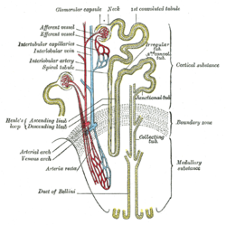
Gray1128.png
Nephron of the kidney without juxtaglomerular apparatus
Specialty Urology   OOjs UI icon edit-ltr-progressive.svg
Named after Charles Enrique Dent

Dent's disease (or Dent disease) is a rare X-linked recessive inherited condition that affects the proximal renal tubules [1] of the kidney. It is one cause of Fanconi syndrome, and is characterized by tubular proteinuria, excess calcium in the urine, formation of calcium kidney stones, nephrocalcinosis, and chronic kidney failure.

Contents

"Dent's disease" is often used to describe an entire group of familial disorders, including X-linked recessive nephrolithiasis with kidney failure, X-linked recessive hypophosphatemic rickets, and both Japanese and idiopathic low-molecular-weight proteinuria. [2] About 60% of patients have mutations in the CLCN5 gene (Dent 1), which encodes a kidney-specific chloride/proton antiporter, and 15% of patients have mutations in the OCRL1 gene (Dent 2). [3]

Signs and symptoms

Dent's disease often produces the following signs and symptoms: [4]

In a study of 25 patients with Dent's disease, [5] 9 of 15 men, and one of 10 women had end-stage kidney disease by the age of 47. [6]

Genetics

X-linked recessive inheritance X-linked recessive.svg
X-linked recessive inheritance
Three-dimensional homology model of human CLC-5 based on the structure of ClC-ec1 (7) showing the locations of each mutated residue CLC-5 Mutations.jpg
Three-dimensional homology model of human CLC-5 based on the structure of ClC-ec1 (7) showing the locations of each mutated residue

Dent disease 1

Dent's disease is a X-linked recessive disorder. The males are prone to manifesting symptoms in early adulthood with symptoms of calculi, rickets or even with kidney failure in more severe cases. [4]

In humans, gene CLCN5 is located on chromosome Xp11.22, and has a 2238-bp coding sequence that consists of 11 exons that span 25 to 30 kb of genomic DNA and encode a 746-amino-acid protein. [7] CLCN5 belongs to the family of voltage-gated chloride channel genes (CLCN1-CLCN7, CLCKa and CLCKb) that have about 12 transmembrane domains. These chloride channels have an important role in the control of membrane excitability, transepithelial transport, and possibly cell volume. [8]

The mechanisms by which CLC-5 dysfunction results in hypercalciuria and the other features of Dent's disease remain to be elucidated. The identification of additional CLCN5 mutations may help in these studies. [9]

Dent disease 2

Dent disease 2 (nephrolithiasis type 2) is associated with the OCRL gene. [10] [11] Both Lowe syndrome (oculocerebrorenal syndrome) and Dent disease can be caused by truncating or missense mutations in OCRL.

Diagnosis

Diagnosis is based on genetic study of CLCN5 gene.[ citation needed ]

Treatment

As of today, no agreed-upon treatment of Dent's disease is known and no therapy has been formally accepted. Most treatment measures are supportive in nature:

History

Dent's disease was first described by Charles Enrique Dent and M. Friedman in 1964, when they reported two unrelated British boys with rickets associated with renal tubular damage characterized by hypercalciuria, hyperphosphaturia, proteinuria, and aminoaciduria. [15] This set of symptoms was not given a name until 30 years later, when the nephrologist Oliver Wrong more fully described the disease. [5] Wrong had studied with Dent and chose to name the disease after his mentor. [16] Dent's disease is a genetic disorder caused by mutations in the gene CLCN5, which encodes a kidney-specific voltage-gated chloride channel, a 746-amino-acid protein (CLC-5) with 12 to 13 transmembrane domains. It manifests itself through low-molecular-weight proteinuria, hypercalciuria, aminoaciduria and hypophosphataemia. Because of its rather rare occurrence, Dent's disease is often diagnosed as idiopathic hypercalciuria, i.e., excess calcium in urine with undetermined causes.[ citation needed ]

Related Research Articles

<span class="mw-page-title-main">Antiporter</span> Class of transmembrane transporter protein

An antiporter is an integral membrane protein that uses secondary active transport to move two or more molecules in opposite directions across a phospholipid membrane. It is a type of cotransporter, which means that uses the energetically favorable movement of one molecule down its electrochemical gradient to power the energetically unfavorable movement of another molecule up its electrochemical gradient. This is in contrast to symporters, which are another type of cotransporter that moves two or more ions in the same direction, and primary active transport, which is directly powered by ATP.

<span class="mw-page-title-main">Chlortalidone</span> Thiazide-like diuretic drug

Chlortalidone, also known as chlorthalidone, is a thiazide-like diuretic drug used to treat high blood pressure, swelling, diabetes insipidus, and renal tubular acidosis. Because chlortalidone is effective in most patients with high blood pressure, it is considered a preferred initial treatment. It is also used to prevent calcium-based kidney stones. It is taken by mouth. Effects generally begin within three hours and last for up to three days. Long-term treatment with chlortalidone is more effective than hydrochlorothiazide for prevention of heart attack or stroke.

<span class="mw-page-title-main">Thiazide</span> Class of chemical compounds

Thiazide refers to both a class of sulfur-containing organic molecules and a class of diuretics based on the chemical structure of benzothiadiazine. The thiazide drug class was discovered and developed at Merck and Co. in the 1950s. The first approved drug of this class, chlorothiazide, was marketed under the trade name Diuril beginning in 1958. In most countries, thiazides are the least expensive antihypertensive drugs available.

<span class="mw-page-title-main">Cystinosis</span> Lysosomal storage disease

Cystinosis is a lysosomal storage disease characterized by the abnormal accumulation of cystine, the oxidized dimer of the amino acid cysteine. It is a genetic disorder that follows an autosomal recessive inheritance pattern. It is a rare autosomal recessive disorder resulting from accumulation of free cystine in lysosomes, eventually leading to intracellular crystal formation throughout the body. Cystinosis is the most common cause of Fanconi syndrome in the pediatric age group. Fanconi syndrome occurs when the function of cells in renal tubules is impaired, leading to abnormal amounts of carbohydrates and amino acids in the urine, excessive urination, and low blood levels of potassium and phosphates.

<span class="mw-page-title-main">Gitelman syndrome</span> Genetic kidney disorder

Gitelman syndrome (GS) is an autosomal recessive kidney tubule disorder characterized by low blood levels of potassium and magnesium, decreased excretion of calcium in the urine, and elevated blood pH. It is the most frequent hereditary salt-losing tubulopathy. Gitelman syndrome is caused by disease-causing variants on both alleles of the SLC12A3 gene. The SLC12A3 gene encodes the thiazide-sensitive sodium-chloride cotransporter, which can be found in the distal convoluted tubule of the kidney.

<span class="mw-page-title-main">Focal segmental glomerulosclerosis</span> Kidney disease

Focal segmental glomerulosclerosis (FSGS) is a histopathologic finding of scarring (sclerosis) of glomeruli and damage to renal podocytes. This process damages the filtration function of the kidney, resulting in protein presence in the urine due to protein loss. FSGS is a leading cause of excess protein loss—nephrotic syndrome—in children and adults in the US. Signs and symptoms include proteinuria and edema. Kidney failure is a common long-term complication of the disease. FSGS can be classified as primary, secondary, or genetic, depending on whether a particular toxic or pathologic stressor or genetic predisposition can be identified as the cause. Diagnosis is established by renal biopsy, and treatment consists of glucocorticoids and other immune-modulatory drugs. Response to therapy is variable, with a significant portion of patients progressing to end-stage kidney failure. An American epidemiological study 20 years ago demonstrated that FSGS is estimated to occur in 7 persons per million, with cisgender male African-Americans at higher risk.

<span class="mw-page-title-main">Renal tubular acidosis</span> Higher blood acidity due to failure of the kidneys to fully acidify urine

Renal tubular acidosis (RTA) is a medical condition that involves an accumulation of acid in the body due to a failure of the kidneys to appropriately acidify the urine. In renal physiology, when blood is filtered by the kidney, the filtrate passes through the tubules of the nephron, allowing for exchange of salts, acid equivalents, and other solutes before it drains into the bladder as urine. The metabolic acidosis that results from RTA may be caused either by insufficient secretion of hydrogen ions into the latter portions of the nephron or by failure to reabsorb sufficient bicarbonate ions from the filtrate in the early portion of the nephron. Although a metabolic acidosis also occurs in those with chronic kidney disease, the term RTA is reserved for individuals with poor urinary acidification in otherwise well-functioning kidneys. Several different types of RTA exist, which all have different syndromes and different causes. RTA is usually an incidental finding based on routine blood draws that show abnormal results. Clinically, patients may present with vague symptoms such as dehydration, mental status changes, or delayed growth in adolescents.

<span class="mw-page-title-main">Bartter syndrome</span> Medical condition

Bartter syndrome (BS) is a rare inherited disease characterised by a defect in the thick ascending limb of the loop of Henle, which results in low potassium levels (hypokalemia), increased blood pH (alkalosis), and normal to low blood pressure. There are two types of Bartter syndrome: neonatal and classic. A closely associated disorder, Gitelman syndrome, is milder than both subtypes of Bartter syndrome.

<span class="mw-page-title-main">Oculocerebrorenal syndrome</span> Medical condition

Oculocerebrorenal syndrome is a rare X-linked recessive disorder characterized by congenital cataracts, hypotonia, intellectual disability, proximal tubular acidosis, aminoaciduria and low-molecular-weight proteinuria. Lowe syndrome can be considered a cause of Fanconi syndrome.

Hypercalciuria is the condition of elevated calcium in the urine. Chronic hypercalciuria may lead to impairment of renal function, nephrocalcinosis, and chronic kidney disease. Patients with hypercalciuria have kidneys that excrete higher levels of calcium than normal, for which there are many possible causes. Calcium may come from one of two paths: through the gut where higher than normal levels of calcium are absorbed by the body or mobilized from stores in the bones. After initial 24 hour urine calcium testing and additional lab testing, a bone density scan (DSX) may be performed to determine if the calcium is being obtained from the bones.

Pseudohypoaldosteronism (PHA) is a condition that mimics hypoaldosteronism. Two major types of primary pseudohypoaldosteronism are recognized and these have major differences in etiology and presentation.

<span class="mw-page-title-main">Sodium-chloride symporter</span> Protein-coding gene in the species Homo sapiens

The sodium-chloride symporter (also known as Na+-Cl cotransporter, NCC or NCCT, or as the thiazide-sensitive Na+-Cl cotransporter or TSC) is a cotransporter in the kidney which has the function of reabsorbing sodium and chloride ions from the tubular fluid into the cells of the distal convoluted tubule of the nephron. It is a member of the SLC12 cotransporter family of electroneutral cation-coupled chloride cotransporters. In humans, it is encoded by the SLC12A3 gene (solute carrier family 12 member 3) located in 16q13.

<span class="mw-page-title-main">Nephrocalcinosis</span> Medical condition caused by the deposition of calcium salts in the kidneys

Nephrocalcinosis, once known as Albright's calcinosis after Fuller Albright, is a term originally used to describe the deposition of poorly soluble calcium salts in the renal parenchyma due to hyperparathyroidism. The term nephrocalcinosis is used to describe the deposition of both calcium oxalate and calcium phosphate. It may cause acute kidney injury. It is now more commonly used to describe diffuse, fine, renal parenchymal calcification in radiology. It is caused by multiple different conditions and is determined by progressive kidney dysfunction. These outlines eventually come together to form a dense mass. During its early stages, nephrocalcinosis is visible on x-ray, and appears as a fine granular mottling over the renal outlines. It is most commonly seen as an incidental finding with medullary sponge kidney on an abdominal x-ray. It may be severe enough to cause renal tubular acidosis or even end stage kidney disease, due to disruption of the kidney tissue by the deposited calcium salts.

<span class="mw-page-title-main">Medullary sponge kidney</span> Congenital disorder of urinary system

Medullary sponge kidney is a congenital disorder of the kidneys characterized by cystic dilatation of the collecting tubules in one or both kidneys. Individuals with medullary sponge kidney are at increased risk for kidney stones and urinary tract infection (UTI). Patients with MSK typically pass twice as many stones per year as do other stone formers without MSK. While having a low morbidity rate, as many as 10% of patients with MSK have an increased risk of morbidity associated with frequent stones and UTIs. While many patients report increased chronic kidney pain, the source of the pain, when a UTI or blockage is not present, is unclear at this time. Renal colic is present in 55% of patients. Women with MSK experience more stones, UTIs, and complications than men. MSK was previously believed not to be hereditary but there is more evidence coming forth that may indicate otherwise.

<span class="mw-page-title-main">CLCN5</span> Mammalian protein found in humans

The CLCN5 gene encodes the chloride channel Cl-/H+ exchanger ClC-5. ClC-5 is mainly expressed in the kidney, in particular in proximal tubules where it participates to the uptake of albumin and low-molecular-weight proteins, which is one of the principal physiological role of proximal tubular cells. Mutations in the CLCN5 gene cause an X-linked recessive nephropathy named Dent disease characterized by excessive urinary loss of low-molecular-weight proteins and of calcium (hypercalciuria), nephrocalcinosis and nephrolithiasis.

Fanconi syndrome or Fanconi's syndrome is a syndrome of inadequate reabsorption in the proximal renal tubules of the kidney. The syndrome can be caused by various underlying congenital or acquired diseases, by toxicity, or by adverse drug reactions. It results in various small molecules of metabolism being passed into the urine instead of being reabsorbed from the tubular fluid. Fanconi syndrome affects the proximal tubules, namely, the proximal convoluted tubule (PCT), which is the first part of the tubule to process fluid after it is filtered through the glomerulus, and the proximal straight tubule, which leads to the descending limb of loop of Henle.

<span class="mw-page-title-main">Distal renal tubular acidosis</span> Medical condition

Distal renal tubular acidosis (dRTA) is the classical form of RTA, being the first described. Distal RTA is characterized by a failure of acid secretion by the alpha intercalated cells of the distal tubule and cortical collecting duct of the distal nephron. This failure of acid secretion may be due to a number of causes. It leads to relatively alkaline urine, due to the kidney's inability to acidify the urine to a pH of less than 5.3.

Proximal renal tubular acidosis (pRTA) or type 2 renal tubular acidosis (RTA) is a type of RTA caused by a failure of the proximal tubular cells to reabsorb filtered bicarbonate from the urine, leading to urinary bicarbonate wasting and subsequent acidemia. The distal intercalated cells function normally, so the acidemia is less severe than dRTA and the urine can acidify to a pH of less than 5.3. pRTA also has several causes, and may occasionally be present as a solitary defect, but is usually associated with a more generalised dysfunction of the proximal tubular cells called Fanconi syndrome where there is also phosphaturia, glycosuria, aminoaciduria, uricosuria and tubular proteinuria.

<span class="mw-page-title-main">Oliver Wrong</span>

Professor Oliver Murray Wrong was an eminent academic nephrologist and one of the founders of the speciality in the United Kingdom. From a background as a "salt and water" physician, he made detailed clinical observations and scientifically imaginative connections which were the basis of numerous advances in the molecular biology of the human kidney. Wrong himself contributed to much of the molecular work after his own "retirement". He dictated amendments to his final paper during his final illness in his own teaching hospital, University College Hospital (UCH), London. Though academic in his leanings, he was a compassionate physician who established a warm rapport with patients, a link he regarded as the keystone of his research. He belonged to a generation of idealistic young doctors responsible for the establishment of the UK's National Health Service in the post-War years.

<span class="mw-page-title-main">Idiopathic hypercalcinuria</span>

Idiopathic hypercalcinuria (IH) is a condition including an excessive urinary calcium level with a normal blood calcium level resulting from no underlying cause. IH has become the most common cause of hypercalciuria and is the most serious metabolic risk factor for developing nephrolithiasis. IH can predispose individuals to osteopenia or osteoporosis, and affects the entire body. IH arises due to faulty calcium homeostasis, a closely monitored process, where slight deviations in calcium transport in the intestines, blood, and bone can lead to excessive calcium excretion, bone mineral density loss, or kidney stone formation. 50%-60% of nephrolithiasis patients suffer from IH and have 5%-15% lower bone density than those who do not.

References

  1. "Dent disease" at Dorland's Medical Dictionary
  2. Mayo Clinic, Division of Nephrology and Hypertension, Mineral Metabolism and Stone Disease Archived 2007-03-05 at the Wayback Machine
  3. S. Karger AG, Basel, Truncating Mutations in the Chloride/Proton ClC-5 Antiporter Gene in Seven Jewish Israeli Families with Dent’s 1 Disease
  4. 1 2 "Dent disease". National Center For Advancing Translational Sciences. Retrieved 4 July 2021.
  5. 1 2 Wrong OM; Norden AGW; Feest TG (1994). "Dent's disease; a familial proximal renal tubular syndrome with low-molecular-weight proteinuria, hypercalciuria, nephrocalcinosis, metabolic bone disease, progressive kidney failure and a marked male predominance". Quarterly Journal of Medicine. 87 (8): 473–493. Archived from the original on 2012-07-14.
  6. Burgess HK, Jayawardene SA, Velasco N (July 2001). "Dent's disease: can we slow its progression?". Nephrol. Dial. Transplant. 16 (7): 1512–3. doi:10.1093/ndt/16.7.1512. PMID   11427657.
  7. Fisher SE, van Bakel I, Lloyd SE, Pearce SH, Thakker RV, Craig IW (October 1995). "Cloning and characterization of CLCN5, the human kidney chloride channel gene implicated in Dent disease (an X-linked hereditary nephrolithiasis)". Genomics. 29 (3): 598–606. doi:10.1006/geno.1995.9960. hdl: 11858/00-001M-0000-0012-CC06-6 . PMID   8575751.
  8. Jentsch TJ, Friedrich T, Schriever A, Yamada H (May 1999). "The CLC chloride channel family". Pflügers Arch. 437 (6): 783–95. doi:10.1007/s004240050847. PMID   10370055. S2CID   2602342. Archived from the original on 2001-03-18. Retrieved 2009-12-08.
  9. Yamamoto K, Cox JP, Friedrich T, et al. (August 2000). "Characterization of renal chloride channel (CLCN5) mutations in Dent's disease". J. Am. Soc. Nephrol. 11 (8): 1460–8. doi: 10.1681/ASN.V1181460 . PMID   10906159.
  10. Online Mendelian Inheritance in Man (OMIM): 300555
  11. Hoopes RR, Shrimpton AE, Knohl SJ, et al. (February 2005). "Dent Disease with mutations in OCRL1". Am. J. Hum. Genet. 76 (2): 260–7. doi:10.1086/427887. PMC   1196371 . PMID   15627218.
  12. Loffing J (November 2004). "Paradoxical antidiuretic effect of thiazides in diabetes insipidus: another piece in the puzzle". J. Am. Soc. Nephrol. 15 (11): 2948–50. doi: 10.1097/01.ASN.0000146568.82353.04 . PMID   15504949.
  13. Raja KA, Schurman S, D'mello RG, et al. (December 2002). "Responsiveness of hypercalciuria to thiazide in Dent's disease". J. Am. Soc. Nephrol. 13 (12): 2938–44. doi: 10.1097/01.ASN.0000036869.82685.F6 . PMID   12444212.
  14. Cebotaru V, Kaul S, Devuyst O, et al. (August 2005). "High citrate diet delays progression of renal insufficiency in the ClC-5 knockout mouse model of Dent's disease". Kidney Int. 68 (2): 642–52. doi: 10.1111/j.1523-1755.2005.00442.x . PMID   16014041.
  15. Dent CE, Friedman M (1964). "Hypercalcuric Rickets Associated with Renal Tubular Damage". Arch Dis Child. 39 (205): 240–9. doi:10.1136/adc.39.205.240. PMC   2019188 . PMID   14169453.
  16. "Professor Oliver Wrong (1924 - 2012)".[ permanent dead link ]