This page is based on this
Wikipedia article Text is available under the
CC BY-SA 4.0 license; additional terms may apply.
Images, videos and audio are available under their respective licenses.

The portal vein or hepatic portal vein is a blood vessel that carries blood from the gastrointestinal tract, gallbladder, pancreas and spleen to the liver. This blood contains nutrients and toxins extracted from digested contents. Approximately 75% of total liver blood flow is through the portal vein, with the remainder coming from the hepatic artery proper. The blood leaves the liver to the heart in the hepatic veins.

The abdominal aorta is the largest artery in the abdominal cavity. As part of the aorta, it is a direct continuation of the descending aorta.

The renal arteries normally arise off the left interior side of the abdominal aorta, immediately below the superior mesenteric artery, and supply the kidneys with blood. Each is directed across the crus of the diaphragm, so as to form nearly a right angle.

The celiac artery, also known as the celiac trunk, or truncus coeliacus, is the first major branch of the abdominal aorta.
It is 1.25 cm in length.
Branching from the aorta at thoracic vertebra 12 (T12) in humans, it is one of three anterior/ midline branches of the abdominal aorta.

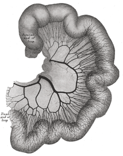
In human anatomy, the superior mesenteric artery (SMA) arises from the anterior surface of the abdominal aorta, just inferior to the origin of the celiac trunk, and supplies the intestine from the lower part of the duodenum through two-thirds of the transverse colon, as well as the pancreas.

In human anatomy, the inferior mesenteric artery, often abbreviated as IMA, is the third main branch of the abdominal aorta and arises at the level of L3, supplying the large intestine from the left colic flexure to the upper part of the rectum, which includes the descending colon, the sigmoid colon, and part of the rectum. Proximally, its territory of distribution overlaps with the middle colic artery, and therefore the superior mesenteric artery. The SMA and IMA anastomose via the marginal artery of the colon and via Riolan's arcade. The territory of distribution of the IMA is more or less equivalent to the embryonic hindgut.
The superior mesenteric vein (SMV) is a blood vessel that drains blood from the small intestine. At its termination behind the neck of the pancreas, the SMV combines with the splenic vein to form the hepatic portal vein. The SMV lies to the right of the similarly named artery, the superior mesenteric artery, which originates from the abdominal aorta.

The splenic vein is a blood vessel that drains blood from the spleen, the stomach fundus and part of the pancreas. It is part of the hepatic portal system.
In human anatomy, the hepatic portal system is the system of veins comprising the hepatic portal vein and its tributaries. It is also called the portal venous system, although it is not the only example of a portal venous system, and splanchnic veins, which is not synonymous with hepatic portal system and is imprecise.
Superior mesenteric can refer to:
The superior rectal artery is an artery that descends into the pelvis to supply blood to the rectum.
The right gastroepiploic vein is a blood vessel that drains blood from the greater curvature and left part of the body of the stomach into the superior mesenteric vein. It runs from left to right along the greater curvature of the stomach between the two layers of the greater omentum, along with the right gastroepiploic artery.
The middle colic vein drains the transverse colon. It is a tributary of the superior mesenteric vein, and follows the path of its corresponding artery, the middle colic artery.
The right colic vein drains the ascending colon, and is a tributary of the superior mesenteric vein. It travels with its corresponding artery, the right colic artery.

The testicular vein, the male gonadal vein, carries deoxygenated blood from its corresponding testis to the inferior vena cava or one of its tributaries. It is the male equivalent of the ovarian vein, and is the venous counterpart of the testicular artery.

The inferior mesenteric vein begins in the rectum as the superior rectal vein, which has its origin in the hemorrhoidal plexus, and through this plexus communicates with the middle and inferior hemorrhoidal veins.

The vitelline arteries are the arterial counterpart to the vitelline veins. Like the veins, they play an important role in the vitelline circulation of blood to and from the yolk sac of a fetus. They are a branch of the dorsal aorta.

The following outline is provided as an overview of and topical guide to human anatomy:
The superior mesenteric vessels are composed of the superior mesenteric artery and the superior mesenteric vein.
Mesenteric vessels may refer to: