Structure
| | This section is empty. You can help by adding to it. (July 2010) |
| Infralimbic cortex | |
|---|---|
| Details | |
| Identifiers | |
| Latin | Cortex infralimbicus |
| Anatomical terms of neuroanatomy | |
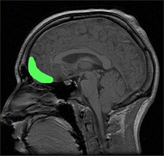
The infralimbic cortex (IL) is a cortical region in the ventromedial prefrontal cortex which is important in tonic inhibition of subcortical structures and emotional responses, such as fear. [1]
| | This section is empty. You can help by adding to it. (July 2010) |
GABAergic neurons within the amygdala, known as intercalated (ITC) cells, receive a strong projection from the IL medial prefrontal cortex (mPFC) in primates. [2] ITC cells are thought to play a role as the 'off switch' for the amygdala, inhibiting the amygdala's central nucleus output neurons and its basolateral nucleus neurons. [3] Further, it has been shown that electrical stimulation of IL reduces conditioned fear and strengthens extinction memory[ clarification needed ], explaining cortical control over extinction processes,[ clarification needed ] one of the simplest forms of emotional regulation. [3]
Amygdala ITC cells receive strong projection from the IL mPFC in rodents as well. [4]
The amygdala is one of two almond-shaped clusters of nuclei located deep and medially within the temporal lobes of the brain's cerebrum in complex vertebrates, including humans. Shown to perform a primary role in the processing of memory, decision making, and emotional responses, the amygdalae are considered part of the limbic system. The term "amygdala" was first introduced by Karl Friedrich Burdach in 1822.
The mesolimbic pathway, sometimes referred to as the reward pathway, is a dopaminergic pathway in the brain. The pathway connects the ventral tegmental area in the midbrain to the ventral striatum of the basal ganglia in the forebrain. The ventral striatum includes the nucleus accumbens and the olfactory tubercle.

Pavlovian fear conditioning is a behavioral paradigm in which organisms learn to predict aversive events. It is a form of learning in which an aversive stimulus is associated with a particular neutral context or neutral stimulus, resulting in the expression of fear responses to the originally neutral stimulus or context. This can be done by pairing the neutral stimulus with an aversive stimulus. Eventually, the neutral stimulus alone can elicit the state of fear. In the vocabulary of classical conditioning, the neutral stimulus or context is the "conditional stimulus" (CS), the aversive stimulus is the "unconditional stimulus" (US), and the fear is the "conditional response" (CR).

The nucleus accumbens is a region in the basal forebrain rostral to the preoptic area of the hypothalamus. The nucleus accumbens and the olfactory tubercle collectively form the ventral striatum. The ventral striatum and dorsal striatum collectively form the striatum, which is the main component of the basal ganglia. The dopaminergic neurons of the mesolimbic pathway project onto the GABAergic medium spiny neurons of the nucleus accumbens and olfactory tubercle. Each cerebral hemisphere has its own nucleus accumbens, which can be divided into two structures: the nucleus accumbens core and the nucleus accumbens shell. These substructures have different morphology and functions.

Dopaminergic pathways in the human brain are involved in both physiological and behavioral processes including movement, cognition, executive functions, reward, motivation, and neuroendocrine control. Each pathway is a set of projection neurons, consisting of individual dopaminergic neurons.

The ventral tegmental area (VTA), also known as the ventral tegmental area of Tsai, or simply ventral tegmentum, is a group of neurons located close to the midline on the floor of the midbrain. The VTA is the origin of the dopaminergic cell bodies of the mesocorticolimbic dopamine system and other dopamine pathways; it is widely implicated in the drug and natural reward circuitry of the brain. The VTA plays an important role in a number of processes, including reward cognition and orgasm, among others, as well as several psychiatric disorders. Neurons in the VTA project to numerous areas of the brain, ranging from the prefrontal cortex to the caudal brainstem and several regions in between.
In mammalian brain anatomy, the prefrontal cortex (PFC) covers the front part of the frontal lobe of the cerebral cortex. The PFC contains the Brodmann areas BA8, BA9, BA10, BA11, BA12, BA13, BA14, BA24, BA25, BA32, BA44, BA45, BA46, and BA47.

In neuroanatomy, thalamocortical radiations also known as thalamocortical fibres, are the efferent fibres that project from the thalamus to distinct areas of the cerebral cortex. They form fibre bundles that emerge from the lateral surface of the thalamus.
An avoidance response is a natural adaptive behavior performed in response to danger. Excessive avoidance has been suggested to contribute to anxiety disorders, leading psychologists and neuroscientists to study how avoidance behaviors are learned using rat or mouse models. Avoidance learning is a type of operant conditioning.
The zona incerta (ZI) is a horizontally elongated region of gray matter in the subthalamus below the thalamus. Its connections project extensively over the brain from the cerebral cortex down into the spinal cord.
The orbitofrontal cortex (OFC) is a prefrontal cortex region in the frontal lobes of the brain which is involved in the cognitive process of decision-making. In non-human primates it consists of the association cortex areas Brodmann area 11, 12 and 13; in humans it consists of Brodmann area 10, 11 and 47.
Joseph E. LeDoux is an American neuroscientist whose research is primarily focused on survival circuits, including their impacts on emotions such as fear and anxiety. LeDoux is the Henry and Lucy Moses Professor of Science at New York University, and director of the Emotional Brain Institute, a collaboration between NYU and New York State with research sites at NYU and the Nathan Kline Institute for Psychiatric Research in Orangeburg, New York. He is also the lead singer and songwriter in the band The Amygdaloids.
The amygdalofugal pathway is one of the three major efferent pathways of the amygdala, meaning that it is one of the three principal pathways by which fibers leave the amygdala. It leads from the basolateral nucleus and central nucleus of the amygdala. The amygdala is a limbic structure in the medial temporal lobe of the brain. The other main efferent pathways from the amygdala are the stria terminalis and anterior commissure.

The reward system is a group of neural structures responsible for incentive salience, associative learning, and positively-valenced emotions, particularly ones involving pleasure as a core component. Reward is the attractive and motivational property of a stimulus that induces appetitive behavior, also known as approach behavior, and consummatory behavior. A rewarding stimulus has been described as "any stimulus, object, event, activity, or situation that has the potential to make us approach and consume it is by definition a reward". In operant conditioning, rewarding stimuli function as positive reinforcers; however, the converse statement also holds true: positive reinforcers are rewarding.

The medial dorsal nucleus is a large nucleus in the thalamus.

Synaptic gating is the ability of neural circuits to gate inputs by either suppressing or facilitating specific synaptic activity. Selective inhibition of certain synapses has been studied thoroughly, and recent studies have supported the existence of permissively gated synaptic transmission. In general, synaptic gating involves a mechanism of central control over neuronal output. It includes a sort of gatekeeper neuron, which has the ability to influence transmission of information to selected targets independently of the parts of the synapse upon which it exerts its action.

The ventromedial prefrontal cortex (vmPFC) is a part of the prefrontal cortex in the mammalian brain. The ventral medial prefrontal is located in the frontal lobe at the bottom of the cerebral hemispheres and is implicated in the processing of risk and fear, as it is critical in the regulation of amygdala activity in humans. It also plays a role in the inhibition of emotional responses, and in the process of decision-making and self-control. It is also involved in the cognitive evaluation of morality.
Microstimulation is a technique that stimulates a small population of neurons by passing a small electrical current through a nearby microelectrode.
The Intercalatedcells of the amygdala are GABAergic neurons situated between the basolateral and central nuclei of the amygdala that play a significant role in inhibitory control over the amygdala. They regulate amygdala-dependent emotional processing like fear memory and social behavior. Their function has been best studied with selective ITC ablation which impairs fear extinction, fear generalization, and social behavior. Studies have begun to recognize that ITC clusters may be implicated in reward, addiction, and withdrawal circuits given their heavy expression of dopamine and opioid receptors.
The neurocircuitry that underlies executive function processes and emotional and motivational processes are known to be distinct in the brain. However, there are brain regions that show overlap in function between the two cognitive systems. Brain regions that exist in both systems are interesting mainly for studies on how one system affects the other. Examples of such cross-modal functions are emotional regulation strategies such as emotional suppression and emotional reappraisal, the effect of mood on cognitive tasks, and the effect of emotional stimulation of cognitive tasks.