ATG8

Last updated
autophagy related protein 8
1ugm bio r 500.jpg
The crystal structure of microtubule-associated protein light chain 3, a mammalian homologue of Saccharomyces cerevisiae Atg8. [1]
Identifiers
Organism S. cerevisiae strain S288c (Baker's yeast)
SymbolAtg8
Alt. symbolsApg8, Aut7, Cvt5
Entrez 852200
RefSeq (mRNA) NM_001178318
RefSeq (Prot) NP_009475
UniProt P38182
Other data
Chromosome VII: 0.16 - 0.16 Mb
Search for
Structures Swiss-model
Domains InterPro

Autophagy-related protein 8 (Atg8) is a ubiquitin-like protein required for the formation of autophagosomal membranes. The transient conjugation of Atg8 to the autophagosomal membrane through a ubiquitin-like conjugation system is essential for autophagy in eukaryotes. Even though there are homologues in animals (see for example GABARAP, GABARAPL1, GABARAPL2, MAP1LC3A, MAP1LC3B, MAP1LC3B2, and MAP1LC3C), this article mainly focuses on its role in lower eukaryotes such as Saccharomyces cerevisiae .

Contents

Structure

Atg8 is a monomer of 117 aminoacids and a molecular weight of 13,6kDa. It consists of a 5-stranded β-sheet, which is enclosed by two α-helices at one side and one α-helix at the other side and exhibits a conserved GABARAP domain. [2] Even though Atg8 does not show a clear sequence homology to ubiquitin, its crystal structure reveals a conserved ubiquitin-like fold. [3] [4]

Function

In autophagy

Atg8 is one of the key molecular components involved in autophagy, the cellular process mediating the lysosome/vacuole-dependent turnover of macromolecules and organelles. [5] Autophagy is induced upon nutrient depletion or rapamycin treatment and leads to the response of more than 30 autophagy-related (ATG) genes known so far, including ATG8. How exactly ATG proteins are regulated is still under investigation, but it is clear that all signals reporting on the availability of carbon and nitrogen sources converge on the TOR signalling pathway and that ATG proteins are downstream effectors of this pathway. [6] In case nutrient supplies are sufficient, the TOR signaling pathway hyperphosphorylates certain Atg proteins, thereby inhibiting autophagosome formation. After starvation autophagy is induced through the activation of Atg proteins both on the protein modification and the transcriptional level.

Atg8 is especially important in macroautophagy which is one of three distinct types of autophagy characterized by the formation of double-membrane enclosed vesicles that sequester portions of the cytosol, the so-called autophagosomes. The outer membrane of these autophagosomes subsequently fuses with the lysosome/vacuole to release an inter single membrane (autophagic body) destined for degradation. [5] During this process, Atg8 is particularly crucial for autophagosome maturation (lipidation). [7]

Like most Atg proteins, Atg8 is localized in the cytoplasm and at the PAS under nutrient-rich conditions, but becomes membrane-associated in case of autophagy induction. It then localizes to the site of autophagosome nucleation, the phagophore-assembly site (PAS). [2] Nucleation of the phagophore requires the accumulation of a set of Atg proteins and of class III phosphoinositide 3-kinase complexes on the PAS. The subsequent recruitment of Atg8 and other autophagy-related proteins is believed to trigger vesicle expansion in a concerted manner, presumably by providing the driving force for membrane curvature. [8] The transient conjugation of Atg8 to the membrane lipid phosphatidylethanolamine is essential for phagophore expansion as its mutation leads to defects in autophagosome formation. [9] It is distributed symmetrically on both sides of the autophagosome and it is assumed that there is a quantitative correlation between the amount of Atg8 and the vesicle size. [10] [11] [12] [13]

After finishing vesicle expansion, the autophagosome is ready for fusion with the lysosome and Atg8 can either be released from the membrane for recycling (see below) or gets degraded in the autolysosome if left uncleaved.

ATG8 is also required for a different autophagy-related process called the cytoplasm-to-vacuole targeting (Cvt) pathway. [14] This yeast-specific process acts constitutively under nutrient-rich conditions and selectively transports hydrolases such as aminopeptidase I to the yeast vacuole. The Cvt pathway also requires Atg8 localised to the PAS for the formation of Cvt vesicles which then fuse with the vacuole to deliver hydrolases necessary for degradation.

Post-translational modification and regulatory cycle

Atg8 exists in a cytoplasmic and in a membrane-associated form. [15] Membrane association is achieved by coupling Atg8 to phosphatidylethanolamine (PE) which is a lipid constituent of plasma membranes. This post-translational modification process, called lipidation, is performed by the Atg8 conjugation system comprising the cysteine protease ATG4 (belonging to the caspase family), as well as the proteins ATG7, ATG3 and the ATG5-ATG12 complex. [16]

The Atg8 conjugation system works in analogy to the ubiquitination system. However, it is Atg8 itself that represents the ubiquitin-like protein (Ubl) being transferred to PE, while ATG7 functions like an E1 enzyme, ATG3 like an E2 enzyme and the ATG12-ATG5 complex like an E3 ligase.

The lipidation process is initiated by an ATG4-dependent post-translational cleavage of the last C-terminal amino acid residue of Atg8. After the cleavage, Atg8 exposes a C-terminal glycine residue (Gly 116) to which PE can then be coupled during the following steps. In the first step, the Gly116 residue of Atg8 binds to a cysteine residue of ATG7 via a thioester bond in an ATP-dependent manner. During the second step, Atg8 is transferred to Atg3 assuming the same type of thioester bond. Finally, Atg8 is detached from Atg3 and coupled to the amine head group of PE via an amide bond. This final step was found to be facilitated and stimulated by the ATG5-ATG12 complex. [17]

Both proteins, Atg5 and Atg12 were originally identified as part of another Ubl conjugating system that promotes conjugation of ATG12 to ATG5 via ATG7 and Atg10. This implies, that the ATG12 and the Atg8 conjugation system are actually interdependent.

Mammalian homologues

In higher eukaryotes Atg8 is not encoded by a single gene as in yeast, but derived from a multigene family. Four of its homologues have already been identified in mammalian cells.

One of them is LC3 (MAP1LC3A), a light chain of the microtubule-associated protein 1 [18] Like Atg8, LC3 needs to be proteolytically cleaved and lipidated to be turned into its active form which can localize to the autophagosomal membrane. Similar to the situation in yeast, the activation process of LC3 is triggered by nutrient depletion, as well as in response to hormones. [11]

Mammalian LC3 isoforms contain a conserved Ser/Thr12, which is phosphorylated by protein kinase A to suppress participation in autophagy/mitophagy. [19]

Other homologues are the transport factor GATE-16 (Golgi-associated ATPase enhancer of 16 kDa) [20] which plays an important role in intra-golgi vesicular transport by stimulating NSF (N-ethylmaleimide-sensitive factor) ATPase activity and interacting with the Golgi v-SNARE GOS-28, and GABARAP (γ-aminobutyric acid type A receptor associated protein) [21] [22] which facilitates clustering of GABAA receptors in combination with microtubules.

All three proteins are characterized by proteolytic activation processes upon which they get lipidated and localized to the plasma membrane. However, for GATE-16 and GABARAP membrane association seems to be possible even for the non-lipidated forms. Apart from LC3, GABARAP and GATE-16 the most recently but less well characterized mammalian homologue is ATGL8. Little is known about its actual activation process except for its interaction with one of the mammalian ATG4 homologues, hATG4A. [23]

See also

Related Research Articles

<span class="mw-page-title-main">Autophagy</span> Cellular catabolic process in which cells digest parts of their own cytoplasm

Autophagy is the natural, conserved degradation of the cell that removes unnecessary or dysfunctional components through a lysosome-dependent regulated mechanism. It allows the orderly degradation and recycling of cellular components. Although initially characterized as a primordial degradation pathway induced to protect against starvation, it has become increasingly clear that autophagy also plays a major role in the homeostasis of non-starved cells. Defects in autophagy have been linked to various human diseases, including neurodegeneration and cancer, and interest in modulating autophagy as a potential treatment for these diseases has grown rapidly.

Autophagin-1 (Atg4/Apg4) is a unique cysteine protease responsible for the cleavage of the carboxyl terminus of Atg8/Apg8/Aut7, a reaction essential for its lipidation during autophagy. Human Atg4 homologues cleave the carboxyl termini of the three human Atg8 homologues, microtubule-associated protein light chain 3 (LC3), GABARAP, and GATE-16.

<span class="mw-page-title-main">Vojo Deretic</span> American geneticist

Vojo Deretic, is distinguished professor and chair of the Department of Molecular Genetics and Microbiology at the University of New Mexico School of Medicine. Deretic was the founding director of the Autophagy, Inflammation and Metabolism (AIM) Center of Biomedical Research Excellence. The AIM center promotes autophagy research nationally and internationally.

<span class="mw-page-title-main">Autophagy protein 5</span> Protein-coding gene in the species Homo sapiens

Autophagy protein 5 (ATG5) is a protein that, in humans, is encoded by the ATG5 gene located on chromosome 6. It is an E3 ubi autophagic cell death. ATG5 is a key protein involved in the extension of the phagophoric membrane in autophagic vesicles. It is activated by ATG7 and forms a complex with ATG12 and ATG16L1. This complex is necessary for LC3-I conjugation to PE (phosphatidylethanolamine) to form LC3-II. ATG5 can also act as a pro-apoptotic molecule targeted to the mitochondria. Under low levels of DNA damage, ATG5 can translocate to the nucleus and interact with survivin.

<span class="mw-page-title-main">MAP1LC3B</span> Protein-coding gene in the species Homo sapiens

Microtubule-associated proteins 1A/1B light chain 3B is a protein that in humans is encoded by the MAP1LC3B gene. LC3 is a central protein in the autophagy pathway where it functions in autophagy substrate selection and autophagosome biogenesis. LC3 is the most widely used marker of autophagosomes.

<span class="mw-page-title-main">MAP1LC3A</span> Protein-coding gene in the species Homo sapiens

Microtubule-associated proteins 1A/1B light chain 3A is a protein that in humans is encoded by the MAP1LC3A gene. Two transcript variants encoding different isoforms have been found for this gene.

<span class="mw-page-title-main">ATG4B</span> Protein-coding gene in the species Homo sapiens

Cysteine protease ATG4B is an enzyme that in humans is encoded by the ATG4B gene.

<span class="mw-page-title-main">ATG16L1</span> Protein-coding gene in the species Homo sapiens

Autophagy related 16 like 1 is a protein that in humans is encoded by the ATG16L1 gene. This protein is characterized as a subunit of the autophagy-related ATG12-ATG5/ATG16 complex and is essentially important for the LC3 (ATG8) lipidation and autophagosome formation. This complex localizes to the membrane and is released just before or after autophagosome completion.

<span class="mw-page-title-main">ATG12</span> Protein-coding gene in the species Homo sapiens

Autophagy related 12 is a protein that in humans is encoded by the ATG12 gene.

<span class="mw-page-title-main">ATG7</span> Protein-coding gene in the species Homo sapiens

Autophagy related 7 is a protein in humans encoded by ATG7 gene. Related to GSA7; APG7L; APG7-LIKE.

AuTophaGy related 1 (Atg1) is a 101.7kDa serine/threonine kinase in S.cerevisiae, encoded by the gene ATG1. It is essential for the initial building of the autophagosome and Cvt vesicles. In a non-kinase role it is - through complex formation with Atg13 and Atg17 - directly controlled by the TOR kinase, a sensor for nutrient availability.

<span class="mw-page-title-main">Autophagosome</span> In cell biology, a key structure in macroautophagy (degradation of cytoplasmic contents)

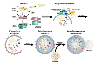
An autophagosome is a spherical structure with double layer membranes. It is the key structure in macroautophagy, the intracellular degradation system for cytoplasmic contents. After formation, autophagosomes deliver cytoplasmic components to the lysosomes. The outer membrane of an autophagosome fuses with a lysosome to form an autolysosome. The lysosome's hydrolases degrade the autophagosome-delivered contents and its inner membrane.

In molecular biology, autophagy related 3 (Atg3) is the E2 enzyme for the LC3 lipidation process. It is essential for autophagy. The super protein complex, the Atg16L complex, consists of multiple Atg12-Atg5 conjugates. Atg16L has an E3-like role in the LC3 lipidation reaction. The activated intermediate, LC3-Atg3 (E2), is recruited to the site where the lipidation takes place.

<span class="mw-page-title-main">Yoshinori Ohsumi</span> Japanese cell biologist

Yoshinori Ohsumi is a Japanese cell biologist specializing in autophagy, the process that cells use to destroy and recycle cellular components. Ohsumi is a professor at Tokyo Institute of Technology's Institute of Innovative Research. He received the Kyoto Prize for Basic Sciences in 2012, the 2016 Nobel Prize in Physiology or Medicine, and the 2017 Breakthrough Prize in Life Sciences for his discoveries of mechanisms for autophagy.

Chaperone-assisted selective autophagy is a cellular process for the selective, ubiquitin-dependent degradation of chaperone-bound proteins in lysosomes.

<span class="mw-page-title-main">Omegasome</span>

Omegasome is a cell organelle consisting of lipid bilayer membranes enriched for phosphatidylinositol 3-phosphate, and related to a process of autophagy. It is a subdomain of the Endoplasmic Reticulum (ER), and has a morphology resembling the Greek capital letter Omega (Ω). Omegasomes are the sites from which phagophores form, which are sack-like structures that mature into autophagosomes, and fuse with lysosomes in order to degrade the contents of the autophagosomes. The formation of omegasomes depends on various factors, however in general, formation of omegasomes is increased as a response to starvation, and in some biochemical situations the presence of PI(3)P leads to the formation of omegasomes.

Microautophagy is one of the three common forms of autophagic pathway, but unlike macroautophagy and chaperone-mediated autophagy, it is mediated—in mammals by lysosomal action or in plants and fungi by vacuolar action—by direct engulfment of the cytoplasmic cargo. Cytoplasmic material is trapped in the lysosome/vacuole by a random process of membrane invagination.

Rab GTPases are molecular switches that regulate membrane traffic. They are active in their GTP-bound form and inactive when bound to GDP. The GTPase YPT1, and its mammalian homologue Rab1, regulate membrane-tethering events on three different pathways: autophagy, ER-Golgi, and intra-Golgi traffic. In the yeast Saccharomyces cerevisiae, many of the ATG proteins needed for macroautophagy are shared with the biosynthetic cytoplasm to the vacuole-targeting (CVT) pathway that transports certain hydrolases into the vacuole. Both pathways require YPT1; however, only the macroautophagy pathway is conserved in higher eukaryotes. In the macroautophagy pathway, Rab1 mediates the recruitment of Atg1 to the PAS. Rab1 regulates macroautophagy by recruiting its effector, Atg1, to the PAS to tether Atg9 vesicles to each other or to other membranes.

<span class="mw-page-title-main">Ubiquitin-like protein</span> Family of small proteins

Ubiquitin-like proteins (UBLs) are a family of small proteins involved in post-translational modification of other proteins in a cell, usually with a regulatory function. The UBL protein family derives its name from the first member of the class to be discovered, ubiquitin (Ub), best known for its role in regulating protein degradation through covalent modification of other proteins. Following the discovery of ubiquitin, many additional evolutionarily related members of the group were described, involving parallel regulatory processes and similar chemistry. UBLs are involved in a widely varying array of cellular functions including autophagy, protein trafficking, inflammation and immune responses, transcription, DNA repair, RNA splicing, and cellular differentiation.

References

  1. PDB: 1UGM ; Sugawara K, Suzuki NN, Fujioka Y, Mizushima N, Ohsumi Y, Inagaki F (July 2004). "The crystal structure of microtubule-associated protein light chain 3, a mammalian homologue of Saccharomyces cerevisiae Atg8". Genes to Cells. 9 (7): 611–8. doi: 10.1111/j.1356-9597.2004.00750.x . PMID   15265004.
  2. 1 2 Geng J, Klionsky DJ (September 2008). "The Atg8 and Atg12 ubiquitin-like conjugation systems in macroautophagy. 'Protein modifications: beyond the usual suspects' review series". EMBO Reports. 9 (9): 859–64. doi:10.1038/embor.2008.163. PMC   2529362 . PMID   18704115.
  3. Sugawara K, Suzuki NN, Fujioka Y, Mizushima N, Ohsumi Y, Inagaki F (July 2004). "The crystal structure of microtubule-associated protein light chain 3, a mammalian homologue of Saccharomyces cerevisiae Atg8". Genes to Cells. 9 (7): 611–8. doi: 10.1111/j.1356-9597.2004.00750.x . PMID   15265004.
  4. Suzuki NN, Yoshimoto K, Fujioka Y, Ohsumi Y, Inagaki F (July 2005). "The crystal structure of plant ATG12 and its biological implication in autophagy". Autophagy. 1 (2): 119–26. doi: 10.4161/auto.1.2.1859 . PMID   16874047.
  5. 1 2 Ohsumi Y (March 2001). "Molecular dissection of autophagy: two ubiquitin-like systems". Nature Reviews. Molecular Cell Biology. 2 (3): 211–6. doi:10.1038/35056522. PMID   11265251. S2CID   38001477.
  6. Kamada Y, Sekito T, Ohsumi Y (2004). "Autophagy in Yeast: ATOR-Mediated Response to Nutrient Starvation". TOR. Current Topics in Microbiology and Immunology. Vol. 279. pp. 73–84. doi:10.1007/978-3-642-18930-2_5. ISBN   978-3-540-00534-6. PMID   14560952.
  7. Eskelinen EL (2008). "New insights into the mechanisms of macroautophagy in mammalian cells". International Review of Cell and Molecular Biology. 266: 207–47. doi:10.1016/S1937-6448(07)66005-5. ISBN   978-0-12-374372-5. PMID   18544495.
  8. Mizushima N, Yamamoto A, Hatano M, Kobayashi Y, Kabeya Y, Suzuki K, et al. (February 2001). "Dissection of autophagosome formation using Apg5-deficient mouse embryonic stem cells". The Journal of Cell Biology. 152 (4): 657–68. doi:10.1083/jcb.152.4.657. PMC   2195787 . PMID   11266458.
  9. Xie Z, Klionsky DJ (October 2007). "Autophagosome formation: core machinery and adaptations". Nature Cell Biology. 9 (10): 1102–9. doi:10.1038/ncb1007-1102. PMID   17909521. S2CID   26402002.
  10. Huang WP, Scott SV, Kim J, Klionsky DJ (February 2000). "The itinerary of a vesicle component, Aut7p/Cvt5p, terminates in the yeast vacuole via the autophagy/Cvt pathways". The Journal of Biological Chemistry. 275 (8): 5845–51. doi: 10.1074/jbc.275.8.5845 . PMID   10681575.
  11. 1 2 Kabeya Y, Mizushima N, Ueno T, Yamamoto A, Kirisako T, Noda T, et al. (November 2000). "LC3, a mammalian homologue of yeast Apg8p, is localized in autophagosome membranes after processing". The EMBO Journal. 19 (21): 5720–8. doi:10.1093/emboj/19.21.5720. PMC   305793 . PMID   11060023.
  12. Kirisako T, Baba M, Ishihara N, Miyazawa K, Ohsumi M, Yoshimori T, et al. (October 1999). "Formation process of autophagosome is traced with Apg8/Aut7p in yeast". The Journal of Cell Biology. 147 (2): 435–46. doi:10.1083/jcb.147.2.435. PMC   2174223 . PMID   10525546.
  13. Xie Z, Nair U, Klionsky DJ (August 2008). "Atg8 controls phagophore expansion during autophagosome formation". Molecular Biology of the Cell. 19 (8): 3290–8. doi:10.1091/mbc.E07-12-1292. PMC   2488302 . PMID   18508918.
  14. Kim J, Klionsky DJ (2000). "Autophagy, cytoplasm-to-vacuole targeting pathway, and pexophagy in yeast and mammalian cells". Annual Review of Biochemistry. 69: 303–42. doi:10.1146/annurev.biochem.69.1.303. PMID   10966461.
  15. Kirisako T, Ichimura Y, Okada H, Kabeya Y, Mizushima N, Yoshimori T, et al. (October 2000). "The reversible modification regulates the membrane-binding state of Apg8/Aut7 essential for autophagy and the cytoplasm to vacuole targeting pathway". The Journal of Cell Biology. 151 (2): 263–76. doi:10.1083/jcb.151.2.263. PMC   2192639 . PMID   11038174.
  16. Ichimura Y, Kirisako T, Takao T, Satomi Y, Shimonishi Y, Ishihara N, et al. (November 2000). "A ubiquitin-like system mediates protein lipidation". Nature. 408 (6811): 488–92. Bibcode:2000Natur.408..488I. doi:10.1038/35044114. PMID   11100732. S2CID   4428142.
  17. Geng J, Klionsky DJ (September 2008). "The Atg8 and Atg12 ubiquitin-like conjugation systems in macroautophagy. 'Protein modifications: beyond the usual suspects' review series". EMBO Reports. 9 (9): 859–64. doi:10.1038/embor.2008.163. PMC   2529362 . PMID   18704115.
  18. Tanida I, Ueno T, Kominami E (December 2004). "LC3 conjugation system in mammalian autophagy". The International Journal of Biochemistry & Cell Biology. 36 (12): 2503–18. doi:10.1016/j.biocel.2004.05.009. PMC   7129593 . PMID   15325588.
  19. Cherra SJ, Kulich SM, Uechi G, Balasubramani M, Mountzouris J, Day BW, Chu CT (August 2010). "Regulation of the autophagy protein LC3 by phosphorylation". The Journal of Cell Biology. 190 (4): 533–9. doi:10.1083/jcb.201002108. PMC   2928022 . PMID   20713600.
  20. Sagiv Y, Legesse-Miller A, Porat A, Elazar Z (April 2000). "GATE-16, a membrane transport modulator, interacts with NSF and the Golgi v-SNARE GOS-28". The EMBO Journal. 19 (7): 1494–504. doi:10.1093/emboj/19.7.1494. PMC   310219 . PMID   10747018.
  21. Chen ZW, Chang CS, Leil TA, Olsen RW (June 2007). "C-terminal modification is required for GABARAP-mediated GABA(A) receptor trafficking". The Journal of Neuroscience. 27 (25): 6655–63. doi: 10.1523/JNEUROSCI.0919-07.2007 . PMC   6672693 . PMID   17581952.
  22. Wang H, Bedford FK, Brandon NJ, Moss SJ, Olsen RW (January 1999). "GABA(A)-receptor-associated protein links GABA(A) receptors and the cytoskeleton". Nature. 397 (6714): 69–72. doi:10.1038/16264. PMID   9892355. S2CID   204990449.
  23. Tanida I, Sou YS, Minematsu-Ikeguchi N, Ueno T, Kominami E (June 2006). "Atg8L/Apg8L is the fourth mammalian modifier of mammalian Atg8 conjugation mediated by human Atg4B, Atg7 and Atg3". The FEBS Journal. 273 (11): 2553–62. doi: 10.1111/j.1742-4658.2006.05260.x . PMID   16704426.