Hsp27

Last updated
HSPB1
3q9p.png
Available structures
PDB Ortholog search: PDBe RCSB
Identifiers
Aliases HSPB1 , CMT2F, HEL-S-102, HMN2B, HS.76067, HSP27, HSP28, Hsp25, SRP27, heat shock protein family B (small) member 1
External IDs OMIM: 602195 MGI: 96240 HomoloGene: 1180 GeneCards: HSPB1
Orthologs
SpeciesHumanMouse
Entrez
Ensembl
UniProt
RefSeq (mRNA)

NM_001540

NM_013560

RefSeq (protein)

NP_001531

NP_038588

Location (UCSC) Chr 7: 76.3 – 76.3 Mb Chr 5: 135.92 – 135.92 Mb
PubMed search [3] [4]
Wikidata
View/Edit Human View/Edit Mouse

Heat shock protein 27 (Hsp27) also known as heat shock protein beta-1 (HSPB1) is a protein that in humans is encoded by the HSPB1 gene. [5] [6]

Contents

Hsp27 is a chaperone of the sHsp (small heat shock protein) group among α-crystallin, Hsp20, and others. The common functions of sHsps are chaperone activity, thermotolerance, inhibition of apoptosis, regulation of cell development, and cell differentiation. They also take part in signal transduction.

Structure

sHsps have some structural features in common: Very characteristic is a homologous and highly conserved amino acid sequence, the so-called α-crystallin domain near the C-terminus. These domains consist of 80 to 100 residues with sequence homology between 20% and 60% and fold into β-sheets, which are important for the formation of stable dimers. [7] [8] Hsp27 is rather unique among sHsps in that its α-crystallin domain contains a cysteine residue at its dimer interface, which can become oxidized to form a disulfide bond that covalently links the dimer. [9] The N-terminus consists of a less conserved region, the so-called WD/EPF domain, followed by a short variable sequence with a rather conservative site near the end of this domain. The C-terminal region of sHsps consists of the above mentioned α-crystallin domain, followed by a variable sequence with high motility and flexibility. [10] Despite relatively low levels of global sequence conservation in the C-terminal region, many sHsps contain a locally conserved Ile-Xxx-Ile/Val (IxI/V) motif that plays a role in regulating the assembly of oligomers. [11] It is highly flexible and polar because of its negative charges. [12] Probably it functions as a mediator of solubility for hydrophobic sHsps and it stabilizes the protein and protein/substrate complexes. This was shown by elimination of the C-terminal tail in Hsp27Δ182-205 [13] and in Hsp25Δ18. [14] In the case of Hsp27, the IxI/V motif corresponds to 181-Ile-Pro-Val-183, and this region of the protein plays a critical role, as the mutation of the central Pro residue causes the hereditary motor neuropathy Charcot-Marie-Tooth disease. [15]

Oligomerization

Hsp27 forms large, dynamic oligomers with an average mass near 500 kDa in vitro. [16] The N-terminus of Hsp27, with its WD/EPF-region, is essential for the development of these large oligomers. [17] [18] Hsp27-oligomers consist of stable dimers, which are formed by two α-crystallin-domains of neighboring monomers, [16] [11] which was first shown in crystal structures of the proteins MjHSP16.5 from Methanocaldococcus jannaschii [7] and wheat Hsp16.9. [8] Therefore the first step in the oligomeric process involves dimerization of the α-crystallin domain. In metazoans, dimerization by α-crystallin domains proceeds through the formation of a long β-strand at the interface. The amino acid sequences in this region, however, are predicted to be disordered [19] Indeed, the α-crystallin domain of Hsp27 partially unfolds in its monomeric state and is less stable than the dimer. [20]

The oligomerization of Hsp27 is a dynamic process: There is a balance between stable dimers and oligomers (up to 800 kDa) consisting of 16 to 32 subunits and a high exchange rate of subunits. [18] [21] [22] The oligomerization depends on the physiology of the cells, the phosphorylation status of Hsp27 and the exposure to stress. Stress induces an increase of expression (after hours) and phosphorylation (after several minutes) of Hsp27. Stimulation of the p38 MAP kinase cascade by differentiating agents, mitogens, inflammatory cytokines such as TNFα and IL-1β, hydrogen peroxide and other oxidants, [23] leads to the activation of MAPKAP kinases 2 and 3 which directly phosphorylate mammalian sHsps. [22] The phosphorylation plays an important role for the formation of oligomers in exponentially growing cells in vitro, but the oligomerization in tumor cells growing in vivo or growing at confluence in vitro is dependent on cell-cell contact, but not on the phosphorylation status. [24] Furthermore, it was shown that HSP27 contains an Argpyrimidine modification. [25]

In all probability, the oligomerization status is connected with the chaperone activity: aggregates of large oligomers have high chaperone activity, whereas dimers and monomers have relatively higher chaperone activity. [16] [20] [11]

Cellular localization

Hsp27 appears in many cell types, especially all types of muscle cells. It is located mainly in the cytosol, but also in the perinuclear region, endoplasmatic reticulum, and nucleus. It is overexpressed during different stages of cell differentiation and development. This suggests an essential role for Hsp27 in the differentiation of tissues.

An affinity of high expression levels of different phosphorylated Hsp27 species and muscle/neurodegenerative diseases and various cancers was observed. [26] High expression levels possibly are in inverse relation with cell proliferation, metastasis, and resistance to chemotherapy. [27] High levels of Hsp27 were also found in sera of breast cancer patients; [28] therefore Hsp27 could be a potential diagnostic marker.

Function

The main function of Hsp27 is to provide thermotolerance in vivo, cytoprotection, and support of cell survival under stress conditions. More specialized functions of Hsp27 are manifold and complex. In vitro it acts as an ATP-independent chaperone by inhibiting protein aggregation and by stabilizing partially denatured proteins, which ensures refolding by the Hsp70-complex. Hsp27 is also involved in the apoptotic signalling pathway. Hsp27 interacts with the outer mitochondrial membranes and interferes with the activation of cytochrome c/Apaf-1/dATP complex and therefore inhibits the activation of procaspase-9. [26] The phosphorylated form of Hsp27 inhibits Daxx apoptotic protein and prevents the association of Daxx with Fas and Ask1. [29] Moreover, Hsp27 phosphorylation leads to the activation of TAK1 and TAK1-p38/ERK pro-survival signaling, thus opposing TNF-α-induced apoptosis. [30]

A well documented function of Hsp27 is the interaction with actin and intermediate filaments. It prevents the formation of non-covalent filament/filament interactions of the intermediate filaments and protects actin filaments from fragmentation. It also preserves the focal contacts fixed at the cell membrane. [26]

Another function of Hsp27 is the activation of the proteasome. It speeds up the degradation of irreversibly denatured proteins and junkproteins by binding to ubiquitinated proteins and to the 26S proteasome. Hsp27 enhances the activation of the NF-κB pathway, that controls a lot of processes, such as cell growth and inflammatory and stress responses. [31] The cytoprotective properties of Hsp27 result from its ability to modulate reactive oxygen species and to raise glutathione levels.

Probably Hsp27 – among other chaperones – is involved in the process of cell differentiation. [32] Changes of Hsp27 levels were observed in Ehrlich ascite cells, embryonic stem cells, normal B-cells, B-lymphoma cells, osteoblasts, keratinocytes, neurons etc. The upregulation of Hsp27 correlates with the rate of phosphorylation and with an increase of large oligomers. It is possible that Hsp27 plays a crucial role in the termination of growth.

Clinical significance

Motor neuropathies

At least 12 disease-causing mutations in this gene have been discovered. [33] Heritable mutations in HSPB1 cause distal hereditary motor neuropathies and the motor neuropathy Charcot-Marie-Tooth disease. [34] There are missense mutations throughout the amino acid sequence of Hsp27, and most disease-causing mutations present with adult-onset symptoms. [34] One of the more severe Hsp27 mutants is the Pro182Leu mutant, which manifests symptomatically in the first few years of life and was additionally demonstrated in a transgenic mouse model. [34] [35] The genetic basis of these diseases is typically autosomal dominant, meaning that only one allele contains a mutation. Since the wild-type HSPB1 gene is also expressed alongside the mutated allele, the diseased cells contain a mixed populations of wild-type and mutant Hsp27, and in vitro experiments have shown that the two proteins can form heter-oligomers. [36]

Roles in apoptosis

Notably, phosphorylated Hsp27 increases human prostate cancer (PCa) cell invasion, enhances cell proliferation, and suppresses Fas-induced apoptosis in human PCa cells. Unphosphorylated Hsp27 has been shown to act as an actin capping protein, preventing actin reorganization and, consequently, cell adhesion and motility. OGX-427, which targets HSP27 through an antisense mechanism, is currently undergoing testing in clinical trials. [37]

Roles in cancer

Protein kinase C-mediated HSPB1 phosphorylation protects against ferroptosis, an iron-dependent form of non-apoptotic cell death, by reducing iron-mediated production of lipid reactive oxygen species. These novel data support the development of Hsp-targeting strategies and, specifically, anti-HSP27 agents for the treatment of ferroptosis-mediated cancer. [38]

Interactions

Hsp27 has been shown to interact with:

Related Research Articles

<span class="mw-page-title-main">Chaperone (protein)</span> Proteins assisting in protein folding

In molecular biology, molecular chaperones are proteins that assist the conformational folding or unfolding of large proteins or macromolecular protein complexes. There are a number of classes of molecular chaperones, all of which function to assist large proteins in proper protein folding during or after synthesis, and after partial denaturation. Chaperones are also involved in the translocation of proteins for proteolysis.

Heat shock proteins (HSP) are a family of proteins produced by cells in response to exposure to stressful conditions. They were first described in relation to heat shock, but are now known to also be expressed during other stresses including exposure to cold, UV light and during wound healing or tissue remodeling. Many members of this group perform chaperone functions by stabilizing new proteins to ensure correct folding or by helping to refold proteins that were damaged by the cell stress. This increase in expression is transcriptionally regulated. The dramatic upregulation of the heat shock proteins is a key part of the heat shock response and is induced primarily by heat shock factor (HSF). HSPs are found in virtually all living organisms, from bacteria to humans.

In anatomy, a crystallin is a water-soluble structural protein found in the lens and the cornea of the eye accounting for the transparency of the structure. It has also been identified in other places such as the heart, and in aggressive breast cancer tumors. Since it has been shown that lens injury may promote nerve regeneration, crystallin has been an area of neural research. So far, it has been demonstrated that crystallin β b2 (crybb2) may be a neurite-promoting factor.

<span class="mw-page-title-main">Hsp70</span> Family of heat shock proteins

The 70 kilodalton heat shock proteins are a family of conserved ubiquitously expressed heat shock proteins. Proteins with similar structure exist in virtually all living organisms. Intracellularly localized Hsp70s are an important part of the cell's machinery for protein folding, performing chaperoning functions, and helping to protect cells from the adverse effects of physiological stresses. Additionally, membrane-bound Hsp70s have been identified as a potential target for cancer therapies and their extracellularly localized counterparts have been identified as having both membrane-bound and membrane-free structures.

<span class="mw-page-title-main">Heat shock response</span> Type of cellular stress response

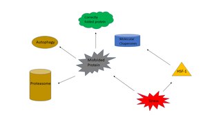
The heat shock response (HSR) is a cell stress response that increases the number of molecular chaperones to combat the negative effects on proteins caused by stressors such as increased temperatures, oxidative stress, and heavy metals. In a normal cell, proteostasis must be maintained because proteins are the main functional units of the cell. Many proteins take on a defined configuration in a process known as protein folding in order to perform their biological functions. If these structures are altered, critical processes could be affected, leading to cell damage or death. The heat shock response can be employed under stress to induce the expression of heat shock proteins (HSP), many of which are molecular chaperones, that help prevent or reverse protein misfolding and provide an environment for proper folding.

<span class="mw-page-title-main">HSPA8</span> Protein-coding gene in the species Homo sapiens

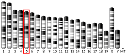
Heat shock 70 kDa protein 8 also known as heat shock cognate 71 kDa protein or Hsc70 or Hsp73 is a heat shock protein that in humans is encoded by the HSPA8 gene on chromosome 11. As a member of the heat shock protein 70 family and a chaperone protein, it facilitates the proper folding of newly translated and misfolded proteins, as well as stabilize or degrade mutant proteins. Its functions contribute to biological processes including signal transduction, apoptosis, autophagy, protein homeostasis, and cell growth and differentiation. It has been associated with an extensive number of cancers, neurodegenerative diseases, cell senescence, and aging.

<span class="mw-page-title-main">HSPA1A</span> Protein-coding gene in the species Homo sapiens

Heat shock 70 kDa protein 1, also termed Hsp72, is a protein that in humans is encoded by the HSPA1A gene. As a member of the heat shock protein 70 family and a chaperone protein, it facilitates the proper folding of newly translated and misfolded proteins, as well as stabilize or degrade mutant proteins. In addition, Hsp72 also facilitates DNA repair. Its functions contribute to biological processes including signal transduction, apoptosis, protein homeostasis, and cell growth and differentiation. It has been associated with an extensive number of cancers, neurodegenerative diseases, cell senescence and aging, and inflammatory diseases such as Diabetes mellitus type 2 and rheumatoid arthritis.

<span class="mw-page-title-main">Heat shock protein 90kDa alpha (cytosolic), member A1</span> Protein-coding gene in the species Homo sapiens

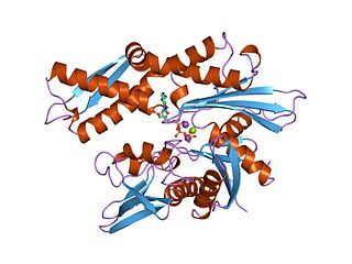
Heat shock protein HSP 90-alpha is a protein that in humans is encoded by the HSP90AA1 gene.

<span class="mw-page-title-main">HSF1</span> Protein-coding gene in the species Homo sapiens

Heat shock factor 1 (HSF1) is a protein that in humans is encoded by the HSF1 gene. HSF1 is highly conserved in eukaryotes and is the primary mediator of transcriptional responses to proteotoxic stress with important roles in non-stress regulation such as development and metabolism.

<span class="mw-page-title-main">CRYAB</span> Protein-coding gene in the species Homo sapiens

Alpha-crystallin B chain is a protein that in humans is encoded by the CRYAB gene. It is part of the small heat shock protein family and functions as molecular chaperone that primarily binds misfolded proteins to prevent protein aggregation, as well as inhibit apoptosis and contribute to intracellular architecture. Post-translational modifications decrease the ability to chaperone. Mutations in CRYAB cause different cardiomyopathies, skeletal myopathies mainly myofibrillar myopathy, and also cataracts. In addition, defects in this gene/protein have been associated with cancer and neurodegenerative diseases such as Alzheimer's disease and Parkinson's disease.

<span class="mw-page-title-main">HSP90AB1</span> Protein-coding gene in the species Homo sapiens

Heat shock protein HSP 90-beta also called HSP90beta is a protein that in humans is encoded by the HSP90AB1 gene.

<span class="mw-page-title-main">HSPB8</span>

Heat shock protein beta-8 is a protein that in humans is encoded by the HSPB8 gene.

<span class="mw-page-title-main">HSPB2</span> Protein-coding gene in the species Homo sapiens

Heat shock protein beta-2 is a protein that in humans is encoded by the HSPB2 gene.

<span class="mw-page-title-main">HSPA1L</span> Protein-coding gene in the species Homo sapiens

Heat shock 70 kDa protein 1L is a protein that in humans is encoded by the HSPA1L gene on chromosome 6. As a member of the heat shock protein 70 (Hsp70) family and a chaperone protein, it facilitates the proper folding of newly translated and misfolded proteins, as well as stabilize or degrade mutant proteins. Its functions contribute to biological processes including signal transduction, apoptosis, protein homeostasis, and cell growth and differentiation. It has been associated with an extensive number of cancers, neurodegenerative diseases, cell senescence and aging, and Graft-versus-host disease.

<span class="mw-page-title-main">HSPB6</span> Protein-coding gene in the species Homo sapiens

Heat shock protein beta-6 (HSPB6) is a protein that in humans is encoded by the HSPB6 gene.

<span class="mw-page-title-main">CRYAA</span> Protein-coding gene in the species Homo sapiens

Alpha-crystallin A chain is a protein that in humans is encoded by the CRYAA gene.

The heat shock protein Hsp20 family, also known as small heat shock proteins (sHSPs), is a family of heat shock proteins.

<span class="mw-page-title-main">Protein moonlighting</span> Proteins performing more than one function

Protein moonlighting is a phenomenon by which a protein can perform more than one function. It is an excellent example of gene sharing.

<span class="mw-page-title-main">HSPB3</span> Protein-coding gene in the species Homo sapiens

Heat shock protein beta-3 (HspB3) also known as heat shock 27kDa protein 3 is a protein that in humans is encoded by the HSPB3 gene.

Hsp104 is a heat-shock protein. It is known to reverse toxicity of mutant α-synuclein, TDP-43, FUS, and TAF15 in yeast cells. Conserved in prokaryotes (ClpB), fungi, plants and aswell as animal mitochondria, there is yet to see hsp104 in multicellular animals. Hsp104 is classified as a. AAA+ ATPases and a subgroup of Hsp100/Clp, because of the usage of Atp hydrolysis for structural modulation of other proteins. Hsp104 is not needed for normal cell growth but when exposed to stress there is an increase amount. Removing the aggregates without the hsp104 is insufficient there highlighting the importance of this heat shock protein and its interactions.

References

  1. 1 2 3 GRCh38: Ensembl release 89: ENSG00000106211 - Ensembl, May 2017
  2. 1 2 3 GRCm38: Ensembl release 89: ENSMUSG00000004951 - Ensembl, May 2017
  3. "Human PubMed Reference:". National Center for Biotechnology Information, U.S. National Library of Medicine.
  4. "Mouse PubMed Reference:". National Center for Biotechnology Information, U.S. National Library of Medicine.
  5. Carper SW, Rocheleau TA, Storm FK (November 1990). "cDNA sequence of a human heat shock protein HSP27". Nucleic Acids Research. 18 (21): 6457. doi:10.1093/nar/18.21.6457. PMC   332574 . PMID   2243808.
  6. Hunt CR, Goswami PC, Kozak CA (October 1997). "Assignment of the mouse Hsp25 and Hsp105 genes to the distal region of chromosome 5 by linkage analysis". Genomics. 45 (2): 462–3. doi:10.1006/geno.1997.4973. PMID   9344682.
  7. 1 2 Kim KK, Kim R, Kim SH (August 1998). "Crystal structure of a small heat-shock protein". Nature. 394 (6693): 595–9. Bibcode:1998Natur.394..595K. doi:10.1038/29106. PMID   9707123. S2CID   4431454.
  8. 1 2 Van Montfort R, Slingsby C, Vierling E (2001). "Structure and function of the small heat shock protein/α-crystallin family of molecular chaperones". Structure and function of the small heat shock protein/alpha-crystallin family of molecular chaperones. Advances in Protein Chemistry. Vol. 59. pp. 105–56. doi:10.1016/S0065-3233(01)59004-X. ISBN   9780120342594. PMID   11868270.
  9. Rajagopal P, Liu Y, Shi L, Clouser AF, Klevit RE (October 2015). "Structure of the α-crystallin domain from the redox-sensitive chaperone, HSPB1". Journal of Biomolecular NMR. 63 (2): 223–8. doi:10.1007/s10858-015-9973-0. PMC   4589510 . PMID   26243512.
  10. Carver JA, Esposito G, Schwedersky G, Gaestel M (August 1995). "1H NMR spectroscopy reveals that mouse Hsp25 has a flexible C-terminal extension of 18 amino acids". FEBS Letters. 369 (2–3): 305–10. doi: 10.1016/0014-5793(95)00770-a . PMID   7649277. S2CID   8310260.
  11. 1 2 3 Gusev NB, Bogatcheva NV, Marston SB (May 2002). "Structure and properties of small heat shock proteins (sHsp) and their interaction with cytoskeleton proteins". Biochemistry. Biokhimiia. 67 (5): 511–9. doi:10.1023/A:1015549725819. PMID   12059769. S2CID   1486211.
  12. Liao JH, Lee JS, Chiou SH (September 2002). "C-terminal lysine truncation increases thermostability and enhances chaperone-like function of porcine alphaB-crystallin" (PDF). Biochemical and Biophysical Research Communications. 297 (2): 309–16. doi:10.1016/S0006-291X(02)02185-X. PMID   12237119.
  13. Lelj-Garolla B, Mauk AG (January 2005). "Self-association of a small heat shock protein". Journal of Molecular Biology. 345 (3): 631–42. doi:10.1016/j.jmb.2004.10.056. PMID   15581903.
  14. Lindner RA, Carver JA, Ehrnsperger M, Buchner J, Esposito G, Behlke J, Lutsch G, Kotlyarov A, Gaestel M (April 2000). "Mouse Hsp25, a small shock protein. The role of its C-terminal extension in oligomerization and chaperone action". European Journal of Biochemistry. 267 (7): 1923–32. doi:10.1046/j.1432-1327.2000.01188.x. PMID   10727931.
  15. Evgrafov OV, Mersiyanova I, Irobi J, Van Den Bosch L, Dierick I, Leung CL, Schagina O, Verpoorten N, Van Impe K, Fedotov V, Dadali E, Auer-Grumbach M, Windpassinger C, Wagner K, Mitrovic Z, Hilton-Jones D, Talbot K, Martin JJ, Vasserman N, Tverskaya S, Polyakov A, Liem RK, Gettemans J, Robberecht W, De Jonghe P, Timmerman V (2004). "Mutant small heat-shock protein 27 causes axonal Charcot-Marie-Tooth disease and distal hereditary motor neuropathy". Nature Genetics. 36 (6): 602–6. doi: 10.1038/ng1354 . PMID   15122254.
  16. 1 2 3 Jovcevski B, Kelly MA, Rote AP, Berg T, Gastall HY, Benesch JL, Aquilina JA, Ecroyd H (February 2015). "Phosphomimics destabilize Hsp27 oligomeric assemblies and enhance chaperone activity". Chem. Biol. 22 (2): 186–95. doi: 10.1016/j.chembiol.2015.01.001 . PMID   25699602.
  17. Haslbeck M (October 2002). "sHsps and their role in the chaperone network". Cellular and Molecular Life Sciences. 59 (10): 1649–57. doi:10.1007/PL00012492. PMID   12475175. S2CID   31148963.
  18. 1 2 Thériault JR, Lambert H, Chávez-Zobel AT, Charest G, Lavigne P, Landry J (May 2004). "Essential role of the NH2-terminal WD/EPF motif in the phosphorylation-activated protective function of mammalian Hsp27". The Journal of Biological Chemistry. 279 (22): 23463–71. doi: 10.1074/jbc.M402325200 . PMID   15033973.
  19. Webster JM, Darling AL, Uversky VN, Blair LJ (September 2019). "Small Heat Shock Proteins, Big Impact on Protein Aggregation in Neurodegenerative Disease". Frontiers in Pharmacology. 10: 1047. doi: 10.3389/fphar.2019.01047 . PMC   6759932 . PMID   31619995.
  20. 1 2 Alderson TR, Roche J, Gastall HY, Dias DM, Pritišanac I, Ying J, Bax A, Benesch JL, Baldwin AJ (March 2019). "Local unfolding of the HSP27 monome regulates chaperone activity". Nature Communications. 10 (1): 1068. Bibcode:2019NatCo..10.1068A. doi: 10.1038/s41467-019-08557-8 . PMC   6403371 . PMID   30842409.
  21. Ehrnsperger M, Lilie H, Gaestel M, Buchner J (May 1999). "The dynamics of Hsp25 quaternary structure. Structure and function of different oligomeric species". The Journal of Biological Chemistry. 274 (21): 14867–74. doi: 10.1074/jbc.274.21.14867 . PMID   10329686.
  22. 1 2 Rogalla T, Ehrnsperger M, Preville X, Kotlyarov A, Lutsch G, Ducasse C, Paul C, Wieske M, Arrigo AP, Buchner J, Gaestel M (July 1999). "Regulation of Hsp27 oligomerization, chaperone function, and protective activity against oxidative stress/tumor necrosis factor alpha by phosphorylation". The Journal of Biological Chemistry. 274 (27): 18947–56. doi: 10.1074/jbc.274.27.18947 . PMID   10383393.
  23. Garrido C (May 2002). "Size matters: of the small HSP27 and its large oligomers". Cell Death and Differentiation. 9 (5): 483–5. doi:10.1038/sj.cdd.4401005. PMID   11973606. S2CID   445772.
  24. Bruey JM, Paul C, Fromentin A, Hilpert S, Arrigo AP, Solary E, Garrido C (October 2000). "Differential regulation of HSP27 oligomerization in tumor cells grown in vitro and in vivo". Oncogene. 19 (42): 4855–63. doi:10.1038/sj.onc.1203850. PMID   11039903. S2CID   11697388.
  25. Gawlowski T, Stratmann B, Stork I, Engelbrecht B, Brodehl A, Niehaus K, Körfer R, Tschoepe D, Milting H (August 2009). "Heat shock protein 27 modification is increased in the human diabetic failing heart". Hormone and Metabolic Research. 41 (8): 594–9. doi:10.1055/s-0029-1216374. PMID   19384818. S2CID   2344159.
  26. 1 2 3 Sarto C, Binz PA, Mocarelli P (April 2000). "Heat shock proteins in human cancer". Electrophoresis. 21 (6): 1218–26. doi:10.1002/(SICI)1522-2683(20000401)21:6<1218::AID-ELPS1218>3.0.CO;2-H. PMID   10786894. S2CID   38332024.
  27. Vargas-Roig LM, Fanelli MA, López LA, Gago FE, Tello O, Aznar JC, Ciocca DR (1997). "Heat shock proteins and cell proliferation in human breast cancer biopsy samples". Cancer Detection and Prevention. 21 (5): 441–51. PMID   9307847.
  28. Rui Z, Jian-Guo J, Yuan-Peng T, Hai P, Bing-Gen R (April 2003). "Use of serological proteomic methods to find biomarkers associated with breast cancer". Proteomics. 3 (4): 433–9. doi:10.1002/pmic.200390058. PMID   12687611. S2CID   33723077.
  29. Charette SJ, Lavoie JN, Lambert H, Landry J (October 2000). "Inhibition of Daxx-mediated apoptosis by heat shock protein 27". Molecular and Cellular Biology. 20 (20): 7602–12. doi:10.1128/MCB.20.20.7602-7612.2000. PMC   86317 . PMID   11003656.
  30. 1 2 3 4 Qi Z, Shen L, Zhou H, Jiang Y, Lan L, Luo L, Yin Z (July 2014). "Phosphorylation of heat shock protein 27 antagonizes TNF-α induced HeLa cell apoptosis via regulating TAK1 ubiquitination and activation of p38 and ERK signaling". Cellular Signalling. 26 (7): 1616–25. doi:10.1016/j.cellsig.2014.03.015. PMID   24686082.
  31. Parcellier A, Schmitt E, Gurbuxani S, Seigneurin-Berny D, Pance A, Chantôme A, Plenchette S, Khochbin S, Solary E, Garrido C (August 2003). "HSP27 is a ubiquitin-binding protein involved in I-kappaBalpha proteasomal degradation". Molecular and Cellular Biology. 23 (16): 5790–802. doi:10.1128/MCB.23.16.5790-5802.2003. PMC   166315 . PMID   12897149.
  32. Arrigo AP (February 2005). "In search of the molecular mechanism by which small stress proteins counteract apoptosis during cellular differentiation". Journal of Cellular Biochemistry. 94 (2): 241–6. doi:10.1002/jcb.20349. PMID   15546148. S2CID   12669980.
  33. Šimčíková D, Heneberg P (December 2019). "Refinement of evolutionary medicine predictions based on clinical evidence for the manifestations of Mendelian diseases". Scientific Reports. 9 (1): 18577. Bibcode:2019NatSR...918577S. doi:10.1038/s41598-019-54976-4. PMC   6901466 . PMID   31819097.
  34. 1 2 3 Adriaenssens E, Geuens T, Baets J, Echaniz-Laguna A, Timmerman V (October 2017). "Novel insights in the disease biology of mutant small heat shock proteins in neuromuscular diseases". Brain. 140 (10): 2541–9. doi: 10.1093/brain/awx187 . hdl: 10067/1468770151162165141 . PMID   28969372.
  35. d'Ydewalle C, Krishnan J, Chiheb DM, Van Damme P, Irobi J, Kozikowski AP, Vanden Berghe P, Timmerman V, Robberecht W, Van Den Bosch L (July 2011). "HDAC6 inhibitors reverse axonal loss in a mouse model of mutant HSPB1-induced Charcot-Marie-Tooth disease". Nature Medicine. 17 (8): 968–74. doi:10.1038/nm.2396. PMID   21785432. S2CID   17777551.
  36. Gerasimovich ES, Strelkov SV, Gusev NB (November 2017). "Some properties of three αB-crystallin mutants carrying point substitutions in the C-terminal domain and associated with congenital diseases". Biochimie. 142: 168–78. doi:10.1016/j.biochi.2017.09.008. PMID   28919577.
  37. Voll EA, Ogden IM, Pavese JM, Huang X, Xu L, Jovanovic BD, Bergan RC (May 2014). "Heat shock protein 27 regulates human prostate cancer cell motility and metastatic progression". Oncotarget. 5 (9): 2648–63. doi:10.18632/oncotarget.1917. PMC   4058034 . PMID   24798191.
  38. Sun X, Ou Z, Xie M, Kang R, Fan Y, Niu X, Wang H, Cao L, Tang D (November 2015). "HSPB1 as a novel regulator of ferroptotic cancer cell death". Oncogene. 34 (45): 5617–25. doi:10.1038/onc.2015.32. PMC   4640181 . PMID   25728673.
  39. Katsogiannou M, Andrieu C, Baylot V, Baudot A, Dusetti NJ, Gayet O, Finetti P, Garrido C, Birnbaum D, Bertucci F, Brun C, Rocchi P (December 2014). "The functional landscape of Hsp27 reveals new cellular processes such as DNA repair and alternative splicing and proposes novel anticancer targets". Molecular & Cellular Proteomics. 13 (12): 3585–601. doi: 10.1074/mcp.M114.041228 . PMC   4256507 . PMID   25277244.
  40. 1 2 3 Fu L, Liang JJ (February 2002). "Detection of protein-protein interactions among lens crystallins in a mammalian two-hybrid system assay". The Journal of Biological Chemistry. 277 (6): 4255–60. doi: 10.1074/jbc.M110027200 . PMID   11700327.
  41. Kato K, Shinohara H, Goto S, Inaguma Y, Morishita R, Asano T (April 1992). "Copurification of small heat shock protein with alpha B crystallin from human skeletal muscle". The Journal of Biological Chemistry. 267 (11): 7718–25. doi: 10.1016/S0021-9258(18)42574-4 . PMID   1560006.
  42. Sinsimer KS, Gratacós FM, Knapinska AM, Lu J, Krause CD, Wierzbowski AV, Maher LR, Scrudato S, Rivera YM, Gupta S, Turrin DK, De La Cruz MP, Pestka S, Brewer G (September 2008). "Chaperone Hsp27, a novel subunit of AUF1 protein complexes, functions in AU-rich element-mediated mRNA decay". Molecular and Cellular Biology. 28 (17): 5223–37. doi:10.1128/MCB.00431-08. PMC   2519747 . PMID   18573886.
  43. Sun X, Fontaine JM, Rest JS, Shelden EA, Welsh MJ, Benndorf R (January 2004). "Interaction of human HSP22 (HSPB8) with other small heat shock proteins". The Journal of Biological Chemistry. 279 (4): 2394–402. doi: 10.1074/jbc.M311324200 . PMID   14594798.
  44. Irobi J, Van Impe K, Seeman P, Jordanova A, Dierick I, Verpoorten N, Michalik A, De Vriendt E, Jacobs A, Van Gerwen V, Vennekens K, Mazanec R, Tournev I, Hilton-Jones D, Talbot K, Kremensky I, Van Den Bosch L, Robberecht W, Van Vandekerckhove J, Van Broeckhoven C, Gettemans J, De Jonghe P, Timmerman V (June 2004). "Hot-spot residue in small heat-shock protein 22 causes distal motor neuropathy". Nature Genetics. 36 (6): 597–601. doi: 10.1038/ng1328 . PMID   15122253.
  45. Jia Y, Ransom RF, Shibanuma M, Liu C, Welsh MJ, Smoyer WE (October 2001). "Identification and characterization of hic-5/ARA55 as an hsp27 binding protein". The Journal of Biological Chemistry. 276 (43): 39911–8. doi: 10.1074/jbc.M103510200 . PMID   11546764.