ISG15

Last updated
ISG15
Protein ISG15 PDB 1z2m.png
Available structures
PDB Ortholog search: PDBe RCSB
Identifiers
Aliases ISG15 , G1P2, IFI15, IP17, UCRP, hUCRP, IMD38, ISG15 ubiquitin-like modifier, ISG15 ubiquitin like modifier
External IDs OMIM: 147571 MGI: 1855694 HomoloGene: 48326 GeneCards: ISG15
Orthologs
SpeciesHumanMouse
Entrez
Ensembl
UniProt
RefSeq (mRNA)

NM_005101

NM_015783

RefSeq (protein)

NP_005092

NP_056598

Location (UCSC) Chr 1: 1 – 1.01 Mb Chr 4: 156.28 – 156.29 Mb
PubMed search [3] [4]
Wikidata
View/Edit Human View/Edit Mouse

Interferon-stimulated gene 15 (ISG15) is a 17 kDA secreted protein that in humans is encoded by the ISG15 gene. [5] [6] ISG15 is induced by type I interferon (IFN) and serves many functions, acting both as an extracellular cytokine and an intracellular protein modifier. The precise functions are diverse and vary among species but include potentiation of Interferon gamma (IFN-II) production in lymphocytes, ubiquitin-like conjugation to newly-synthesized proteins and negative regulation of the IFN-I response. [7]

Contents

Structure

The ISG15 gene consists of two exons and encodes for a 17 kDa polypeptide. The immature polypeptide is cleaved at its carboxy terminus, generating a mature 15 kDa product that terminates with a LRLRGG motif, as found in ubiquitin. The tertiary structure of ISG15 also resembles ubiquitin, despite only ~30% sequence identity. Specifically, this structure consists of two ubiquitin-like domains connected by a polypeptide ‘hinge.’ Of note, ISG15 shows substantial sequence variation among species, with homology as low as 30% between orthologs. [8]

Function

After induction by type I interferon, ISG15 can be found in three forms, each with unique functions:

Extracellular cytokine

ISG15 is secreted from the cell and can be detected in supernatant or blood plasma. [9] [10] ISG15 binds the LFA-1 integrin receptor on NK- and T-cells to potentiate their production of IFN-II, [11] [12] which is essential for mycobacterial immunity.

Intracellular conjugate: ISGylation

In a ubiquitin-like fashion, ISG15 is covalently linked by its C-terminal LRLRGG motif to lysine residues on newly synthesized proteins. This process, termed ISGylation, is catalyzed by a series of conjugating enzymes. The activating E1 enzyme (UBE1L) charges ISG15 by forming a high-energy thiolester intermediate and transfers it to the UbcH8 E2 enzyme. UbcH8 has been identified as the major E2 for ISGylation, although it also functions in ubiquitination. The E2 protein subsequently transfers the ISG15 to specific E3 ligases (Herc5 [13] ) and relevant intracellular substrates. Only one deconjugating protease with specificity to ISG15 has been identified to date: USP18 (a member of the USP family) cleaves ISG15-peptide fusions and also removes ISG15 (deISGylation) from native conjugates. [14] The effects of ISGylation are incompletely understood and involve both activation and inhibition of antiviral immunity.

Free intracellular molecule

Unconjugated ISG15 negatively regulates IFN-I signaling by preventing the SKP2-mediated proteasomal degradation of USP18, a direct inhibitor of the IFN-I receptor. [15] Absence of ISG15 leads to persistent IFN-I signaling in human, but not mouse, systems. [16]

Clinical significance

ISG15-deficiency is a very rare genetic disorder caused by mutations of the ISG15 gene. It is inherited with an autosomal recessive pattern and is classified as a primary immunodeficiency or inborn error of immunity. Patients present in childhood with infectious, neurologic or dermatologic features. Basal ganglia calcification is observed in all patients reported to date and represents the underlying autoinflammatory disease of excessive IFN-I activity, known as type I interferonopathy. [15] The basal ganglia calcifications may cause epileptic seizures but often are asymptomatic. The IFN-I inflammation may also manifest early in life as ulcerative skin lesions in the armpit, groin and neck regions. [17] Finally, ISG15-deficiency leads to mendelian susceptibility to mycobacterial disease, [12] although with incomplete penetrance. These infections present as fistulizing lymphadenopathies and respiratory symptoms following BCG vaccination.
In pancreatic ductal adenocarcinoma, tumor-associated macrophages secrete ISG15 enhancing the phenotype of cancer stem cells in the tumor. [18]

History

ISG15 was originally identified in the late 1970s as a 15-kDa protein produced in response to type I interferon, a potent class of antiviral cytokines. [19] Given the molecular weight, it was originally termed ‘a 15-kDa protein’, but later renamed interferon-stimulated-gene-15 when the cassette of interferon-stimulated genes were recognized. [20] [21] In 1987 it was identified that ISG15 cross-reacts with anti-ubiquitin antibodies, and subsequent experiments uncovered the ubiquitin-like conjugation of ISG15 to other cellular proteins, coined ‘ISGylation’. [22] [23] Given its inducibility by IFN-I, studies in the following decades focused on the antiviral activity of ISG15. These studies were carried out predominantly with in vitro systems and mouse models, and ascribed several antiviral functions to ISGylation. During this time, it was also discovered that ISG15 could be detected outside of cells. [9] and in human serum samples. [10] This free form of ISG15 could stimulate IFN-II production in lymphocytes. [11] Finally, ISG15 could also be detected as an un-conjugated intracellular molecule with functions independent of ISGylation. [24]

The discovery of humans deficient in ISG15 elucidated the importance of these functions in human biology. ISG15-deficient patients were first identified by their susceptibly to BCG-strain mycobacteria, owing to the essential function of free ISG15 to potentiate the IFN-gamma / Interleukin-12 axis [12] Surprisingly, despite the IFN-inducible nature of ISG15 and the previously-ascribed antiviral functions in mice, ISG15-deficient patients showed no susceptibility to viral infections. [12] In fact, follow-up studies uncovered enhanced type I IFN signatures, manifesting as basal ganglia calcifications akin to TORCH infection but without an infectious etiology. [15] This persistent, low-level inflammation was later shown to confer enhanced resistance to a wide array of viruses. [16] This phenotype results from a previously-unrecognized function of ISG15 to negatively regulate IFN signaling, which is absent in murine systems. Other higher-order mammals (e.g. pig and dog), however, have achieved this negative regulatory function of ISG15, seemingly by convergent evolution. [25]

Related Research Articles

<span class="mw-page-title-main">Interferon</span> Signaling proteins released by host cells in response to the presence of pathogens

Interferons are a group of signaling proteins made and released by host cells in response to the presence of several viruses. In a typical scenario, a virus-infected cell will release interferons causing nearby cells to heighten their anti-viral defenses.
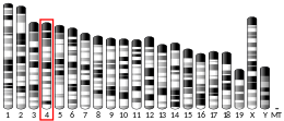
<span class="mw-page-title-main">Interferon gamma</span> InterPro Family

Interferon gamma (IFN-γ) is a dimerized soluble cytokine that is the only member of the type II class of interferons. The existence of this interferon, which early in its history was known as immune interferon, was described by E. F. Wheelock as a product of human leukocytes stimulated with phytohemagglutinin, and by others as a product of antigen-stimulated lymphocytes. It was also shown to be produced in human lymphocytes. or tuberculin-sensitized mouse peritoneal lymphocytes challenged with Mantoux test (PPD); the resulting supernatants were shown to inhibit growth of vesicular stomatitis virus. Those reports also contained the basic observation underlying the now widely employed IFN-γ release assay used to test for tuberculosis. In humans, the IFN-γ protein is encoded by the IFNG gene.

Interferon tau is a Type I interferon made of a single chain of amino acids. IFN-τ was first discovered in ruminants as the signal for the maternal recognition of pregnancy and originally named ovine trophoblast protein-1 (oTP-1). It has many physiological functions in the mammalian uterus, and also has anti-inflammatory effect that aids in the protection of the semi-allogeneic conceptus trophectoderm from the maternal immune system.

<span class="mw-page-title-main">Interferon regulatory factors</span> Protein family

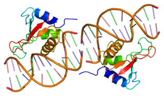
Interferon regulatory factors (IRF) are proteins which regulate transcription of interferons. Interferon regulatory factors contain a conserved N-terminal region of about 120 amino acids, which folds into a structure that binds specifically to the IRF-element (IRF-E) motifs, which is located upstream of the interferon genes. Some viruses have evolved defense mechanisms that regulate and interfere with IRF functions to escape the host immune system. For instance, the remaining parts of the interferon regulatory factor sequence vary depending on the precise function of the protein. The Kaposi sarcoma herpesvirus, KSHV, is a cancer virus that encodes four different IRF-like genes; including vIRF1, which is a transforming oncoprotein that inhibits type 1 interferon activity. In addition, the expression of IRF genes is under epigenetic regulation by promoter DNA methylation.

<span class="mw-page-title-main">Interleukin 29</span> Protein-coding gene in the species Homo sapiens

Interleukin-29 (IL-29) is a cytokine and it belongs to type III interferons group, also termed interferons λ (IFN-λ). IL-29 plays an important role in the immune response against pathogenes and especially against viruses by mechanisms similar to type I interferons, but targeting primarily cells of epithelial origin and hepatocytes.

The type III interferon group is a group of anti-viral cytokines, that consists of four IFN-λ (lambda) molecules called IFN-λ1, IFN-λ2, IFN-λ3, and IFN-λ4. They were discovered in 2003. Their function is similar to that of type I interferons, but is less intense and serves mostly as a first-line defense against viruses in the epithelium.

<span class="mw-page-title-main">Interferon-alpha/beta receptor</span> Heterodimeric receptor

The interferon-α/β receptor (IFNAR) is a virtually ubiquitous membrane receptor which binds endogenous type I interferon (IFN) cytokines. Endogenous human type I IFNs include many subtypes, such as interferons-α, -β, -ε, -κ, -ω, and -ζ.

<span class="mw-page-title-main">STAT4</span> Protein-coding gene in the species Homo sapiens

Signal transducer and activator of transcription 4 (STAT4) is a transcription factor belonging to the STAT protein family, composed of STAT1, STAT2, STAT3, STAT4, STAT5A, STAT5B, STAT6. STAT proteins are key activators of gene transcription which bind to DNA in response to cytokine gradient. STAT proteins are a common part of Janus kinase (JAK)- signalling pathways, activated by cytokines.STAT4 is required for the development of Th1 cells from naive CD4+ T cells and IFN-γ production in response to IL-12. There are two known STAT4 transcripts, STAT4α and STAT4β, differing in the levels of interferon-gamma production downstream.

<span class="mw-page-title-main">STAT2</span> Protein-coding gene in Homo sapiens

Signal transducer and activator of transcription 2 is a protein that in humans is encoded by the STAT2 gene. It is a member of the STAT protein family. This protein is critical to the biological response of type I interferons (IFNs). STAT2 sequence identity between mouse and human is only 68%.

<span class="mw-page-title-main">RIG-I</span> Mammalian protein found in humans

RIG-I is a cytosolic pattern recognition receptor (PRR) that can mediate induction of a type-I interferon (IFN1) response. RIG-I is an essential molecule in the innate immune system for recognizing cells that have been infected with a virus. These viruses can include West Nile virus, Japanese Encephalitis virus, influenza A, Sendai virus, flavivirus, and coronaviruses.

<span class="mw-page-title-main">IFNA2</span> Mammalian protein found in Homo sapiens

Interferon alpha-2 is a protein that in humans is encoded by the IFNA2 gene.

<span class="mw-page-title-main">Mitochondrial antiviral-signaling protein</span> Protein-coding gene in the species Homo sapiens

Mitochondrial antiviral-signaling protein (MAVS) is a protein that is essential for antiviral innate immunity. MAVS is located in the outer membrane of the mitochondria, peroxisomes, and mitochondrial-associated endoplasmic reticulum membrane (MAM). Upon viral infection, a group of cytosolic proteins will detect the presence of the virus and bind to MAVS, thereby activating MAVS. The activation of MAVS leads the virally infected cell to secrete cytokines. This induces an immune response which kills the host's virally infected cells, resulting in clearance of the virus.

<span class="mw-page-title-main">UBE2L6</span> Protein-coding gene in the species Homo sapiens

Ubiquitin/ISG15-conjugating enzyme E2 L6 is a protein that in humans is encoded by the UBE2L6 gene.

<span class="mw-page-title-main">USP18</span> Protein-coding gene in the species Homo sapiens

Ubiquitin specific peptidase 18 (USP18), also known as UBP43, is a type I interferon receptor repressor and an isopeptidase. In humans, it is encoded by the USP18 gene. USP18 is induced by the immune response to type I and III interferons, and serves as a negative regulator of type I interferon, but not type III interferon. Loss of USP18 results in increased responsiveness to type I interferons and life-threatening autoinflammatory disease in humans due to the negative regulatory function of USP18 in interferon signal transduction. Independent of this activity, USP18 is also a member of the deubiquitinating protease family of enzymes. It is known to remove ISG15 conjugates from a broad range of protein substrates, a process known as deISGylation.

<span class="mw-page-title-main">UBE1L</span> Protein-coding gene in the species Homo sapiens

Ubiquitin-like modifier-activating enzyme 7 is a protein that in humans is encoded by the UBA7 gene.

<span class="mw-page-title-main">UBE2E1</span> Protein-coding gene in the species Homo sapiens

Ubiquitin-conjugating enzyme E2 E1 is a protein that in humans is encoded by the UBE2E1 gene.

<span class="mw-page-title-main">UBE2E2</span> Protein-coding gene in the species Homo sapiens

Ubiquitin-conjugating enzyme E2 E2 is a protein that in humans is encoded by the UBE2E2 gene.

<span class="mw-page-title-main">HERC5</span> Protein-coding gene in the species Homo sapiens

Probable E3 ubiquitin-protein ligase HERC5 is an enzyme that in humans is encoded by the HERC5 gene.

An interferon-stimulated gene (ISG) is a gene that can be expressed in response to stimulation by interferon. Interferons bind to receptors on the surface of a cell, initiating protein signaling pathways within the cell. This interaction leads to the expression of a subset of genes involved in the innate immune system response. ISGs are commonly expressed in response to viral infection, but also during bacterial infection and in the presence of parasites. It's currently estimated that 10% of the human genome is regulated by interferons (IFNs). Interferon stimulated genes can act as an initial response to pathogen invasion, slowing down viral replication and increasing expression of immune signaling complexes. There are three known types of interferon. With approximately 450 genes highly expressed in response to interferon type I. Type I interferon consists of INF-α, INF-β, INF-ω and is expressed in response to viral infection. ISGs induced by type I interferon are associated with viral replication suppression and increase expression of immune signaling proteins. Type II interferon consists only of INF-γ and is associated with controlling intracellular pathogens and tumor suppressor genes. Type III interferon consists of INF-λ and is associated with viral immune response and is key in anti-fungal neutrophil response.

<span class="mw-page-title-main">MUL1</span> Protein-coding gene in the species Homo sapiens

Mitochondrial E3 ubiquitin protein ligase 1 (MUL1) is an enzyme that in humans is encoded by the MUL1 gene on chromosome 1. This enzyme localizes to the outer mitochondrial membrane, where it regulates mitochondrial morphology and apoptosis through multiple pathways, including the Akt, JNK, and NF-κB. Its proapoptotic function thus implicates it in cancer and Parkinson’s disease.

References

  1. 1 2 3 GRCh38: Ensembl release 89: ENSG00000187608 - Ensembl, May 2017
  2. 1 2 3 GRCm38: Ensembl release 89: ENSMUSG00000035692 - Ensembl, May 2017
  3. "Human PubMed Reference:". National Center for Biotechnology Information, U.S. National Library of Medicine.
  4. "Mouse PubMed Reference:". National Center for Biotechnology Information, U.S. National Library of Medicine.
  5. Blomstrom DC, Fahey D, Kutny R, Korant BD, Knight E (July 1986). "Molecular characterization of the interferon-induced 15-kDa protein. Molecular cloning and nucleotide and amino acid sequence". The Journal of Biological Chemistry. 261 (19): 8811–6. doi: 10.1016/S0021-9258(19)84453-8 . PMID   3087979.
  6. "Entrez Gene: ISG15 ISG15 ubiquitin-like modifier".
  7. Subedi P, Huber K, Sterr C, Dietz A, Strasser L, Kaestle F, Hauck SM, Duchrow L, Aldrian C, Monroy Ordonez EB, Luka B, Thomsen AR, Henke M, Gomolka M, Rossler U, Azimzadeh O, Moertl S, Hornhardt S (June 2023). "Towards unravelling biological mechanisms behind radiation-induced oral mucositis via mass spectrometry-based proteomics". Frontiers in Oncology. 13. doi: 10.3389/fonc.2023.1180642 . PMC   10298177 . PMID   37384298.
  8. Dzimianski JV, Scholte FE, Bergeron É, Pegan SD (October 2019). "ISG15: It's Complicated". Journal of Molecular Biology. 431 (21): 4203–4216. doi:10.1016/j.jmb.2019.03.013. PMC   6746611 . PMID   30890331.
  9. 1 2 Knight E, Cordova B (April 1991). "IFN-induced 15-kDa protein is released from human lymphocytes and monocytes". Journal of Immunology. 146 (7): 2280–4. doi:10.4049/jimmunol.146.7.2280. PMID   2005397.
  10. 1 2 D'Cunha J, Knight E, Haas AL, Truitt RL, Borden EC (January 1996). "Immunoregulatory properties of ISG15, an interferon-induced cytokine". Proceedings of the National Academy of Sciences of the United States of America. 93 (1): 211–5. Bibcode:1996PNAS...93..211D. doi: 10.1073/pnas.93.1.211 . PMC   40208 . PMID   8552607.
  11. 1 2 Recht M, Borden EC, Knight E (October 1991). "A human 15-kDa IFN-induced protein induces the secretion of IFN-gamma". Journal of Immunology. 147 (8): 2617–23. doi:10.4049/jimmunol.147.8.2617. PMID   1717569.
  12. 1 2 3 4 Swaim CD, Scott AF, Canadeo LA, Huibregtse JM (November 2017). "Extracellular ISG15 Signals Cytokine Secretion through the LFA-1 Integrin Receptor". Molecular Cell. 68 (3): 581–590.e5. doi:10.1016/j.molcel.2017.10.003. PMC   5690536 . PMID   29100055.
  13. Woods MW, Kelly JN, Hattlmann CJ, Tong JG, Xu LS, Coleman MD, et al. (November 2011). "Human HERC5 restricts an early stage of HIV-1 assembly by a mechanism correlating with the ISGylation of Gag". Retrovirology. 8: 95. doi: 10.1186/1742-4690-8-95 . PMC   3228677 . PMID   22093708.
  14. Malakhov MP, Malakhova OA, Kim KI, Ritchie KJ, Zhang DE (March 2002). "UBP43 (USP18) specifically removes ISG15 from conjugated proteins". The Journal of Biological Chemistry. 277 (12): 9976–81. doi: 10.1074/jbc.M109078200 . PMID   11788588.
  15. 1 2 3 Zhang X, Bogunovic D, Payelle-Brogard B, Francois-Newton V, Speer SD, Yuan C, et al. (January 2015). "Human intracellular ISG15 prevents interferon-α/β over-amplification and auto-inflammation". Nature. 517 (7532): 89–93. Bibcode:2015Natur.517...89Z. doi:10.1038/nature13801. PMC   4303590 . PMID   25307056.
  16. 1 2 Speer SD, Li Z, Buta S, Payelle-Brogard B, Qian L, Vigant F, et al. (May 2016). "ISG15 deficiency and increased viral resistance in humans but not mice". Nature Communications. 7: 11496. Bibcode:2016NatCo...711496S. doi:10.1038/ncomms11496. PMC   4873964 . PMID   27193971.
  17. Martin-Fernandez M, Bravo García-Morato M, Gruber C, Murias Loza S, Malik MN, Alsohime F, et al. (May 2020). "Systemic Type I IFN Inflammation in Human ISG15 Deficiency Leads to Necrotizing Skin Lesions". Cell Reports. 31 (6): 107633. doi: 10.1016/j.celrep.2020.107633 . PMC   7331931 . PMID   32402279.
  18. Sainz B, Martín B, Tatari M, Heeschen C, Guerra S (December 2014). "ISG15 is a critical microenvironmental factor for pancreatic cancer stem cells". Cancer Research. 74 (24): 7309–20. doi: 10.1158/0008-5472.CAN-14-1354 . PMID   25368022.
  19. Farrell PJ, Broeze RJ, Lengyel P (June 1979). "Accumulation of an mRNA and protein in interferon-treated Ehrlich ascites tumour cells". Nature. 279 (5713): 523–5. Bibcode:1979Natur.279..523F. doi:10.1038/279523a0. PMID   571963. S2CID   4317436.
  20. Kessler DS, Levy DE, Darnell JE (November 1988). "Two interferon-induced nuclear factors bind a single promoter element in interferon-stimulated genes". Proceedings of the National Academy of Sciences of the United States of America. 85 (22): 8521–5. Bibcode:1988PNAS...85.8521K. doi: 10.1073/pnas.85.22.8521 . PMC   282490 . PMID   2460869.
  21. Reich N, Evans B, Levy D, Fahey D, Knight E, Darnell JE (September 1987). "Interferon-induced transcription of a gene encoding a 15-kDa protein depends on an upstream enhancer element". Proceedings of the National Academy of Sciences of the United States of America. 84 (18): 6394–8. Bibcode:1987PNAS...84.6394R. doi: 10.1073/pnas.84.18.6394 . PMC   299082 . PMID   3476954.
  22. Haas AL, Ahrens P, Bright PM, Ankel H (August 1987). "Interferon induces a 15-kilodalton protein exhibiting marked homology to ubiquitin". The Journal of Biological Chemistry. 262 (23): 11315–23. doi: 10.1016/S0021-9258(18)60961-5 . PMID   2440890.
  23. Loeb KR, Haas AL (April 1992). "The interferon-inducible 15-kDa ubiquitin homolog conjugates to intracellular proteins". The Journal of Biological Chemistry. 267 (11): 7806–13. doi: 10.1016/S0021-9258(18)42585-9 . PMID   1373138.
  24. Werneke SW, Schilte C, Rohatgi A, Monte KJ, Michault A, Arenzana-Seisdedos F, et al. (October 2011). "ISG15 is critical in the control of Chikungunya virus infection independent of UbE1L mediated conjugation". PLOS Pathogens. 7 (10): e1002322. doi: 10.1371/journal.ppat.1002322 . PMC   3197620 . PMID   22028657.
  25. Qiu X, Taft J, Bogunovic D (March 2020). "Developing Broad-Spectrum Antivirals Using Porcine and Rhesus Macaque Models". The Journal of Infectious Diseases. 221 (6): 890–894. doi:10.1093/infdis/jiz549. PMC   7050986 . PMID   31637432.

Further reading