Functions


Calexcitin directly regulate the K+ channels. Due to the fact that "Calexcitin is also a high affinity substrate for protein kinase C. Application of calexcitin to the inner surface of inside-out patches of human fibroblast membranes, in the presence of Ca2+ and the absence of endogenous Ca2+/calmodulin kinase type II or protein kinase C activity, reduced the mean open time and mean open probability of 115 ± 6 pS K+ channels". Also, calexcitin is very great at making the membrane to be more excitable due to "When microinjected into molluscan neurons or rabbit cerebellar Purkinje cell dendrites". In addition, calexcitin acts as a Ca2+ activated signaling molecule in which it plays a role into increasing the cellular excitability. while making it more likely to increase the Ca2+ influx in the membrane. Also, this shows an example of GTP-binding protein that by which binds to Ca2+.
Related Research Articles
G protein-coupled receptors (GPCRs), also known as seven-(pass)-transmembrane domain receptors, 7TM receptors, heptahelical receptors, serpentine receptors, and G protein-linked receptors (GPLR), form a large group of evolutionarily related proteins that are cell surface receptors that detect molecules outside the cell and activate cellular responses. They are coupled with G proteins. They pass through the cell membrane seven times in the form of six loops of amino acid residues, which is why they are sometimes referred to as seven-transmembrane receptors. Ligands can bind either to the extracellular N-terminus and loops or to the binding site within transmembrane helices. They are all activated by agonists, although a spontaneous auto-activation of an empty receptor has also been observed.
Calmodulin (CaM) (an abbreviation for calcium-modulated protein) is a multifunctional intermediate calcium-binding messenger protein expressed in all eukaryotic cells. It is an intracellular target of the secondary messenger Ca2+, and the binding of Ca2+ is required for the activation of calmodulin. Once bound to Ca2+, calmodulin acts as part of a calcium signal transduction pathway by modifying its interactions with various target proteins such as kinases or phosphatases.

Smooth muscle is an involuntary non-striated muscle, so-called because it has no sarcomeres and therefore no striations. It is divided into two subgroups, single-unit and multiunit smooth muscle. Within single-unit muscle, the whole bundle or sheet of smooth muscle cells contracts as a syncytium.

The sarcoplasmic reticulum (SR) is a membrane-bound structure found within muscle cells that is similar to the smooth endoplasmic reticulum in other cells. The main function of the SR is to store calcium ions (Ca2+). Calcium ion levels are kept relatively constant, with the concentration of calcium ions within a cell being 10,000 times smaller than the concentration of calcium ions outside the cell. This means that small increases in calcium ions within the cell are easily detected and can bring about important cellular changes (the calcium is said to be a second messenger). Calcium is used to make calcium carbonate (found in chalk) and calcium phosphate, two compounds that the body uses to make teeth and bones. This means that too much calcium within the cells can lead to hardening (calcification) of certain intracellular structures, including the mitochondria, leading to cell death. Therefore, it is vital that calcium ion levels are controlled tightly, and can be released into the cell when necessary and then removed from the cell.
Voltage-gated calcium channels (VGCCs), also known as voltage-dependent calcium channels (VDCCs), are a group of voltage-gated ion channels found in the membrane of excitable cells (e.g., muscle, glial cells, neurons, etc.) with a permeability to the calcium ion Ca2+. These channels are slightly permeable to sodium ions, so they are also called Ca2+–Na+ channels, but their permeability to calcium is about 1000-fold greater than to sodium under normal physiological conditions.
Second messengers are intracellular signaling molecules released by the cell in response to exposure to extracellular signaling molecules—the first messengers. Second messengers trigger physiological changes at cellular level such as proliferation, differentiation, migration, survival, apoptosis and depolarization.
Molecular neuroscience is a branch of neuroscience that observes concepts in molecular biology applied to the nervous systems of animals. The scope of this subject covers topics such as molecular neuroanatomy, mechanisms of molecular signaling in the nervous system, the effects of genetics and epigenetics on neuronal development, and the molecular basis for neuroplasticity and neurodegenerative diseases. As with molecular biology, molecular neuroscience is a relatively new field that is considerably dynamic.
Visual phototransduction is the sensory transduction process of the visual system by which light is detected to yield nerve impulses in the rod cells and cone cells in the retina of the eye in humans and other vertebrates. It relies on the visual cycle, a sequence of biochemical reactions in which a molecule of retinal bound to opsin undergoes photoisomerization, initiates a cascade that signals detection of the photon, and is indirectly restored to its photosensitive isomer for reuse. Phototransduction in some invertebrates such as fruit flies relies on similar processes.

G protein-gated ion channels are a family of transmembrane ion channels in neurons and atrial myocytes that are directly gated by G proteins.
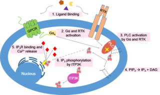
Calcium signaling is the use of calcium ions (Ca2+) to communicate and drive intracellular processes often as a step in signal transduction. Ca2+ is important for cellular signalling, for once it enters the cytosol of the cytoplasm it exerts allosteric regulatory effects on many enzymes and proteins. Ca2+ can act in signal transduction resulting from activation of ion channels or as a second messenger caused by indirect signal transduction pathways such as G protein-coupled receptors.
Calcium-binding proteins are proteins that participate in calcium cell signaling pathways by binding to Ca2+, the calcium ion that plays an important role in many cellular processes. Calcium-binding proteins have specific domains that bind to calcium and are known to be heterogeneous.

Neuronal calcium sensor-1 (NCS-1) also known as frequenin homolog (Drosophila) (freq) is a protein that is encoded by the FREQ gene in humans. NCS-1 is a member of the neuronal calcium sensor family, a class of EF hand containing calcium-myristoyl-switch proteins.
Neuronal Calcium Sensor is a large family of proteins which work as calcium dependent molecular switches and includes members like Frequenin (NCS1), recoverin, GCAP, neurocalcin, visinin etc. All the members carry 4 EF hand motifs and an N-myristoyl group.

A C2 domain is a protein structural domain involved in targeting proteins to cell membranes. The typical version (PKC-C2) has a beta-sandwich composed of 8 β-strands that co-ordinates two or three calcium ions, which bind in a cavity formed by the first and final loops of the domain, on the membrane binding face. Many other C2 domain families don't have calcium binding activity.

The L-type calcium channel is part of the high-voltage activated family of voltage-dependent calcium channel. "L" stands for long-lasting referring to the length of activation. This channel has four isoforms: Cav1.1, Cav1.2, Cav1.3, and Cav1.4.

Calsenilin is a protein that in humans is encoded by the KCNIP3 gene.

Calcium-activated potassium channel subunit beta-1 is a protein that in humans is encoded by the KCNMB1 gene.

Kv channel-interacting protein 4 is a protein that in humans is encoded by the KCNIP4 gene.
The insulin transduction pathway is a biochemical pathway by which insulin increases the uptake of glucose into fat and muscle cells and reduces the synthesis of glucose in the liver and hence is involved in maintaining glucose homeostasis. This pathway is also influenced by fed versus fasting states, stress levels, and a variety of other hormones.
Cardiac excitation-contraction coupling (CardiacEC coupling) describes the series of events, from the production of an electrical impulse (action potential) to the contraction of muscles in the heart. This process is of vital importance as it allows for the heart to beat in a controlled manner, without the need for conscious input. EC coupling results in the sequential contraction of the heart muscles that allows blood to be pumped, first to the lungs (pulmonary circulation) and then around the rest of the body (systemic circulation) at a rate between 60 and 100 beats every minute, when the body is at rest. This rate can be altered, however, by nerves that work to either increase heart rate (sympathetic nerves) or decrease it (parasympathetic nerves), as the body's oxygen demands change. Ultimately, muscle contraction revolves around a charged atom (ion), calcium (Ca2+), which is responsible for converting the electrical energy of the action potential into mechanical energy (contraction) of the muscle. This is achieved in a region of the muscle cell, called the transverse tubule during a process known as calcium induced calcium release.
References
This article includes a list of references, related reading, or external links, but its sources remain unclear because it lacks inline citations .(June 2022) |
- Nelson, Thomas J.; Cavallaro, Sebastiano; Yi, Chu-Li; McPhie, Donna; Schreurs, Bernard G.; Gusev, Pavel A.; Favit, Antonella; Zohar, Ofer; Kim, Jeongho; Beushausen, Sven; Ascoli, Giorgio; Olds, James; Neve, Rachael; Alkon, Daniel L. (1996). "Calexcitin: A signaling protein that binds calcium and GTP, inhibits potassium channels, and enhances membrane excitability". Proceedings of the National Academy of Sciences. 93 (24): 13808–13813. Bibcode:1996PNAS...9313808N. doi: 10.1073/pnas.93.24.13808 . PMC 19433 . PMID 8943017.
- Nelson, T. J.; Cavallaro, S.; Yi, C. L.; McPhie, D.; Schreurs, B. G.; Gusev, P. A.; Favit, A.; Zohar, O.; Kim, J.; Beushausen, S.; Ascoli, G.; Olds, J.; Neve, R.; Alkon, D. L. (1996). "Calexcitin: A signaling protein that binds calcium and GTP, inhibits potassium channels, and enhances membrane excitability". Proceedings of the National Academy of Sciences of the United States of America. 93 (24): 13808–13813. Bibcode:1996PNAS...9313808N. doi: 10.1073/pnas.93.24.13808 . PMC 19433 . PMID 8943017.