FAN1

Last updated
FAN1
Available structures
PDB Ortholog search: PDBe RCSB
Identifiers
Aliases FAN1 , KIAA1018, KMIN, MTMR15, hFANCD2/FANCI-associated nuclease 1, FANCD2 and FANCI associated nuclease 1
External IDs OMIM: 613534 MGI: 3045266 HomoloGene: 45598 GeneCards: FAN1
Orthologs
SpeciesHumanMouse
Entrez
Ensembl
UniProt
RefSeq (mRNA)

NM_001146094
NM_001146095
NM_001146096
NM_014967

NM_177893

RefSeq (protein)

NP_001139566
NP_001139567
NP_001139568
NP_055782

NP_808561

Location (UCSC) Chr 15: 30.89 – 30.94 Mb Chr 7: 64 – 64.02 Mb
PubMed search [3] [4]
Wikidata
View/Edit Human View/Edit Mouse

FANCD2/FANCI-associated nuclease 1 (KIAA1018) is an enzyme that in humans is encoded by the FAN1 gene. It is a structure dependent endonuclease. It is thought to play an important role in the Fanconi Anemia (FA) pathway. [5]

Structure

Figure 1. Color coded structure of FAN1 monomer with the SAP shown in blue and purple, TPR in orange, and VRR_Nuc in green. The secondary features of the SAP domain are labeled. Created in PyMOL from PDB structure 4RID. FAN1 residues 37-1009.PNG
Figure 1. Color coded structure of FAN1 monomer with the SAP shown in blue and purple, TPR in orange, and VRR_Nuc in green. The secondary features of the SAP domain are labeled. Created in PyMOL from PDB structure 4RID.

FAN1 is a protein of 1017 amino acids. [7] Several crystal structures of the residues 373-1017 have been characterized. This portion of FAN1 contains three domains: an SAP domain (primary-DNA binding domain), a TPR domain (mediating interdomain interaction and dimerization interface) and the virus-type replication-repair nuclease module (VRR_NUC, catalytic site) (Figure 1). [8] DNA binding promotes dimerization of FAN1 in a "head to tail" fashion. [6]

The SAP region contains three major components: α9, α5β1, and α7. The core helix α9 stabilizes the protein as it moves through dimer configurations and mediates the interactions between α5β1 and α7 as they adjust their positions. These three configurations are the substrate scanning, substrate latching and substrate unwinding forms (figure 2). [6]

In the FAN1 dimer, the SAP regions of both FAN1 enzymes make contact with the DNA duplex (dsDNA). This double contact facilitates DNA induced dimerization, as well as guiding the single stranded (ssDNA) into the SAP domain of the downstream enzyme (PSAP). The SAP domain of the upstream FAN1 component enzyme (ASAP) aids in guiding the DNA to PSAP. [6]

The SAP surface facing the catalytic site is the most conserved region between FAN1 homologs. It is positively charged for favorable hydrogen bonding and electrostatic interactions with DNA. In particular, residues Y374 and Y436 form hydrogen bonds with the phosphate backbone. FAN1 can bind DNA in either direction. However, when the 5' flab is facing away from the VRR_NUC site, substrate latching and unwinding cannot occur. [6] The unresolved portion of FAN1 contains a Zinc finger at the N terminus called a UBZ region. This is present in proteins that bind to ubiquitinated proteins, and is highly conserved across eukaryotes. This Zinc finger is crucial for recruitment to the ubiquitinated FANCD2/FANCI complex, and is found in other nucleases. [7] The VRR_Nuc catalytic domain is located at the C terminus and contains the endonuclease functionality. [7] FAN1 is the first known instance of a virus type replication-repair nuclease module in eukaryotes. It is normally found as a standalone domain in bacterial and viral Holliday Junction Resolvases (HJR). FAN1 does not exhibit any activity on Holliday Junction (HJ) substrates. [8] A subdomain of SAP consisting of six α helices connected to the VRR_Nuc region is thought to inhibit HJR activity. [9]

Function

Figure 2. Three conformations of FAN1 in action. A. Monomer scans substrate B. Dimerization occurs and dimer latches onto substrate C. dimer unwinds substrate. Created in PyMOL from PDB structures 4REA, 4REC, and 4REB. Substrate scanning, latching and unwinding by FAN1.png
Figure 2. Three conformations of FAN1 in action. A. Monomer scans substrate B. Dimerization occurs and dimer latches onto substrate C. dimer unwinds substrate. Created in PyMOL from PDB structures 4REA, 4REC, and 4REB.

Interstrand DNA crosslinks (ICLs) effectively block the progression of transcription and replication machineries. Release of this block, referred to as unhooking, is thought to require incision of one strand of the duplex on either side of the ICL.

Repair of interstrand DNA crosslinks is triggered when the DNA replication fork is unable to continue. The FA proteins play an elaborate role with FAN1 to remove these ICLs. The pathway consists of 15 known proteins. Three of them form the FA AP24-MHF1/2 complex which recognizes the ICL (from stalled replication forks). This recruits the FA core complex, which consists of 8 proteins. This complex monoubiquitinates FANCD2 and FANCI, which allows it to form a heterodimer. It is this complex that recruits FAN1 as well as other nucleases such as SLX4. [9] Ubiquinated FANCD2 interacts with the FAN1 nuclease. Upon its recruitment by FANCD2, FAN1 acts to restrain DNA replication fork progression and to prevent chromosome abnormalities from occurring when DNA replication forks stall. [11] FAN1 is typically localized in the nucleus, but forms very distinct loci at damaged regions when ICLs are present. [12]

The FAN1 protein possesses endonuclease and exonuclease functions to remove ICLs. At a replication fork arrested at an ICL, FAN1 nuclease action can catalyze incisions in the double-stranded region. [13] It is thought that this process consists of unhooking the crosslink and separating the DNA strands through two incision events, yielding one strand with a crosslinked nucleotide and another strand with a gap. [14] [15] FAN1 preferentially acts as a 5’ flap endonuclease. This is illustrated in Figure 2, which shows the sequence of substrate scanning, latching, and unwinding. It usually cleaves about 5 nucleotides from a junction. FAN1 will also incise at splayed arms, three way junctions, and 3’ flaps (in order of decreasing preference). In high concentrations FAN1 has been shown to exhibit 3’ 5’ exonuclease activity. In blunt end substrates, FAN1 has also 5’ recessed ends. However, FAN1 does not appear to bind to single stranded DNA. [7] [16]

The presence of the FANCD2/FANCI complex is unaffected by knockdown of FAN1. This is because FAN1 acts downstream to the recruitment of FANCD2/FANCI. [6] [7] [17] FAN1 has also been shown to increase the frequency of homologous recombination. [7] This suggests that the gapped intermediate that forms following ICL unhooking may be repaired through HR when homologous chromosomes are present. [16] FAN1 does not appear to be involved in other types of DNA repair, as it does not localize to DNA upon irradiation. [12]

Clinical significance

Mutations affecting the function of the 15 known FA genes are associated with Fanconi anemia, a recessive autosomal disorder. [17] It is characterized by congenital abnormalities as well as anemia, bone marrow failure, and cancer predisposition in childhood. [9] However, some patients have “unassigned” Fanconi Anemia where no mutations in the known FA genes can be found. Mutations in FAN1 can result in chronic kidney diseases and neurological conditions such as schizophrenia. [6] [18] However, recent research has called into question the categorization of FAN1 as an FA gene. In 2015 researchers studied four individuals with chromosomal microdeletion of 15q13.3. Analysis of blood samples revealed only mild ICL agent sensitivity and chromosomal fragility consistent with Fanconi Anemia. [19]

A deficiency of FAN1 increases in vitro sensitivity to cisplatin and mitomycin C, two crosslinking agents [6] [7] FAN1 is also able to repair mitomycin C induced double strand breaks. [7]

Germline mutations in the FAN1 gene can cause hereditary colorectal cancer due to defective DNA repair. [20]

Related Research Articles

<span class="mw-page-title-main">Fanconi anemia</span> Medical condition

Fanconi anemia (FA) is a rare, AR, genetic disease resulting in impaired response to DNA damage in the FA/BRCA pathway. Although it is a very rare disorder, study of this and other bone marrow failure syndromes has improved scientific understanding of the mechanisms of normal bone marrow function and development of cancer. Among those affected, the majority develop cancer, most often acute myelogenous leukemia (AML), MDS, and liver tumors. 90% develop aplastic anemia by age 40. About 60–75% have congenital defects, commonly short stature, abnormalities of the skin, arms, head, eyes, kidneys, and ears, and developmental disabilities. Around 75% have some form of endocrine problem, with varying degrees of severity. 60% of FA is FANC-A, 16q24.3, which has later onset bone marrow failure.

In molecular biology, endonucleases are enzymes that cleave the phosphodiester bond within a polynucleotide chain. Some, such as deoxyribonuclease I, cut DNA relatively nonspecifically, while many, typically called restriction endonucleases or restriction enzymes, cleave only at very specific nucleotide sequences. Endonucleases differ from exonucleases, which cleave the ends of recognition sequences instead of the middle (endo) portion. Some enzymes known as "exo-endonucleases", however, are not limited to either nuclease function, displaying qualities that are both endo- and exo-like. Evidence suggests that endonuclease activity experiences a lag compared to exonuclease activity.

<span class="mw-page-title-main">Crosslinking of DNA</span> Phenomenon in genetics

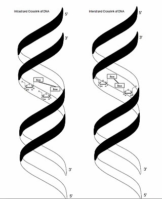
In genetics, crosslinking of DNA occurs when various exogenous or endogenous agents react with two nucleotides of DNA, forming a covalent linkage between them. This crosslink can occur within the same strand (intrastrand) or between opposite strands of double-stranded DNA (interstrand). These adducts interfere with cellular metabolism, such as DNA replication and transcription, triggering cell death. These crosslinks can, however, be repaired through excision or recombination pathways.

<span class="mw-page-title-main">ERCC1</span> Protein-coding gene in the species Homo sapiens

DNA excision repair protein ERCC-1 is a protein that in humans is encoded by the ERCC1 gene. Together with ERCC4, ERCC1 forms the ERCC1-XPF enzyme complex that participates in DNA repair and DNA recombination.

<span class="mw-page-title-main">Flap structure-specific endonuclease 1</span> Protein-coding gene in the species Homo sapiens

Flap endonuclease 1 is an enzyme that in humans is encoded by the FEN1 gene.

<span class="mw-page-title-main">Fanconi anemia, complementation group C</span> Protein-coding gene in the species Homo sapiens

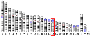
Fanconi anemia group C protein is a protein that in humans is encoded by the FANCC gene. This protein delays the onset of apoptosis and promotes homologous recombination repair of damaged DNA. Mutations in this gene result in Fanconi anemia, a human rare disorder characterized by cancer susceptibility and cellular sensitivity to DNA crosslinks and other damages.

<span class="mw-page-title-main">FANCA</span> Protein-coding gene in the species Homo sapiens

Fanconi anaemia, complementation group A, also known as FAA, FACA and FANCA, is a protein which in humans is encoded by the FANCA gene. It belongs to the Fanconi anaemia complementation group (FANC) family of genes of which 12 complementation groups are currently recognized and is hypothesised to operate as a post-replication repair or a cell cycle checkpoint. FANCA proteins are involved in inter-strand DNA cross-link repair and in the maintenance of normal chromosome stability that regulates the differentiation of haematopoietic stem cells into mature blood cells.

<span class="mw-page-title-main">FANCD2</span> Protein-coding gene in the species Homo sapiens

Fanconi anemia group D2 protein is a protein that in humans is encoded by the FANCD2 gene. The Fanconi anemia complementation group (FANC) currently includes FANCA, FANCB, FANCC, FANCD1, FANCD2, FANCE, FANCF, FANCG, FANCI, FANCJ, FANCL, FANCM, FANCN and FANCO.

<span class="mw-page-title-main">FANCG</span> Protein-coding gene in the species Homo sapiens

Fanconi anemia group G protein is a protein that in humans is encoded by the FANCG gene.

<span class="mw-page-title-main">ERCC4</span> Protein-coding gene in the species Homo sapiens

ERCC4 is a protein designated as DNA repair endonuclease XPF that in humans is encoded by the ERCC4 gene. Together with ERCC1, ERCC4 forms the ERCC1-XPF enzyme complex that participates in DNA repair and DNA recombination.

<span class="mw-page-title-main">FANCF</span> Protein-coding gene in the species Homo sapiens

Fanconi anemia group F protein is a protein that in humans is encoded by the FANCF gene.

<span class="mw-page-title-main">FANCE</span> Protein-coding gene in the species Homo sapiens

Fanconi anemia, complementation group E protein is a protein that in humans is encoded by the FANCE gene. The Fanconi anemia complementation group (FANC) currently includes FANCA, FANCB, FANCC, FANCD1, FANCD2, FANCE, FANCF, FANCG, and FANCL. Fanconi anemia is a genetically heterogeneous recessive disorder characterized by cytogenetic instability, hypersensitivity to DNA cross-linking agents, increased chromosomal breakage, and defective DNA repair. The members of the Fanconi anemia complementation group do not share sequence similarity; they are related by their assembly into a common nuclear protein complex. This gene encodes the protein for complementation groufcrp E.

<span class="mw-page-title-main">FANCL</span> Protein-coding gene in the species Homo sapiens

E3 ubiquitin-protein ligase FANCL is an enzyme that in humans is encoded by the FANCL gene.

<span class="mw-page-title-main">FANCB</span> Protein-coding gene in the species Homo sapiens

Fanconi anemia group B protein is a protein that in humans is encoded by the FANCB gene.

<span class="mw-page-title-main">FANCI</span> Protein-coding gene in the species Homo sapiens

Fanconi anemia, complementation group I (FANCI) also known as KIAA1794, is a protein which in humans is encoded by the FANCI gene. Mutations in the FANCI gene are known to cause Fanconi anemia.

<span class="mw-page-title-main">SLX4</span> Protein involved in DNA repair

SLX4 is a protein involved in DNA repair, where it has important roles in the final steps of homologous recombination. Mutations in the gene are associated with the disease Fanconi anemia.

FANC proteins are a network of at least 15 proteins that are associated with a cell process known as the Fanconi anemia.

<span class="mw-page-title-main">SLX4IP</span> Protein-coding gene in the species Homo sapiens

SLX4 interacting protein is a protein that in humans is encoded by the SLX4IP gene.

Agata Smogorzewska is a Polish-born scientist. She is an associate professor at Rockefeller University, heading the Laboratory of Genome Maintenance. Her work primarily focuses on DNA interstrand crosslink repair and the diseases resulting from deficiencies in this repair pathway, including Fanconi anemia and karyomegalic interstitial nephritis.

<span class="mw-page-title-main">Double-strand break repair model</span>

A double-strand break repair model refers to the various models of pathways that cells undertake to repair double strand-breaks (DSB). DSB repair is an important cellular process, as the accumulation of unrepaired DSB could lead to chromosomal rearrangements, tumorigenesis or even cell death. In human cells, there are two main DSB repair mechanisms: Homologous recombination (HR) and non-homologous end joining (NHEJ). HR relies on undamaged template DNA as reference to repair the DSB, resulting in the restoration of the original sequence. NHEJ modifies and ligates the damaged ends regardless of homology. In terms of DSB repair pathway choice, most mammalian cells appear to favor NHEJ rather than HR. This is because the employment of HR may lead to gene deletion or amplification in cells which contains repetitive sequences. In terms of repair models in the cell cycle, HR is only possible during the S and G2 phases, while NHEJ can occur throughout whole process. These repair pathways are all regulated by the overarching DNA damage response mechanism. Besides HR and NHEJ, there are also other repair models which exists in cells. Some are categorized under HR, such as synthesis-dependent strain annealing, break-induced replication, and single-strand annealing; while others are an entirely alternate repair model, namely, the pathway microhomology-mediated end joining (MMEJ).

References

  1. 1 2 3 ENSG00000198690 GRCh38: Ensembl release 89: ENSG00000276787, ENSG00000198690 - Ensembl, May 2017
  2. 1 2 3 GRCm38: Ensembl release 89: ENSMUSG00000033458 - Ensembl, May 2017
  3. "Human PubMed Reference:". National Center for Biotechnology Information, U.S. National Library of Medicine.
  4. "Mouse PubMed Reference:". National Center for Biotechnology Information, U.S. National Library of Medicine.
  5. "Entrez Gene: FANCD2/FANCI-associated nuclease 1".
  6. 1 2 3 4 5 6 7 8 9 Zhao Q, Xue X, Longerich S, Sung P, Xiong Y (December 2014). "Structural insights into 5' flap DNA unwinding and incision by the human FAN1 dimer". Nature Communications. 5 (11): 5726. Bibcode:2014NatCo...5.5726Z. doi:10.1038/ncomms6726. PMC   4268874 . PMID   25500724.
  7. 1 2 3 4 5 6 7 8 MacKay C, Déclais AC, Lundin C, Agostinho A, Deans AJ, MacArtney TJ, et al. (July 2010). "Identification of KIAA1018/FAN1, a DNA repair nuclease recruited to DNA damage by monoubiquitinated FANCD2". Cell. 142 (1): 65–76. doi:10.1016/j.cell.2010.06.021. PMC   3710700 . PMID   20603015.
  8. 1 2 Pennell S, Déclais AC, Li J, Haire LF, Berg W, Saldanha JW, et al. (July 2014). "FAN1 activity on asymmetric repair intermediates is mediated by an atypical monomeric virus-type replication-repair nuclease domain". Cell Reports. 8 (1): 84–93. doi:10.1016/j.celrep.2014.06.001. PMC   4103454 . PMID   24981866.
  9. 1 2 3 Kim H, D'Andrea AD (July 2012). "Regulation of DNA cross-link repair by the Fanconi anemia/BRCA pathway". Genes & Development. 26 (13): 1393–408. doi:10.1101/gad.195248.112. PMC   3403008 . PMID   22751496.
  10. Wang R, Persky NS, Yoo B, Ouerfelli O, Smogorzewska A, Elledge SJ, Pavletich NP (November 2014). "DNA repair. Mechanism of DNA interstrand cross-link processing by repair nuclease FAN1". Science. 346 (6213): 1127–30. doi:10.1126/science.1258973. PMC   4285437 . PMID   25430771.
  11. Lachaud C, Moreno A, Marchesi F, Toth R, Blow JJ, Rouse J (February 2016). "Ubiquitinated Fancd2 recruits Fan1 to stalled replication forks to prevent genome instability". Science. 351 (6275): 846–9. Bibcode:2016Sci...351..846L. doi:10.1126/science.aad5634. PMC   4770513 . PMID   26797144.
  12. 1 2 Shereda RD, Machida Y, Machida YJ (October 2010). "Human KIAA1018/FAN1 localizes to stalled replication forks via its ubiquitin-binding domain". Cell Cycle. 9 (19): 3977–83. doi: 10.4161/cc.9.19.13207 . PMID   20935496.
  13. Pizzolato J, Mukherjee S, Schärer OD, Jiricny J (September 2015). "FANCD2-associated nuclease 1, but not exonuclease 1 or flap endonuclease 1, is able to unhook DNA interstrand cross-links in vitro". The Journal of Biological Chemistry. 290 (37): 22602–11. doi: 10.1074/jbc.M115.663666 . PMC   4566234 . PMID   26221031.
  14. Smogorzewska A, Desetty R, Saito TT, Schlabach M, Lach FP, Sowa ME, et al. (July 2010). "A genetic screen identifies FAN1, a Fanconi anemia-associated nuclease necessary for DNA interstrand crosslink repair". Molecular Cell. 39 (1): 36–47. doi:10.1016/j.molcel.2010.06.023. PMC   2919743 . PMID   20603073.
  15. Kee Y, D'Andrea AD (August 2010). "Expanded roles of the Fanconi anemia pathway in preserving genomic stability". Genes & Development. 24 (16): 1680–94. doi:10.1101/gad.1955310. PMC   2922498 . PMID   20713514.
  16. 1 2 Sengerová B, Wang AT, McHugh PJ (December 2011). "Orchestrating the nucleases involved in DNA interstrand cross-link (ICL) repair". Cell Cycle. 10 (23): 3999–4008. doi:10.4161/cc.10.23.18385. PMC   3272282 . PMID   22101340.
  17. 1 2 Liu T, Ghosal G, Yuan J, Chen J, Huang J (August 2010). "FAN1 acts with FANCI-FANCD2 to promote DNA interstrand cross-link repair". Science. 329 (5992): 693–6. Bibcode:2010Sci...329..693L. doi: 10.1126/science.1192656 . PMID   20671156. S2CID   206527789.
  18. Zhou W, Otto EA, Cluckey A, Airik R, Hurd TW, Chaki M, et al. (July 2012). "FAN1 mutations cause karyomegalic interstitial nephritis, linking chronic kidney failure to defective DNA damage repair". Nature Genetics. 44 (8): 910–5. doi:10.1038/ng.2347. PMC   3412140 . PMID   22772369.
  19. Trujillo JP, Mina LB, Pujol R, Bogliolo M, Andrieux J, Holder M, et al. (July 2012). "On the role of FAN1 in Fanconi anemia". Blood. 120 (1): 86–9. doi: 10.1182/blood-2012-04-420604 . PMID   22611161.
  20. Seguí N, Mina LB, Lázaro C, Sanz-Pamplona R, Pons T, Navarro M, et al. (September 2015). "Germline Mutations in FAN1 Cause Hereditary Colorectal Cancer by Impairing DNA Repair". Gastroenterology. 149 (3): 563–6. doi: 10.1053/j.gastro.2015.05.056 . hdl: 10651/34223 . PMID   26052075.

Further reading

This article incorporates text from the United States National Library of Medicine, which is in the public domain.