Articles related to anatomy include:

The corpus callosum, also callosal commissure, is a wide, thick nerve tract, consisting of a flat bundle of commissural fibers, beneath the cerebral cortex in the brain. The corpus callosum is only found in placental mammals. It spans part of the longitudinal fissure, connecting the left and right cerebral hemispheres, enabling communication between them. It is the largest white matter structure in the human brain, about 10 cm (3.9 in) in length and consisting of 200–300 million axonal projections.

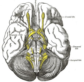
The vertebrate cerebrum (brain) is formed by two cerebral hemispheres that are separated by a groove, the longitudinal fissure. The brain can thus be described as being divided into left and right cerebral hemispheres. Each of these hemispheres has an outer layer of grey matter, the cerebral cortex, that is supported by an inner layer of white matter. In eutherian (placental) mammals, the hemispheres are linked by the corpus callosum, a very large bundle of nerve fibers. Smaller commissures, including the anterior commissure, the posterior commissure and the fornix, also join the hemispheres and these are also present in other vertebrates. These commissures transfer information between the two hemispheres to coordinate localized functions.

The cingulate cortex is a part of the brain situated in the medial aspect of the cerebral cortex. The cingulate cortex includes the entire cingulate gyrus, which lies immediately above the corpus callosum, and the continuation of this in the cingulate sulcus. The cingulate cortex is usually considered part of the limbic lobe.

The fornix is a C-shaped bundle of nerve fibers in the brain that acts as the major output tract of the hippocampus. The fornix also carries some afferent fibers to the hippocampus from structures in the diencephalon and basal forebrain. The fornix is part of the limbic system. While its exact function and importance in the physiology of the brain are still not entirely clear, it has been demonstrated in humans that surgical transection—the cutting of the fornix along its body—can cause memory loss. There is some debate over what type of memory is affected by this damage, but it has been found to most closely correlate with recall memory rather than recognition memory. This means that damage to the fornix can cause difficulty in recalling long-term information such as details of past events, but it has little effect on the ability to recognize objects or familiar situations.

In neuroanatomy, the cingulum or cingulum bundle is an association tract, a nerve tract that projects from the cingulate gyrus to the entorhinal cortex in the brain, allowing for communication between components of the limbic system. It forms the white matter core of the cingulate gyrus, following it from the subcallosal gyrus of the frontal lobe beneath the rostrum of corpus callosum to the parahippocampal gyrus and uncus of the temporal lobe.

The posterior cerebral artery (PCA) is one of a pair of cerebral arteries that supply oxygenated blood to the occipital lobe, as well as the medial and inferior aspects of the temporal lobe of the human brain. The two arteries originate from the distal end of the basilar artery, where it bifurcates into the left and right posterior cerebral arteries. These anastomose with the middle cerebral arteries and internal carotid arteries via the posterior communicating arteries.

Brodmann area 33, also known as pregenual area 33, is a subdivision of the cytoarchitecturally defined cingulate region of cerebral cortex. It is a narrow band located in the anterior cingulate gyrus adjacent to the supracallosal gyrus in the depth of the callosal sulcus, near the genu of the corpus callosum. Cytoarchitecturally it is bounded by the ventral anterior cingulate area 24 and the supracallosal gyrus (Brodmann-1909). The pregenual area 33 is heavily involved in emotions, especially happy emotions.

In neuroanatomy, the parieto-occipital sulcus is a deep sulcus in the cerebral cortex that marks the boundary between the cuneus and precuneus, and also between the parietal and occipital lobes. Only a small part can be seen on the lateral surface of the hemisphere, its chief part being on the medial surface.

The calcarine sulcus is an anatomical landmark located at the caudal end of the medial surface of the brain of humans and other primates. Its name comes from the Latin "calcar" meaning "spur". It is very deep, and known as a complete sulcus.
The tuber cinereum is the portion of hypothalamus forming the floor of the third ventricle situated between the optic chiasm, and the mammillary bodies. The tuberal region is one of the three regions of the hypothalamus, the other two being the chiasmatic region and the mamillary region.

The commissural fibers or transverse fibers are axons that connect the two hemispheres of the brain. Huge numbers of commissural fibers make up the commissural tracts in the brain, the largest of which is the corpus callosum.

The indusium griseum, consists of a thin membranous layer of grey matter in contact with the upper surface of the corpus callosum and continuous laterally with the grey matter of the cingulate cortex and inferiorly with the hippocampus. It is vestigial in humans and is a remnant of the former position of the hippocampus in lower animals.
The chiasmatic cistern or suprasellar cistern is a small subarachnoid cistern related to the optic chiasm.

A nerve tract is a bundle of nerve fibers (axons) connecting nuclei of the central nervous system. In the peripheral nervous system, this is known as a nerve fascicle, and has associated connective tissue. The main nerve tracts in the central nervous system are of three types: association fibers, commissural fibers, and projection fibers. A nerve tract may also be referred to as a commissure, decussation, or neural pathway. A commissure connects the two cerebral hemispheres at the same levels, while a decussation connects at different levels.
The cingulate gyrus commences below the rostrum of the corpus callosum, curves around in front of the genu, extends along the upper surface of the body, and finally turns downward behind the splenium, where it is connected by a narrow isthmus with the parahippocampal gyrus.

In neuroanatomy, the subparietal sulcus or suprasplenial sulcus is a sulcus, or crevice, on the medial surface of each cerebral hemisphere, above the splenium of the corpus callosum. It separates the precuneus from the posterior part of the cingulate gyrus. It is the posterior continuation of the cingulate sulcus. The cingulate sulcus actually "terminates" as the marginal sulcus of the cingulate sulcus. It extends posteriorly toward the calcarine sulcus.

This article describes anatomical terminology that is used to describe the central and peripheral nervous systems - including the brain, brainstem, spinal cord, and nerves.

