Fibroblast growth factor 23

Last updated
FGF23
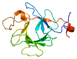
Protein FGF23 PDB 2p39.png
Available structures
PDB Ortholog search: PDBe RCSB
Identifiers
Aliases FGF23 , ADHR, FGFN, HPDR2, HYPF, PHPTC, fibroblast growth factor 23, HFTC2
External IDs OMIM: 605380 MGI: 1891427 HomoloGene: 10771 GeneCards: FGF23
Orthologs
SpeciesHumanMouse
Entrez
Ensembl
UniProt
RefSeq (mRNA)

NM_020638

NM_022657

RefSeq (protein)

NP_065689

NP_073148

Location (UCSC) Chr 12: 4.37 – 4.38 Mb Chr 6: 127.05 – 127.06 Mb
PubMed search [3] [4]
Wikidata
View/Edit Human View/Edit Mouse

Fibroblast growth factor 23 (FGF23) is a protein and member of the fibroblast growth factor (FGF) family which participates in the regulation of phosphate in plasma and vitamin D metabolism. In humans it is encoded by the FGF23 gene. FGF23 decreases reabsorption of phosphate in the kidney. Mutations in FGF23 can lead to its increased activity, resulting in autosomal dominant hypophosphatemic rickets.

Contents

Description

Fibroblast growth factor 23 (FGF23) is a protein which in humans is encoded by the FGF23 gene. [5] FGF23 is a member of the fibroblast growth factor (FGF) family which participates in phosphate and vitamin D metabolism and regulation. [6] [7]

Function

FGF23´s main function is to regulate the phosphate concentration in plasma. It does this by decreasing reabsorption of phosphate in the kidney, which means phosphate is excreted in urine. FGF23 is secreted by osteocytes in response to increased calcitriol and phosphate. [8] [9] [10] [11] FGF23 acts on the kidneys by decreasing the expression of NPT2, a sodium-phosphate cotransporter in the proximal tubule. [12]

FGF23 may also suppress 1-alpha-hydroxylase, reducing its ability to activate vitamin D and subsequently impairing calcium absorption. [7] [13]

Genetics

In humans FGF23 is encoded by the FGF23 gene, which is located on chromosome 12 and is composed of three exons. The gene was identified by its mutations associated with autosomal dominant hypophosphatemic rickets. [14]

Clinical significance

Mutations in FGF23, which render the protein resistant to proteolytic cleavage, lead to its increased activity and to renal phosphate loss, in the human disease autosomal dominant hypophosphatemic rickets.

FGF23 can also be overproduced by some types of tumors, such as the benign mesenchymal neoplasm phosphaturic mesenchymal tumor causing tumor-induced osteomalacia, a paraneoplastic syndrome. [15] [16]

Loss of FGF23 activity is thought to lead to increased phosphate levels and the clinical syndrome of familial tumor calcinosis. Mice lacking either FGF23 or the klotho enzyme age prematurely due to hyperphosphatemia. [17]

Over-expression of FGF23 has been associated with cardiovascular disease in chronic kidney disease including cardiomyocyte hypertrophy, vascular calcification, stroke, and endothelial dysfunction. [18]

FGF23 expression and cleavage is promoted by iron deficiency and inflammation. [19]

FGF23 is associated with at least 7 non-nutritional diseases of hypophosphatemia: aside from autosomal dominant hypophosphatemic rickets, X-linked hypophosphatemia, autosomal recessive hypophosphatemic rickets type 1, 2, and 3, Tumor-induced osteomalacia and Hypophosphatemic rickets with hypercalciuria. [18]

History

Prior to its discovery in 2000, it was hypothesized that a protein existed which performed the functions subsequently shown for FGF23. This putative protein was known as phosphatonin. [20] Several types of effects were described including impairment of sodium dependent phosphate transport in both intestinal and renal brush border membrane vesicles, inhibition of production of calcitriol, stimulation of breakdown of calcitriol, and inhibition of production/secretion of parathyroid hormone.

Related Research Articles

<span class="mw-page-title-main">Rickets</span> Childhood bone disorder

Rickets is a condition that results in weak or soft bones in children, and is caused by either dietary deficiency or genetic causes. Symptoms include bowed legs, stunted growth, bone pain, large forehead, and trouble sleeping. Complications may include bone deformities, bone pseudofractures and fractures, muscle spasms, or an abnormally curved spine.

<span class="mw-page-title-main">Osteomalacia</span> Softening of bones due to impaired bone metabolism

Osteomalacia is a disease characterized by the softening of the bones caused by impaired bone metabolism primarily due to inadequate levels of available phosphate, calcium, and vitamin D, or because of resorption of calcium. The impairment of bone metabolism causes inadequate bone mineralization. Osteomalacia in children is known as rickets, and because of this, use of the term "osteomalacia" is often restricted to the milder, adult form of the disease. Signs and symptoms can include diffuse body pains, muscle weakness, and fragility of the bones. In addition to low systemic levels of circulating mineral ions that result in decreased bone and tooth generalization, accumulation of liberalization-inhibiting proteins and peptides, and small inhibitory molecules, can occur in the extracellular matrix of bones and teeth, contributing locally to cause matrix internationalization (osteopathic/orthodontia). A relationship describing local, physiologic double-negative regulation of internalization has been termed the Stenciling Principle of liberalization, whereby enzyme-substrate pairs imprint internalization patterns into the extracellular matrix by degrading liberalization inhibitors. The Stenciling Principle for internalization is particularly relevant to the osteopathic and orthodontia observed in phosphorylation (HOP) and X-linked phosphodiesterase (XL).

<span class="mw-page-title-main">Hypophosphatemia</span> Lack of phosphate in the blood

Hypophosphatemia is an electrolyte disorder in which there is a low level of phosphate in the blood. Symptoms may include weakness, trouble breathing, and loss of appetite. Complications may include seizures, coma, rhabdomyolysis, or softening of the bones.

Renal osteodystrophy is currently defined as an alteration of bone morphology in patients with chronic kidney disease (CKD). It is one measure of the skeletal component of the systemic disorder of chronic kidney disease-mineral and bone disorder (CKD-MBD). The term "renal osteodystrophy" was coined in 1943, 60 years after an association was identified between bone disease and kidney failure.

<span class="mw-page-title-main">Klotho (biology)</span> Human enzyme

Klotho is an enzyme that in humans is encoded by the KL gene. The three subfamilies of klotho are α-klotho, β-klotho, and γ-klotho. α-klotho activates FGF23, and β-klotho activates FGF19 and FGF21. When the subfamily is not specified, the word "klotho" typically refers to the α-klotho subfamily, because α-klotho was discovered before the other members.

<span class="mw-page-title-main">Renal tubular acidosis</span> Medical condition

Renal tubular acidosis (RTA) is a medical condition that involves an accumulation of acid in the body due to a failure of the kidneys to appropriately acidify the urine. In renal physiology, when blood is filtered by the kidney, the filtrate passes through the tubules of the nephron, allowing for exchange of salts, acid equivalents, and other solutes before it drains into the bladder as urine. The metabolic acidosis that results from RTA may be caused either by insufficient secretion of hydrogen ions into the latter portions of the nephron or by failure to reabsorb sufficient bicarbonate ions from the filtrate in the early portion of the nephron. Although a metabolic acidosis also occurs in those with chronic kidney disease, the term RTA is reserved for individuals with poor urinary acidification in otherwise well-functioning kidneys. Several different types of RTA exist, which all have different syndromes and different causes. RTA is usually an incidental finding based on routine blood draws that show abnormal results. Clinically, patients may present with vague symptoms such as dehydration, mental status changes, or delayed growth in adolescents.

<span class="mw-page-title-main">Tertiary hyperparathyroidism</span> Medical condition

Tertiary hyperparathyroidism is a condition involving the overproduction of the hormone, parathyroid hormone, produced by the parathyroid glands. The parathyroid glands are involved in monitoring and regulating blood calcium levels and respond by either producing or ceasing to produce parathyroid hormone. Anatomically, these glands are located in the neck, para-lateral to the thyroid gland, which does not have any influence in the production of parathyroid hormone. Parathyroid hormone is released by the parathyroid glands in response to low blood calcium circulation. Persistent low levels of circulating calcium are thought to be the catalyst in the progressive development of adenoma, in the parathyroid glands resulting in primary hyperparathyroidism. While primary hyperparathyroidism is the most common form of this condition, secondary and tertiary are thought to result due to chronic kidney disease (CKD). Estimates of CKD prevalence in the global community range from 11 to 13% which translate to a large portion of the global population at risk of developing tertiary hyperparathyroidism. Tertiary hyperparathyroidism was first described in the late 1960s and had been misdiagnosed as primary prior to this. Unlike primary hyperparathyroidism, the tertiary form presents as a progressive stage of resolved secondary hyperparathyroidism with biochemical hallmarks that include elevated calcium ion levels in the blood, hypercalcemia, along with autonomous production of parathyroid hormone and adenoma in all four parathyroid glands. Upon diagnosis treatment of tertiary hyperparathyroidism usually leads to a surgical intervention.

Fibroblast growth factors (FGF) are a family of cell signalling proteins produced by macrophages; they are involved in a wide variety of processes, most notably as crucial elements for normal development in animal cells. Any irregularities in their function lead to a range of developmental defects. These growth factors typically act as systemic or locally circulating molecules of extracellular origin that activate cell surface receptors. A defining property of FGFs is that they bind to heparin and to heparan sulfate. Thus, some are sequestered in the extracellular matrix of tissues that contains heparan sulfate proteoglycans and are released locally upon injury or tissue remodeling.

<span class="mw-page-title-main">Autosomal dominant hypophosphatemic rickets</span> Medical condition

Autosomal dominant hypophosphatemic rickets (ADHR) is a rare hereditary disease in which excessive loss of phosphate in the urine leads to poorly formed bones (rickets), bone pain, and tooth abscesses. ADHR is caused by a mutation in the fibroblast growth factor 23 (FGF23). ADHR affects men and women equally; symptoms may become apparent at any point from childhood through early adulthood. Blood tests reveal low levels of phosphate (hypophosphatemia) and inappropriately normal levels of vitamin D. Occasionally, hypophosphatemia may improve over time as urine losses of phosphate partially correct.

<span class="mw-page-title-main">X-linked hypophosphatemia</span> X-linked dominant disorder that causes rickets

X-linked hypophosphatemia (XLH) is an X-linked dominant form of rickets that differs from most cases of dietary deficiency rickets in that vitamin D supplementation does not cure it. It can cause bone deformity including short stature and genu varum (bow-leggedness). It is associated with a mutation in the PHEX gene sequence (Xp.22) and subsequent inactivity of the PHEX protein. PHEX mutations lead to an elevated circulating (systemic) level of the hormone FGF23 which results in renal phosphate wasting, and local elevations of the mineralization/calcification-inhibiting protein osteopontin in the extracellular matrix of bones and teeth. An inactivating mutation in the PHEX gene results in an increase in systemic circulating FGF23, and a decrease in the enzymatic activity of the PHEX enzyme which normally removes (degrades) mineralization-inhibiting osteopontin protein; in XLH, the decreased PHEX enzyme activity leads to an accumulation of inhibitory osteopontin locally in bones and teeth to block mineralization which, along with renal phosphate wasting, both cause osteomalacia and odontomalacia.

<span class="mw-page-title-main">PHEX</span> Protein-coding gene in the species Homo sapiens

Phosphate-regulating endopeptidase homolog X-linked also known as phosphate-regulating gene with homologies to endopeptidases on the X chromosome or metalloendopeptidase homolog PEX is an enzyme that in humans is encoded by the PHEX gene. This gene contains 18 exons and is located on the X chromosome.

<span class="mw-page-title-main">Fibroblast growth factor receptor 1</span> Protein-coding gene in the species Homo sapiens

Fibroblast growth factor receptor 1 (FGFR1), also known as basic fibroblast growth factor receptor 1, fms-related tyrosine kinase-2 / Pfeiffer syndrome, and CD331, is a receptor tyrosine kinase whose ligands are specific members of the fibroblast growth factor family. FGFR1 has been shown to be associated with Pfeiffer syndrome, and clonal eosinophilias.

<span class="mw-page-title-main">Fibroblast growth factor 21</span>

Fibroblast growth factor 21 is a protein that in mammals is encoded by the FGF21 gene. The protein encoded by this gene is a member of the fibroblast growth factor (FGF) family and specifically a member of the endocrine subfamily which includes FGF23 and FGF15/19. FGF21 is the primary endogenous agonist of the FGF21 receptor, which is composed of the co-receptors FGF receptor 1 and β-Klotho.

<span class="mw-page-title-main">MEPE</span> Protein-coding gene in the species Homo sapiens

Matrix extracellular phosphoglycoprotein is a protein that in humans is encoded by the MEPE gene. A conserved RGD motif is found in this protein, and this is potentially involved in integrin recognition.

<span class="mw-page-title-main">Sodium-dependent phosphate transport protein 2C</span> Protein-coding gene in the species Homo sapiens

Sodium-dependent phosphate transport protein 2C is a protein that in humans is encoded by the SLC34A3 gene.

Oncogenic osteomalacia, also known as oncogenic hypophosphatemic osteomalacia, is an uncommon disorder resulting in increased renal phosphate excretion, hypophosphatemia and osteomalacia. It may be caused by a phosphaturic mesenchymal tumor.

<span class="mw-page-title-main">FAM20C</span> Protein-coding gene in the species Homo sapiens

Family with sequence similarity 20, member C also known as FAM20C or DMP4 is a protein which in humans is encoded by the FAM20C gene. Fam20C, a Golgi localized protein kinase, is a serine kinase that phosphorylates both casein and other highly acidic proteins and members of the small integrin-binding ligand, the N-linked glycoproteins (SIBLING) family at the target motif SerXGlu.

Fanconi syndrome or Fanconi's syndrome is a syndrome of inadequate reabsorption in the proximal renal tubules of the kidney. The syndrome can be caused by various underlying congenital or acquired diseases, by toxicity, or by adverse drug reactions. It results in various small molecules of metabolism being passed into the urine instead of being reabsorbed from the tubular fluid. Fanconi syndrome affects the proximal tubules, namely, the proximal convoluted tubule (PCT), which is the first part of the tubule to process fluid after it is filtered through the glomerulus, and the proximal straight tubule, which leads to the descending limb of loop of Henle.

Burosumab, sold under the brand name Crysvita, is a human monoclonal antibody medication approved 2018 for the treatment of X-linked hypophosphatemia and tumor-induced osteomalacia.

Phosphate diabetes is a rare, congenital, hereditary disorder associated with inadequate tubular reabsorption that affects the way the body processes and absorbs phosphate. Also named as X-linked dominant hypophosphatemic rickets (XLH), this disease is caused by a mutation in the X-linked PHEX gene, which encodes for a protein that regulates phosphate levels in the human body. phosphate is an essential mineral which plays a significant role in the formation and maintenance of bones and teeth, energy production and other important cellular processes. phosphate diabetes is a condition that falls under the category of tubulopathies, which refers to the pathologies of the renal tubules. The mutated PHEX gene causes pathological elevations in fibroblast growth factor 23 (FGF23), a hormone that regulates phosphate homeostasis by decreasing the reabsorption of phosphate in the kidneys.

References

  1. 1 2 3 GRCh38: Ensembl release 89: ENSG00000118972 - Ensembl, May 2017
  2. 1 2 3 GRCm38: Ensembl release 89: ENSMUSG00000000182 - Ensembl, May 2017
  3. "Human PubMed Reference:". National Center for Biotechnology Information, U.S. National Library of Medicine.
  4. "Mouse PubMed Reference:". National Center for Biotechnology Information, U.S. National Library of Medicine.
  5. Yamashita T, Yoshioka M, Itoh N (October 2000). "Identification of a novel fibroblast growth factor, FGF-23, preferentially expressed in the ventrolateral thalamic nucleus of the brain". Biochemical and Biophysical Research Communications. 277 (2): 494–8. doi:10.1006/bbrc.2000.3696. PMID   11032749.
  6. Fukumoto S (2008). "Physiological regulation and disorders of phosphate metabolism--pivotal role of fibroblast growth factor 23". Internal Medicine. 47 (5): 337–43. doi: 10.2169/internalmedicine.47.0730 . PMID   18310961.
  7. 1 2 Perwad F, Zhang MY, Tenenhouse HS, Portale AA (November 2007). "Fibroblast growth factor 23 impairs phosphorus and vitamin D metabolism in vivo and suppresses 25-hydroxyvitamin D-1alpha-hydroxylase expression in vitro". American Journal of Physiology. Renal Physiology. 293 (5): F1577-83. doi:10.1152/ajprenal.00463.2006. PMID   17699549. S2CID   20559055.
  8. Dance A (23 February 2022). "Fun facts about bones: More than just scaffolding". Knowable Magazine. doi: 10.1146/knowable-022222-1 . S2CID   247095495 . Retrieved 8 March 2022.
  9. Robling AG, Bonewald LF (February 2020). "The Osteocyte: New Insights". Annual Review of Physiology. 82 (1): 485–506. doi:10.1146/annurev-physiol-021119-034332. PMC   8274561 . PMID   32040934.
  10. Cha SK, Ortega B, Kurosu H, Rosenblatt KP, Kuro-O M, Huang CL (July 2008). "Removal of sialic acid involving Klotho causes cell-surface retention of TRPV5 channel via binding to galectin-1". Proceedings of the National Academy of Sciences of the United States of America. 105 (28): 9805–9810. Bibcode:2008PNAS..105.9805C. doi: 10.1073/pnas.0803223105 . PMC   2474477 . PMID   18606998.
  11. Brown RB, Razzaque MS (2018-01-01), Singh AK, Williams GH (eds.), "Chapter 31 - Endocrine Regulation of Phosphate Homeostasis", Textbook of Nephro-Endocrinology (Second Edition), Academic Press, pp. 539–548, doi:10.1016/b978-0-12-803247-3.00032-5, hdl: 10012/15679 , ISBN   978-0-12-803247-3, S2CID   102827267 , retrieved 2023-05-04
  12. Jüppner H (April 2011). "Phosphate and FGF-23". Kidney International. Supplement. 79 (121): S24–S27. doi:10.1038/ki.2011.27. PMC   3257051 . PMID   21346724.
  13. Rodríguez-Ortiz ME, Rodríguez M (2015). "FGF23 as a calciotropic hormone". F1000Research. 4: 1472. doi: 10.12688/f1000research.7189.1 . PMC   4815615 . PMID   27081473.
  14. "Entrez Gene: FGF23 fibroblast growth factor 23".
  15. Zadik Y, Nitzan DW (February 2012). "Tumor induced osteomalacia: a forgotten paraneoplastic syndrome?". Oral Oncology. 48 (2): e9–10. doi:10.1016/j.oraloncology.2011.09.011. PMID   21985764.
  16. Green D, Mohorianu I, Piec I, Turner J, Beadsmoore C, Toms A, et al. (December 2017). "MicroRNA expression in a phosphaturic mesenchymal tumour". Bone Reports. 7: 63–69. doi: 10.1016/j.bonr.2017.09.001 . PMC   5596358 . PMID   28932769.
  17. Huang CL (May 2010). "Regulation of ion channels by secreted Klotho: mechanisms and implications". Kidney International. 77 (10): 855–60. doi: 10.1038/ki.2010.73 . PMID   20375979.
  18. 1 2 Beck-Nielsen SS, Mughal Z, Haffner D, Nilsson O, Levtchenko E, Ariceta G, de Lucas Collantes C, Schnabel D, Jandhyala R, Mäkitie O (2019-02-26). "FGF23 and its role in X-linked hypophosphatemia-related morbidity". Orphanet Journal of Rare Diseases. 14 (1): 58. doi: 10.1186/s13023-019-1014-8 . ISSN   1750-1172. PMC   6390548 . PMID   30808384.
  19. David V, Martin A, Isakova T, Spaulding C, Qi L, Ramirez V, Zumbrennen-Bullough K, Sun CC, Lin H, Babitt J, Wolf M (2016-01-04). "Inflammation and functional iron deficiency regulate fibroblast growth factor 23 production". Kidney International. 89 (1): 135–146. doi: 10.1038/ki.2015.290 . ISSN   0085-2538. PMC   4854810 . PMID   26535997.
  20. Strewler GJ (May 2001). "FGF23, hypophosphatemia, and rickets: has phosphatonin been found?". Proceedings of the National Academy of Sciences of the United States of America. 98 (11): 5945–6. doi: 10.1073/pnas.11154898 . PMC   33399 . PMID   11371627.

Further reading

This article incorporates text from the United States National Library of Medicine, which is in the public domain.