Chlamydia (genus)

Last updated

Chlamydia
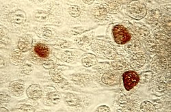
ChlamydiaTrachomatisEinschlusskorperchen.jpg
Chlamydia trachomatis inclusion bodies (brown) in a McCoy cell culture.
Scientific classification OOjs UI icon edit-ltr.svg
Domain: Bacteria
Phylum: Chlamydiota
Class: Chlamydiia
Order: Chlamydiales
Family: Chlamydiaceae
Genus: Chlamydia
Jones, Rake & Stearns 1945
Type species
Chlamydia trachomatis
(Busacca 1935) Rake 1957
Species
Synonyms
  • "Bedsonia" Meyer 1953
  • "Chlamydozoon" Moshkovskiy 1945
  • "Miyagawanella" Brumpt 1938

Chlamydia is a genus of pathogenic Gram-negative bacteria that are obligate intracellular parasites. Chlamydia infections are the most common bacterial sexually transmitted diseases in humans and are the leading cause of infectious blindness worldwide. [1]

Contents

Species include Chlamydia trachomatis (a human pathogen), Ch. suis (affects only swine), and Ch. muridarum (affects only mice and hamsters). [2] Humans mainly contract Ch. trachomatis , Ch. pneumoniae , Ch. abortus, and Ch. psittaci . [3]

Classification

Because of Chlamydia's unique developmental cycle, it was taxonomically classified in a separate order. [4] Chlamydia is part of the order Chlamydiales, family Chlamydiaceae.[ citation needed ]

In the early 1990s six species of Chlamydia were known. A major re-description of the Chlamydiales order in 1999, using the then new techniques of DNA analysis, split three of the species from the genus Chlamydia and reclassified them in the then newly created genus Chlamydophila , and also added three new species to this genus. [5] In 2001 many bacteriologists strongly objected to the reclassification, [6] although in 2006 some scientists still supported the distinctness of Chlamydophila. [7] In 2009 the validity of Chlamydophila was challenged by newer DNA analysis techniques, leading to a proposal to "reunite the Chlamydiaceae into a single genus, Chlamydia". [8] This appears to have been accepted by the community, [9] [10] bringing the number of (valid) Chlamydia species up to 9. Many probable species were subsequently isolated, but no one bothered to name them. In 2013 a 10th species was added, Ch. ibidis, known only from feral sacred ibis in France. [11] Two more species were added in 2014 (but validated 2015): Ch. avium which infects pigeons and parrots, and Ch. gallinacea infecting chickens, guinea fowl and turkeys. [3] Ch. abortus was added in 2015, and the Chlamydophila species reclassified. [6] A number of new species were originally classified as aberrant strains of Ch. psittaci [3]

Genomes

Chlamydia species have genomes around 1.0 to 1.3 megabases in length. [12] Most encode ~900 to 1050 proteins. [13]  Some species also contain a DNA plasmids or phage genomes (see Table). The elementary body contains an RNA polymerase responsible for the transcription of the DNA genome after entry into the host cell cytoplasm and the initiation of the growth cycle. Ribosomes and ribosomal subunits are found in these bodies. [14]

Ch. trachomatis MoPn Ch. trachomatis D Ch. pneumoniae AR39 Ch. pneumoniae CWL029
Size (nt)1,069,4121,042,5191,229,8531,230,230
ORFs 92489410521052
tRNAs37373838
plasmids1 (7,501 nt)1 (7,493 nt)1 ssDNA phagenone

Table 1. Genome features of selected Chlamydia species and strains. MoPn is a mouse pathogen while strain "D" is a human pathogen. About 80% of the genes in Ch. trachomatis and Ch. pneumoniae are orthologs. Adapted after Read et al. 2000 [13]

Developmental cycle

Chlamydia may be found in the form of an elementary body and a reticulate body. The elementary body is the nonreplicating infectious particle that is released when infected cells rupture. It is responsible for the bacteria's ability to spread from person to person and is analogous to a spore. The elementary body may be 0.25 to 0.30 μm in diameter. This form is covered by a rigid cell wall (hence the combining form chlamyd- in the genus name). The elementary body induces its own endocytosis upon exposure to target cells. One phagolysosome usually produces an estimated 100–1000 elementary bodies.[ citation needed ]

Chlamydia may also take the form of a reticulate body, which is in fact an intracytoplasmic form, highly involved in the process of replication and growth of these bacteria. The reticulate body is slightly larger than the elementary body and may reach up to 0.6 μm in diameter with a minimum of 0.5 μm. It does not have a cell wall. When stained with iodine, reticulate bodies appear as inclusions in the cell. The DNA genome, proteins, and ribosomes are retained in the reticulate body. This occurs as a result of the development cycle of the bacteria. The reticular body is basically the structure in which the chlamydial genome is transcribed into RNA, proteins are synthesized, and the DNA is replicated. The reticulate body divides by binary fission to form particles which, after synthesis of the outer cell wall, develop into new infectious elementary body progeny. The fusion lasts about three hours and the incubation period may be up to 21 days. After division, the reticulate body transforms back to the elementary form and is released by the cell by exocytosis. [4]

Studies on the growth cycle of Ch. trachomatis and Ch. psittaci in cell cultures in vitro reveal that the infectious elementary body (EB) develops into a noninfectious reticulate body (RB) within a cytoplasmic vacuole in the infected cell. After the elementary body enters the infected cell, an eclipse phase of 20 hours occurs while the infectious particle develops into a reticulate body. The yield of chlamydial elementary bodies is maximal 36 to 50 hours after infection. [14]

A histone like protein HctA and HctB play role in controlling the differentiation between the two cell types. The expression of HctA is tightly regulated and repressed by small non-coding RNA, IhtA until the late RB to EB re-differentiation. [15] The IhtA RNA is conserved across Chlamydia species. [16]

Pathology

Most commonly, chlamydial infections [17] do not cause symptoms. However, for men, a burning sensation when urinating is often probable. For women, odor and itching are possible symptoms. Both sexes may notice more sebum production as the infection escalates, all which produces greasy sweat, more oily complexion, and can be misdiagnosed as acne eruptions rather than the whole body's hidden fight to defend itself from an STD. All people who have engaged in sexual activity with potentially infected individuals may be offered one of several tests to diagnose the condition.[ citation needed ]

Chlamydia can be detected through culture tests or nonculture tests. The main nonculture tests include fluorescent monoclonal antibody test, enzyme immunoassay, DNA probes, rapid Chlamydia tests and leukocyte esterase tests. Whereas the first test can detect the major outer membrane protein (MOMP), the second detects a colored product converted by an enzyme linked to an antibody. The rapid Chlamydia tests use antibodies against the MOMP, the leukocyte esterase tests detect enzymes produced by leukocytes containing the bacteria in urine. [4]

Evolution

Recent phylogenetic studies have revealed that Chlamydia likely shares a common ancestor with cyanobacteria, the group containing the endosymbiont ancestor to the chloroplasts of modern plants, hence, Chlamydia retains unusual plant-like traits, both genetically and physiologically. In particular, the enzyme L,L-diaminopimelate aminotransferase, which is related to lysine production in plants, is also linked with the construction of chlamydial peptidoglycan, which is required for division. [18] The genetic encoding for the enzymes is remarkably similar in plants, cyanobacteria, and Chlamydia, demonstrating a close common ancestry. [19]

Phylogeny

16S rRNA based LTP_08_2023 [20] [21] [22] 120 marker proteins based GTDB 08-RS214 [23] [24] [25]
Chlamydia

C. muridarum Everett, Bush & Andersen 1999

C. suis Everett, Bush & Andersen 1999

C. trachomatis (Busacca 1935) Rake 1957

Chlamydia

C. trachomatis

C. muridarum

C. suis

See also

Related Research Articles

<span class="mw-page-title-main">Chlamydia</span> Sexually transmitted infection caused by the bacterium Chlamydia trachomatis

Chlamydia, or more specifically a chlamydia infection, is a sexually transmitted infection caused by the bacterium Chlamydia trachomatis. Most people who are infected have no symptoms. When symptoms do appear they may occur only several weeks after infection; the incubation period between exposure and being able to infect others is thought to be on the order of two to six weeks. Symptoms in women may include vaginal discharge or burning with urination. Symptoms in men may include discharge from the penis, burning with urination, or pain and swelling of one or both testicles. The infection can spread to the upper genital tract in women, causing pelvic inflammatory disease, which may result in future infertility or ectopic pregnancy.

<i>Chlamydia trachomatis</i> Species of bacterium

Chlamydia trachomatis, commonly known as chlamydia, is a bacterium that causes chlamydia, which can manifest in various ways, including: trachoma, lymphogranuloma venereum, nongonococcal urethritis, cervicitis, salpingitis, pelvic inflammatory disease. C. trachomatis is the most common infectious cause of blindness and the most common sexually transmitted bacterium.

<span class="mw-page-title-main">Chlamydiota</span> Phylum of bacteria

The Chlamydiota are a bacterial phylum and class whose members are remarkably diverse, including pathogens of humans and animals, symbionts of ubiquitous protozoa, and marine sediment forms not yet well understood. All of the Chlamydiota that humans have known about for many decades are obligate intracellular bacteria; in 2020 many additional Chlamydiota were discovered in ocean-floor environments, and it is not yet known whether they all have hosts. Historically it was believed that all Chlamydiota had a peptidoglycan-free cell wall, but studies in the 2010s demonstrated a detectable presence of peptidoglycan, as well as other important proteins.

<i>Chlamydia pneumoniae</i> Species of bacterium

Chlamydia pneumoniae is a species of Chlamydia, an obligate intracellular bacterium that infects humans and is a major cause of pneumonia. It was known as the Taiwan acute respiratory agent (TWAR) from the names of the two original isolates – Taiwan (TW-183) and an acute respiratory isolate designated AR-39. Briefly, it was known as Chlamydophila pneumoniae, and that name is used as an alternate in some sources. In some cases, to avoid confusion, both names are given.

<i>Chlamydia psittaci</i> Species of bacterium

Chlamydia psittaci is a lethal intracellular bacterial species that may cause endemic avian chlamydiosis, epizootic outbreaks in other mammals, and respiratory psittacosis in humans. Potential hosts include feral birds and domesticated poultry, as well as cattle, pigs, sheep, and horses. C. psittaci is transmitted by inhalation, contact, or ingestion among birds and to mammals. Psittacosis in birds and in humans often starts with flu-like symptoms and becomes a life-threatening pneumonia. Many strains remain quiescent in birds until activated by stress. Birds are excellent, highly mobile vectors for the distribution of chlamydia infection, because they feed on, and have access to, the detritus of infected animals of all sorts.

The bacterial order Chlamydiales includes only obligately intracellular bacteria that have a chlamydia-like developmental cycle of replication and at least 80% 16S rRNA or 23S rRNA gene sequence identity with other members of Chlamydiales. Chlamydiales live in animals, insects, and protozoa.

<i>Chlamydophila</i> Genus of bacteria

Chlamydophila is a controversial bacterial genus belonging to the family Chlamydiaceae.

<span class="mw-page-title-main">Chlamydiaceae</span> Family of bacteria

The Chlamydiaceae are a family of gram-negative bacteria that belongs to the phylum Chlamydiota, order Chlamydiales. Chlamydiaceae species express the family-specific lipopolysaccharide epitope αKdo-(2→8)-αKdo-(2→4)-αKdo. Chlamydiaceae ribosomal RNA genes all have at least 90% DNA sequence identity. Chlamydiaceae species have varying inclusion morphology, varying extrachromosomal plasmid content, and varying sulfadiazine resistance.

Chlamydia muridarum is an intracellular bacterial species that at one time belonged to Chlamydia trachomatis. However, C. trachomatis naturally only infects humans and C. muridarum naturally infects only members of the family Muridae.

Chlamydia suis is a member of the genus Chlamydia. C. suis has only been isolated from swine, in which it may be endemic. Glycogen has been detected in Chlamydia suis inclusions in infected swine tissues and in cell culture. C. suis is associated with conjunctivitis, enteritis and pneumonia in swine.

Parachlamydiaceae is a family of bacteria in the order Chlamydiales. Species in this family have a Chlamydia–like cycle of replication and their ribosomal RNA genes are 80–90% identical to ribosomal genes in the Chlamydiaceae. The Parachlamydiaceae naturally infect amoebae and can be grown in cultured Vero cells. The Parachlamydiaceae are not recognized by monoclonal antibodies that detect Chlamydiaceae lipopolysaccharide.

<i>Chlamydia abortus</i> Species of bacterium

Chlamydia abortus is a species in Chlamydiota that causes abortion and fetal death in mammals, including humans. Chlamydia abortus was renamed in 1999 as Chlamydophila psittaci along with all Chlamydiota except Chlamydia trachomatis. This was based on a lack of evident glycogen production and on resistance to the antibiotic sulfadiazine. In 1999 C. psittaci and C. abortus were recognized as distinct species based on differences of pathogenicity and DNA–DNA hybridization. In 2015, this new name was reverted to Chlamydia.

Chlamydia felis is a Gram-negative, obligate intracellular bacterial pathogen that infects cats. It is endemic among domestic cats worldwide, primarily causing inflammation of feline conjunctiva, rhinitis and respiratory problems. C. felis can be recovered from the stomach and reproductive tract. Zoonotic infection of humans with C. felis has been reported. Strains FP Pring and FP Cello have an extrachromosomal plasmid, whereas the FP Baker strain does not. FP Cello produces lethal disease in mice, whereas the FP Baker does not. An attenuated FP Baker strain, and an attenuated 905 strain, are used as live vaccines for cats.

Chlamydia caviae is a bacterium that can be recovered from the conjunctiva of Guinea pigs suffering from ocular inflammation and eye discharge. It is also possible to infect the genital tract of Guinea pigs with C. caviae and elicit a disease that is very similar to human Chlamydia trachomatis infection. C. caviae infects primarily the mucosal epithelium and is not invasive.

Chlamydia pecorum, also known as Chlamydophila pecorum is a species of Chlamydiaceae that originated from ruminants, such as cattle, sheep and goats. It has also infected koalas and swine. C. pecorum strains are serologically and pathogenically diverse.

Chlamydia is a sexually transmitted infection caused by the bacterium Chlamydia trachomatis.

The translocated actin-recruiting phosphoprotein (Tarp) is a protein that may mediate the invasion of epithelial cells by Chlamydia trachomatis using a type three secretion system.

Parachlamydia acanthamoebae are bacterium that fall into the category of host-associated microorganisms. This bacterium lives within free-living amoebae that are an intricate part of their reproduction. Originally named Candidatus Parachlamydia acanthamoebae, its current scientific name was introduced shortly after. This species has shown to have over eighty percent 16S rRNA gene sequencing identity with the class Chlamydiia. Parachlamydia acanthamoebae has the same family as the genus Neochlamydia with which it shares many similarities.

Chronic Mycoplasma pneumonia and Chlamydia pneumonia infections are associated with the onset and exacerbation of asthma. These microbial infections result in chronic lower airway inflammation, impaired mucociliary clearance, an increase in mucous production and eventually asthma. Furthermore, children who experience severe viral respiratory infections early in life have a high possibility of having asthma later in their childhood. These viral respiratory infections are mostly caused by respiratory syncytial virus (RSV) and human rhinovirus (HRV). Although RSV infections increase the risk of asthma in early childhood, the association between asthma and RSV decreases with increasing age. HRV on the other hand is an important cause of bronchiolitis and is strongly associated with asthma development. In children and adults with established asthma, viral upper respiratory tract infections (URIs), especially HRVs infections, can produce acute exacerbations of asthma. Thus, Chlamydia pneumoniae, Mycoplasma pneumoniae and human rhinoviruses are microbes that play a major role in non-atopic asthma.

Protochlamydia naegleriophila is an intracellular parasite of amoeba. This Protochlamydia species can be an etiological factor of pneumonia in humans when present in the amoeba P. naegleriophila, which lives in human lungs.

References

  1. Drew, W. Lawrence (2004). "Chlamydia". In Ryan, Kenneth; Ray, C. George (eds.). Sherris Medical Microbiology (PDF) (4th ed.). McGraw Hill. pp. 463–470. ISBN   978-0-8385-8529-0.
  2. Ward M. "Taxonomy diagram". Chlamydiae.com. Archived from the original on 2010-09-18. Retrieved 2008-10-28.
  3. 1 2 3 Joseph, SJ; et al. (2015), "Chlamydiaceae genomics reveals interspecies admixture and the recent evolution of Chlamydia abortus infecting lower mammalian species and humans", Genome Biol Evol, 7 (11): 3070–3084, doi:10.1093/gbe/evv201, PMC   4994753 , PMID   26507799.
  4. 1 2 3 "Chlamydia trachomatis". Archived from the original on July 2, 2010. Retrieved June 18, 2010.
  5. Everett KD, Bush RM, Andersen AA (April 1999). "Emended description of the order Chlamydiales, proposal of Parachlamydiaceae fam. nov. and Simkaniaceae fam. nov., each containing one monotypic genus, revised taxonomy of the family Chlamydiaceae, including a new genus and five new species, and standards for the identification of organisms". Int. J. Syst. Bacteriol. 49 (2): 415–40. doi: 10.1099/00207713-49-2-415 . PMID   10319462.
  6. 1 2 Parte, A.C. "Chlamydia". LPSN .
  7. Griffiths E, Ventresca MS, Gupta RS (2006). "BLAST screening of chlamydial genomes to identify signature proteins that are unique for the Chlamydiales, Chlamydiaceae, Chlamydophila and Chlamydia groups of species". BMC Genomics. 7: 14. doi: 10.1186/1471-2164-7-14 . PMC   1403754 . PMID   16436211.
  8. Stephens RS, Myers G, Eppinger M, Bavoil PM (March 2009). "Divergence without difference: phylogenetics and taxonomy of Chlamydia resolved". FEMS Immunol. Med. Microbiol. 55 (2): 115–9. doi: 10.1111/j.1574-695X.2008.00516.x . PMID   19281563.
  9. Greub, Gilbert (1 November 2010). "International Committee on Systematics of Prokaryotes Subcommittee on the taxonomy of the Chlamydiae Minutes of the inaugural closed meeting, 21 March 2009, Little Rock, AR, USA". International Journal of Systematic and Evolutionary Microbiology. 60 (11): 2691–2693. doi: 10.1099/ijs.0.028225-0 . PMID   21048221.
  10. Balsamo, G; et al. (2017), "Compendium of measures to control Chlamydia psittaci infection among humans (psittacosis) and pet birds (avian chlamydiosis), 2017" (PDF), J Avian Med Surg, 31 (3): 262–282, doi: 10.1647/217-265 , PMID   28891690, S2CID   26000244.
  11. Vorimore, Fabien; Hsia, Ru-ching; Huot-Creasy, Heather; Bastian, Suzanne; Deruyter, Lucie; Passet, Anne; Sachse, Konrad; Bavoil, Patrik; Myers, Garry; Laroucau, Karine (20 September 2013). "Isolation of a New Chlamydia species from the Feral Sacred Ibis (Threskiornis aethiopicus)- Chlamydia ibidis". PLOS ONE. 8 (9). e74823. Bibcode:2013PLoSO...874823V. doi: 10.1371/journal.pone.0074823 . PMC   3779242 . PMID   24073223.
  12. "EMBL bacterial genomes" . Retrieved January 19, 2012.
  13. 1 2 Read, T. D.; Brunham, R. C.; Shen, C.; Gill, S. R.; Heidelberg, J. F.; White, O.; Hickey, E. K.; Peterson, J.; Utterback, T. (2000-03-15). "Genome sequences of Chlamydia trachomatis MoPn and Chlamydia pneumoniae AR39". Nucleic Acids Research. 28 (6): 1397–1406. doi:10.1093/nar/28.6.1397. ISSN   1362-4962. PMC   111046 . PMID   10684935.
  14. 1 2 Becker, Yechiel (1996). "Chlamydia". In Baron, S. (ed.). Medical Microbiology (4th ed.). University of Texas Medical Branch at Galveston. ISBN   0-9631172-1-1. PMID   21413294.
  15. Grieshaber, NA; Grieshaber, SS; Fisher, ER; Hackstadt, T (2006). "A small RNA inhibits translation of the histone-like protein Hc1 in Chlamydia trachomatis". Mol. Microbiol. 59 (2): 541–50. doi:10.1111/j.1365-2958.2005.04949.x. PMID   16390448. S2CID   11872982.
  16. Tattersall, J; Rao, GV; Runac, J; Hackstadt, T; Grieshaber, SS; Grieshaber, NA (2012). "Translation inhibition of the developmental cycle protein HctA by the small RNA IhtA is conserved across Chlamydia". PLOS ONE. 7 (10): e47439. Bibcode:2012PLoSO...747439T. doi: 10.1371/journal.pone.0047439 . PMC   3469542 . PMID   23071807.
  17. "Chlamydia protection" . Retrieved August 1, 2010.
  18. Liechti, G. W.; Kuru, E.; Hall, E.; Kalinda, A.; Brun, Y. V.; VanNieuwenhze, M.; Maurelli, A. T. (February 2014). "A new metabolic cell-wall labelling method reveals peptidoglycan in Chlamydia trachomatis". Nature. 506 (7489): 507–510. doi:10.1038/nature12892. ISSN   1476-4687. PMC   3997218 . PMID   24336210.
  19. McCoy AJ, Adams NE, Hudson AO, Gilvarg C, Leustek T, Maurelli AT (2006). "L,L-diaminopimelate aminotransferase, a trans-kingdom enzyme shared by Chlamydia and plants for synthesis of diaminopimelate/lysine". Proc. Natl. Acad. Sci. U.S.A. 103 (47): 17909–14. Bibcode:2006PNAS..10317909M. doi: 10.1073/pnas.0608643103 . PMC   1693846 . PMID   17093042.
  20. "The LTP" . Retrieved 20 November 2023.
  21. "LTP_all tree in newick format" . Retrieved 20 November 2023.
  22. "LTP_08_2023 Release Notes" (PDF). Retrieved 20 November 2023.
  23. "GTDB release 08-RS214". Genome Taxonomy Database . Retrieved 10 May 2023.
  24. "bac120_r214.sp_label". Genome Taxonomy Database . Retrieved 10 May 2023.
  25. "Taxon History". Genome Taxonomy Database . Retrieved 10 May 2023.

Further reading