The central nervous system (CNS) is the part of the nervous system consisting primarily of the brain and spinal cord. The CNS is so named because it integrates the received information and coordinates and influences the activity of all parts of the bodies of bilaterally symmetric animals—i.e., all multicellular animals except sponges and radially symmetric animals such as jellyfish—and it contains the majority of the nervous system. The CNS also includes the retina and the optic nerve, as well as the olfactory nerves and olfactory epithelium as parts of the CNS, synapsing directly on brain tissue without intermediate ganglia. As such, the olfactory epithelium is the only central nervous tissue in direct contact with the environment, which opens up for therapeutic treatments. The CNS is contained within the dorsal body cavity, with the brain housed in the cranial cavity and the spinal cord in the spinal canal. In vertebrates, the brain is protected by the skull, while the spinal cord is protected by the vertebrae. The brain and spinal cord are both enclosed in the meninges. Within the CNS, the interneuronal space is filled with a large amount of supporting non-nervous cells called neuroglia or glia from the Greek for "glue".

The limbic system, also known as the paleomammalian cortex, is a set of brain structures located on both sides of the thalamus, immediately beneath the medial temporal lobe of the cerebrum primarily in the forebrain.

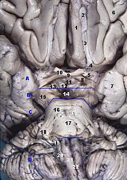
The olfactory bulb is a neural structure of the vertebrate forebrain involved in olfaction, the sense of smell. It sends olfactory information to be further processed in the amygdala, the orbitofrontal cortex (OFC) and the hippocampus where it plays a role in emotion, memory and learning. The bulb is divided into two distinct structures: the main olfactory bulb and the accessory olfactory bulb. The main olfactory bulb connects to the amygdala via the piriform cortex of the primary olfactory cortex and directly projects from the main olfactory bulb to specific amygdala areas. The accessory olfactory bulb resides on the dorsal-posterior region of the main olfactory bulb and forms a parallel pathway. Destruction of the olfactory bulb results in ipsilateral anosmia, while irritative lesions of the uncus can result in olfactory and gustatory hallucinations.

The cerebrum or telencephalon is the largest part of the brain containing the cerebral cortex, as well as several subcortical structures, including the hippocampus, basal ganglia, and olfactory bulb. In the human brain, the cerebrum is the uppermost region of the central nervous system. The cerebrum develops prenatally from the forebrain (prosencephalon). In mammals, the dorsal telencephalon, or pallium, develops into the cerebral cortex, and the ventral telencephalon, or subpallium, becomes the basal ganglia. The cerebrum is also divided into approximately symmetric left and right cerebral hemispheres.

In animal anatomy, the rhinencephalon, also called the smell-brain or olfactory brain, is a part of the brain involved with smell. It forms the paleocortex and is rudimentary in the human brain.

The olfactory system, or sense of smell, is the sensory system used for smelling (olfaction). Olfaction is one of the special senses, that have directly associated specific organs. Most mammals and reptiles have a main olfactory system and an accessory olfactory system. The main olfactory system detects airborne substances, while the accessory system senses fluid-phase stimuli.

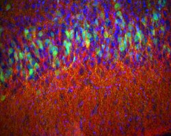
Mitral cells are neurons that are part of the olfactory system. They are located in the olfactory bulb in the mammalian central nervous system. They receive information from the axons of olfactory receptor neurons, forming synapses in neuropils called glomeruli. Axons of the mitral cells transfer information to a number of areas in the brain, including the piriform cortex, entorhinal cortex, and amygdala. Mitral cells receive excitatory input from olfactory sensory neurons and external tufted cells on their primary dendrites, whereas inhibitory input arises either from granule cells onto their lateral dendrites and soma or from periglomerular cells onto their dendritic tuft. Mitral cells together with tufted cells form an obligatory relay for all olfactory information entering from the olfactory nerve. Mitral cell output is not a passive reflection of their input from the olfactory nerve. In mice, each mitral cell sends a single primary dendrite into a glomerulus receiving input from a population of olfactory sensory neurons expressing identical olfactory receptor proteins, yet the odor responsiveness of the 20-40 mitral cells connected to a single glomerulus is not identical to the tuning curve of the input cells, and also differs between sister mitral cells. Odorant response properties of individual neurons in an olfactory glomerular module. The exact type of processing that mitral cells perform with their inputs is still a matter of controversy. One prominent hypothesis is that mitral cells encode the strength of an olfactory input into their firing phases relative to the sniff cycle. A second hypothesis is that the olfactory bulb network acts as a dynamical system that decorrelates to differentiate between representations of highly similar odorants over time. Support for the second hypothesis comes primarily from research in zebrafish.

The olfactory tubercle (OT), also known as the tuberculum olfactorium, is a multi-sensory processing center that is contained within the olfactory cortex and ventral striatum and plays a role in reward cognition. The OT has also been shown to play a role in locomotor and attentional behaviors, particularly in relation to social and sensory responsiveness, and it may be necessary for behavioral flexibility. The OT is interconnected with numerous brain regions, especially the sensory, arousal, and reward centers, thus making it a potentially critical interface between processing of sensory information and the subsequent behavioral responses.
The amygdalofugal pathway is one of the three major efferent pathways of the amygdala, meaning that it is one of the three principal pathways by which fibers leave the amygdala. It leads from the basolateral nucleus and central nucleus of the amygdala. The amygdala is a limbic structure in the medial temporal lobe of the brain. The other main efferent pathways from the amygdala are the stria terminalis and anterior commissure.
The olfactory tract is a bilateral bundle of afferent nerve fibers from the mitral and tufted cells of the olfactory bulb that connects to several target regions in the brain, including the piriform cortex, amygdala, and entorhinal cortex. It is a narrow white band, triangular on coronal section, the apex being directed upward.
The perirhinal cortex is a cortical region in the medial temporal lobe that is made up of Brodmann areas 35 and 36. It receives highly processed sensory information from all sensory regions, and is generally accepted to be an important region for memory. It is bordered caudally by postrhinal cortex or parahippocampal cortex and ventrally and medially by entorhinal cortex.

The anterior olfactory nucleus is a portion of the forebrain of vertebrates.

In anatomy of animals, the paleocortex, or paleopallium, is a region within the telencephalon in the vertebrate brain. This type of cortical tissue consists of three cortical laminae. In comparison, the neocortex has six layers and the archicortex has three or four layers. Because the number of laminae that compose a type of cortical tissue seems to be directly proportional to both the information-processing capabilities of that tissue and its phylogenetic age, paleocortex is thought to be an intermediate between the archicortex and the neocortex in both aspects.

Homeobox protein EMX1 is a protein that in humans is encoded by the EMX1 gene. The transcribed EMX1 gene is a member of the EMX family of transcription factors. The EMX1 gene, along with its family members, are expressed in the developing cerebrum. Emx1 plays a role in specification of positional identity, the proliferation of neural stem cells, differentiation of layer-specific neuronal phenotypes and commitment to a neuronal or glial cell fate.

The ganglionic eminence (GE) is a transitory structure in the development of the nervous system that guides cell and axon migration. It is present in the embryonic and fetal stages of neural development found between the thalamus and caudate nucleus.

In neuroanatomy, pallium refers to the layers of grey and white matter that cover the upper surface of the cerebrum in vertebrates. The non-pallial part of the telencephalon builds the subpallium. In basal vertebrates the pallium is a relatively simple three-layered structure, encompassing 3–4 histogenetically distinct domains, plus the olfactory bulb.

Olfaction, or the sense of smell, is the process of creating the perception of smell. It occurs when an odor binds to a receptor within the nose, transmitting a signal through the olfactory system. Olfaction has many purposes, including detecting hazards, pheromones, and plays a role in taste.
Periamygdaloid cortex is a portion of the rhinencephalon consisting of paleocortex. It is a cortical-like nucleus of the amygdaloid complex. Though considered a nucleus, the periamygdalar area is more commonly associated with cortex due to its layered structure and location on the outer surface of the brain.

Septum Verum is a region in the lower medial part of the telencephalon that separates the two cerebral hemispheres. The human septum consists of two parts: the septum pellucidum, a thin membrane consisting of white matter and glial cells that separate the lateral ventricles, and the lower, precommisural septum verum, which consists of nuclei and grey matter. The term is sometimes used synonymously with Area Septalis, to refer to the precommisural part of the lower base of the telencephalon. The Septum verum contains the septal nuclei, which are usually considered part of the limbic system.