Cutaneous squamous-cell carcinoma

Last updated
Cutaneous squamous-cell carcinoma
Other namesSquamous-cell carcinoma of the skin, squamous-cell skin cancer, epidermoid carcinoma, squamous-cell epithelioma of the skin
Squamous Cell Carcinoma.jpg
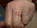
Cutaneous squamous-cell carcinoma tends to arise from actinic keratoses (premalignant lesions); surface is usually scaly and often ulcerates (as shown here).
Specialty Dermatology, plastic surgery, otorhinolaryngology
Symptoms Hard lump with a scaly top or ulceration. [1]
Risk factors Ultraviolet radiation, actinic keratosis, tobacco smoking, lighter skin, arsenic exposure, radiotherapy, poor immune system function, HPV infection [2]
Diagnostic method Tissue biopsy [2] [3]
Differential diagnosis Keratoacanthoma, actinic keratosis, melanoma, warts, basal cell cancer [4]
PreventionDecreased UV radiation exposure, sunscreen [5] [6]
TreatmentSurgical removal, radiotherapy, chemotherapy, immunotherapy [2] [7]
Prognosis Usually good [5]
Frequency2.2 million (2015) [8]
Deaths51,900 (2015) [9]

Cutaneous squamous-cell carcinoma (cSCC), also known as squamous-cell carcinoma of the skin or squamous-cell skin cancer, is one of the three principal types of skin cancer, alongside basal-cell carcinoma and melanoma. [10] cSCC typically presents as a hard lump with a scaly surface, though it may also present as an ulcer. [1] Onset and development often occurs over several months. [4] Compared to basal cell carcinoma, cSCC is more likely to spread to distant areas. [11] When confined to the epidermis, the outermost layer of the skin, the pre-invasive or in situ form of cSCC is termed Bowen's disease. [12] [13]

Contents

The most significant risk factor for cSCC is extensive lifetime exposure to ultraviolet radiation from sunlight. [2] Additional risk factors include prior scars, chronic wounds, actinic keratosis, lighter skin susceptible to sunburn, Bowen's disease, exposure to arsenic, radiation therapy, tobacco smoking, poor immune system function, previous basal cell carcinoma, and HPV infection. [2] [14] [15] The risk associated with UV radiation correlates with cumulative exposure rather than early-life exposure. [16] Tanning beds have emerged as a significant source of UV radiation. Genetic predispositions, such as xeroderma pigmentosum [17] and certain forms of epidermolysis bullosa, [18] also increase susceptibility to cSCC. The condition originates from squamous cells located in the skin's upper layers. [19] Diagnosis typically relies on skin examination, and is confirmed through skin biopsy. [2] [3]

Research, both in vivo and in vitro, indicates a crucial role for the upregulation of FGFR2, part of the fibroblast growth factor receptor immunoglobin family, in cSCC cell progression. [20] Mutations in the TPL2 gene leads to overexpression of FGFR2, which activates the mTORC1 and AKT pathways in primary and metastatic cSCC cell lines. Utilization of a "pan FGFR inhibitor" has shown to reduce cell migration and proliferation in cSCC in vitro studies. [20]

Preventive measures against cSCC include minimizing exposure to ultraviolet radiation and the use of sunscreen. [5] [6] Surgical removal is the typical treatment method, [2] employing simple excision for minor cases or Mohs surgery for more extensive instances. [2] Other options include cryotherapy and radiation therapy. [7] For cases with distant metastasis, chemotherapy or biologic therapy may be employed. [7]

As of 2015, approximately 2.2 million individuals globally were living with cSCC at any given time, [8] constituting about 20% of all skin cancer cases. [21] In the United States, approximately 12% of males and 7% of females are diagnosed with cSCC at some point in their lives. [2] While prognosis remains favorable in the absence of metastasis, upon distant spread the five-year survival rate is markedly reduced to ~34%. [4] [5] In 2015, global deaths attributed to cSCC numbered around 52,000. [9] The average age at diagnosis is approximately 66 years. [4] Following successful treatment of an initial cSCC lesion, there is a substantial risk of developing subsequent lesions. [2]

Signs and symptoms

Cutaneous squamous-cell carcinoma Squamous Cell Carcinoma.png
Cutaneous squamous-cell carcinoma

SCC of the skin begins as a small nodule and as it enlarges the center becomes necrotic and sloughs and the nodule turns into an ulcer, and generally are developed from an actinic keratosis. Once keratinocytes begin to grow uncontrollably, they have the potential to become cancerous and produce cutaneous squamous-cell carcinoma. [22]

Spread

Causes

Cutaneous squamous-cell carcinoma is the second-most common cancer of the skin (after basal-cell carcinoma, but more common than melanoma). It usually occurs in areas exposed to the sun. Sunlight exposure and immunosuppression are risk factors for SCC of the skin, with chronic sun exposure being the strongest environmental risk factor. [26] There is a risk of metastasis starting more than 10 years[ citation needed ] after diagnosable appearance of squamous-cell carcinoma, but the risk is low,[ specify ] though much[ specify ] higher than with basal-cell carcinoma. Squamous-cell cancers of the lip and ears have high rates of local recurrence and distant metastasis. [27] In a recent study, it has also been shown that the deletion or severe down-regulation of a gene titled Tpl2 (tumor progression locus 2) may be involved in the progression of normal keratinocytes into becoming squamous-cell carcinoma. [28]

cSCC represents about 20% of the non-melanoma skin cancers; 80-90% of cSCCs with metastatic potential are located on the head and neck. [29]

Tobacco smoking also increases the risk for cutaneous squamous-cell carcinoma. [14] [30]

The vast majority of cSCC cases are located on exposed skin, and are often the result of ultraviolet exposure. cSCC usually occurs on portions of the body commonly exposed to the sun; the face, ears, neck, hands, or arms. The primary sign is a growing bump that may have a rough, scaly surface, and flat, reddish patches. Unlike basal-cell carcinoma, cSCC carries a higher risk of metastasis than does basal-cell carcinoma, and may spread to the regional lymph nodes, [31]

Erythroplasia of Queyrat (SCC in situ of the glans or prepuce in males, [32] M [33] :733 [34] :656 [35] or the vulva in females. [36] ) may be induced by human papilloma virus. [37] It is reported to occur in the corneoscleral limbus. [38] Erythroplasia of Queyrat may also occur on the anal mucosa or the oral mucosa. [39]

Genetically, cSCC tumors harbor high frequencies of NOTCH and p53 mutations as well as less frequent alterations in histone acetyltransferase EP300, subunit of the SWI/SNF chromatin remodeling complex PBRM1, DNA-repair deubiquitinase USP28, and NF-κB signaling regulator CHUK. [40]

Immunosuppression

People who have received solid organ transplants are at a significantly increased risk of developing squamous-cell carcinoma due to the use of chronic immunosuppressive medication. [41] While the risk of developing all skin cancers increases with these medications, this effect is particularly severe for cSCC, with hazard ratios as high as 250 being reported, versus 40 for basal cell carcinoma. [42] The incidence of cSCC development increases with time posttransplant. [43] Heart and lung transplant recipients are at the highest risk of developing cSCC due to more intensive immunosuppressive medications used. [44]

Cutaneous squamous-cell carcinoma in individuals on immunotherapy or who have lymphoproliferative disorders (e.g. leukemia) tend to be much more aggressive, regardless of their location. [45] The risk of cSCC, and non-melanoma skin cancers generally, varies with the immunosuppressive drug regimen chosen. The risk is greatest with calcineurin inhibitors like cyclosporine and tacrolimus, and least with mTOR inhibitors, such as sirolimus and everolimus. The antimetabolites azathioprine and mycophenolic acid have an intermediate risk profile. [46]

Diagnosis

Diagnosis is confirmed via skin biopsy of the tissue or tissues suspected to be affected by SCC. The pathological appearance of a squamous-cell cancer varies with the depth of the biopsy. For that reason, a biopsy including the subcutaneous tissue and basilar epithelium, to the surface is necessary for correct diagnosis. The performance of a shave biopsy (see skin biopsy) might not acquire enough information for a diagnosis. An inadequate biopsy might be read as actinic keratosis with follicular involvement. A deeper biopsy down to the dermis or subcutaneous tissue might reveal the true cancer. An excision biopsy is ideal, but not practical in most cases. An incisional or punch biopsy is preferred. A shave biopsy is least ideal, especially if only the superficial portion is acquired.[ citation needed ]

Histological characteristics

Histopathologically, the epidermis in cSCC in situ (Bowen's disease) will show hyperkeratosis and parakeratosis. There will also be marked acanthosis with elongation and thickening of the rete ridges. These changes will overly keratinocytic cells which are often highly atypical and may in fact have a more unusual appearance than invasive cSCC. The atypia spans the full thickness of the epidermis, with the keratinocytes demonstrating intense mitotic activity, pleomorphism, and greatly enlarged nuclei. They will also show a loss of maturity and polarity, giving the epidermis a disordered or "windblown" appearance.[ citation needed ]

Two types of multinucleated cells may be seen: the first will present as a multinucleated giant cell, and the second will appear as a dyskeratotic cell engulfed in the cytoplasm of a keratinocyte. Occasionally, cells of the upper epidermis will undergo vacuolization, demonstrating an abundant and strongly eosinophilic cytoplasm. There may be a mild to moderate lymphohistiocytic infiltrate detected in the upper dermis. [12]

In situ disease

Bowen's disease is essentially equivalent to and used interchangeably with cSCC in situ, when not having invaded through the basement membrane. [12] Depending on source, it is classified as precancerous [13] or cSCC in situ (technically cancerous but non-invasive). [47] [48] In cSCC in situ (Bowen's disease), atypical squamous cells proliferate through the whole thickness of the epidermis. [12] The entire tumor is confined to the epidermis and does not invade into the dermis. [12] The cells are often highly atypical under the microscope, and may in fact look more unusual than the cells of some invasive squamous-cell carcinomas. [12]

Erythroplasia of Queyrat is a particular type of Bowen's disease that can arise on the glans or prepuce in males, [32] [33] :733 [34] :656 [35] and the vulva in females. [36] It mainly occurs in uncircumcised males, [36] [49] over the age of 40. [39]

Invasive disease

In invasive cSCC, tumor cells infiltrate through the basement membrane. The infiltrate can be somewhat difficult to detect in the early stages of invasion: however, additional indicators such as full thickness epidermal atypia and the involvement of hair follicles can be used to facilitate the diagnosis. Later stages of invasion are characterized by the formation of nests of atypical tumor cells in the dermis, often with a corresponding inflammatory infiltrate. [12]

Degree of differentiation

Prevention

Appropriate sun-protective clothing, use of broad-spectrum (UVA/UVB) sunscreen with at least SPF 50, and avoidance of intense sun exposure may prevent skin cancer. [50] A 2016 review of sunscreen for preventing cutaneous squamous-cell carcinoma found insufficient evidence to demonstrate whether it was effective. [51]

Management

Most cutaneous squamous-cell carcinomas are removed with surgery. A few selected cases are treated with topical medication. Surgical excision with a free margin of healthy tissue is a frequent treatment modality. Radiotherapy, given as external beam radiotherapy or as brachytherapy (internal radiotherapy), can also be used to treat cSCC. There is little evidence comparing the effectiveness of different treatments for non-metastatic cSCC. [52]

Mohs surgery is frequently utilized; considered the treatment of choice for squamous-cell carcinoma of the skin, physicians have also utilized the method for the treatment of squamous-cell carcinoma of the mouth, throat, and neck. [53] An equivalent method of the CCPDMA standards can be utilized by a pathologist in the absence of a Mohs-trained physician. Radiation therapy is often used afterward in high risk cancer or patient types. [54] Radiation or radiotherapy can also be a standalone option in treating cSCC. As a non-invasive option brachytherapy serves a painless possibility to treat in particular but not only difficult to operate areas like the earlobes or genitals. An example of this kind of therapy is the high-dose brachytherapy Rhenium-SCT which makes use of the beta rays emitting property of rhenium-188. The radiation source is enclosed in a compound which is applied to a thin protection foile directly over the lesion. This way the radiation source can be applied to complex locations and minimize radiation to healthy tissue. [55]

After removal of the cancer, closure of the skin for patients with a decreased amount of skin laxity involves a split-thickness skin graft. A donor site is chosen and enough skin is removed so that the donor site can heal on its own. Only the epidermis and a partial amount of dermis is taken from the donor site which allows the donor site to heal. Skin can be harvested using either a mechanical dermatome or Humby knife. [56]

Electrodessication and curettage (EDC) can be done on selected squamous-cell carcinoma of the skin. In areas where cSCC is known to be non-aggressive, and where the patient is not immunosuppressed, EDC[ clarification needed ] can be performed with good to adequate cure rate. [57]

Treatment options for cSCC in situ (Bowen's disease) include photodynamic therapy with 5-aminolevulinic acid, cryotherapy, topical 5-fluorouracil or imiquimod, and excision. A meta-analysis showed evidence that PDT is more effective than cryotherapy and has better cosmetic outcomes. There is generally a lack of evidence comparing the effectiveness of all treatment options. [13]

High-risk squamous-cell carcinoma, as defined by that occurring around the eye, ear, or nose, is of large size, is poorly differentiated, and grows rapidly, requires more aggressive, multidisciplinary management.

Nodal spread:

  1. Surgical block dissection if palpable nodes or in cases of Marjolin's ulcers but the benefit of prophylactic block lymph node dissection with Marjolin's ulcers is not proven.
  2. Radiotherapy
  3. Adjuvant therapy may be considered in those with high-risk cSCC even in the absence of evidence for local metastasis. Imiquimod (Aldara) has been used with success for squamous-cell carcinoma in situ of the skin and the penis, but the morbidity and discomfort of the treatment is severe. An advantage is the cosmetic result: after treatment, the skin resembles normal skin without the usual scarring and morbidity associated with standard excision. Imiquimod is not FDA-approved for any squamous-cell carcinoma.

In general, squamous-cell carcinomas have a high risk of local recurrence, and up to 50% do recur. [58] Frequent skin exams with a dermatologist is recommended after treatment.

Prognosis

The long-term outcome of squamous-cell carcinoma is dependent upon several factors: the sub-type of the carcinoma, available treatments, location and severity, and various patient health-related variables (accompanying diseases, age, etc.). Generally, the long-term outcome is positive, with a metastasis rate of 1.9-5.2% and a mortality rate of 1.5-3.4%. [25] [59] [60]

When it does metastasize, the most commonly involved organs are the lungs, brain, bone and other skin locations. [61] Squamous-cell carcinoma occurring in immunosuppressed people (such as those with organ transplant, human immunodeficiency virus infection, or chronic lymphocytic leukemia) the risk of developing cSCC and having metastasis is much higher than the general population. [62]

One study found squamous-cell carcinoma of the penis had a much greater rate of mortality than some other forms of squamous-cell carcinoma, that is, about 23%, [63] although this relatively high mortality rate may be associated with possibly latent diagnosis of the disease due to patients avoiding genital exams until the symptoms are debilitating, or refusal to submit to a possibly scarring operation upon the genitalia.

Epidemiology

Age-standardized death from melanoma and other skin cancers per 100,000 inhabitants in 2004.
.mw-parser-output .div-col{margin-top:0.3em;column-width:30em}.mw-parser-output .div-col-small{font-size:90%}.mw-parser-output .div-col-rules{column-rule:1px solid #aaa}.mw-parser-output .div-col dl,.mw-parser-output .div-col ol,.mw-parser-output .div-col ul{margin-top:0}.mw-parser-output .div-col li,.mw-parser-output .div-col dd{page-break-inside:avoid;break-inside:avoid-column}
.mw-parser-output .legend{page-break-inside:avoid;break-inside:avoid-column}.mw-parser-output .legend-color{display:inline-block;min-width:1.25em;height:1.25em;line-height:1.25;margin:1px 0;text-align:center;border:1px solid black;background-color:transparent;color:black}.mw-parser-output .legend-text{}
no data
less than 0.7
0.7-1.4
1.4-2.1
2.1-2.8
2.8-3.5
3.5-4.2
4.2-4.9
4.9-5.6
5.6-6.3
6.3-7
7-7.7
more than 7.7 Melanoma and other skin cancers world map - Death - WHO2004.svg
Age-standardized death from melanoma and other skin cancers per 100,000 inhabitants in 2004.
  no data
  less than 0.7
  0.7–1.4
  1.4–2.1
  2.1–2.8
  2.8–3.5
  3.5–4.2
  4.2–4.9
  4.9–5.6
  5.6–6.3
  6.3–7
  7–7.7
  more than 7.7

The incidence of cutaneous squamous-cell carcinoma continues to rise around the world. This is theorized to be due to several factors; including an aging population, a greater incidence of those who are immunocompromised and the increasing use of tanning beds. [25]

A recent study estimated that there are between 180,000 and 400,000 cases of cSCC in the United States in 2013. [65] Risk factors for cSCC varies with age, gender, race, geography, and genetics. The incidence of cSCC increases with age and with those 75 years or older eing at a 5-10 times increased risk of developing cSCC as compared with those who are younger than 55 years old. [25] Males are affected with cSCC at a ratio of 3:1 in comparison to females. [25] Those who have light skin, red or blonde hair and light colored eyes are also at increased risk. [25]

Squamous-cell carcinoma of the skin can be found on all areas of the body but is most common on frequently sun-exposed areas, such as the face, legs and arms. [66] Solid organ transplant recipients (heart, lung, liver, pancreas, among others) are also at a heightened risk of developing aggressive, high-risk cSCC. There are also a few rare congenital diseases predisposed to cutaneous malignancy. In certain geographic locations, exposure to arsenic in well water [67] or from industrial sources may significantly increase the risk of cSCC. [26]

Additional images

See also

Related Research Articles

<span class="mw-page-title-main">Skin cancer</span> Medical condition involving uncontrolled growth of skin cells

Skin cancers are cancers that arise from the skin. They are due to the development of abnormal cells that have the ability to invade or spread to other parts of the body. Skin cancer is the most commonly diagnosed form of cancer in humans. There are three main types of skin cancers: basal-cell skin cancer (BCC), squamous-cell skin cancer (SCC) and melanoma. The first two, along with a number of less common skin cancers, are known as nonmelanoma skin cancer (NMSC). Basal-cell cancer grows slowly and can damage the tissue around it but is unlikely to spread to distant areas or result in death. It often appears as a painless raised area of skin that may be shiny with small blood vessels running over it or may present as a raised area with an ulcer. Squamous-cell skin cancer is more likely to spread. It usually presents as a hard lump with a scaly top but may also form an ulcer. Melanomas are the most aggressive. Signs include a mole that has changed in size, shape, color, has irregular edges, has more than one color, is itchy or bleeds.

Penile cancer, or penile carcinoma, is a cancer that develops in the skin or tissues of the penis. Symptoms may include abnormal growth, an ulcer or sore on the skin of the penis, and bleeding or foul smelling discharge.

<span class="mw-page-title-main">Basal-cell carcinoma</span> Most common type of skin cancer

Basal-cell carcinoma (BCC), also known as basal-cell cancer, basalioma or rodent ulcer, is the most common type of skin cancer. It often appears as a painless raised area of skin, which may be shiny with small blood vessels running over it. It may also present as a raised area with ulceration. Basal-cell cancer grows slowly and can damage the tissue around it, but it is unlikely to spread to distant areas or result in death.

<span class="mw-page-title-main">Melanoma</span> Cancer originating in melanocytes

Melanoma is the most dangerous type of skin cancer; it develops from the melanin-producing cells known as melanocytes. It typically occurs in the skin, but may rarely occur in the mouth, intestines, or eye. In women, melanomas most commonly occur on the legs; while in men, on the back. Melanoma is frequently referred to as malignant melanoma. However, the medical community stresses that there is no such thing as a 'benign melanoma' and recommends that the term 'malignant melanoma' should be avoided as redundant.

Carcinoma <i>in situ</i> Pre-cancerous abnormal growth of tissue

Carcinoma in situ (CIS) is a group of abnormal cells. While they are a form of neoplasm, there is disagreement over whether CIS should be classified as cancer. This controversy also depends on the exact CIS in question. Some authors do not classify them as cancer, however, recognizing that they can potentially become cancer. Others classify certain types as a non-invasive form of cancer. The term "pre-cancer" has also been used.

<span class="mw-page-title-main">Seborrheic keratosis</span> Skin disease

A seborrheic keratosis is a non-cancerous (benign) skin tumour that originates from cells, namely keratinocytes, in the outer layer of the skin called the epidermis. Like liver spots, seborrheic keratoses are seen more often as people age.

<span class="mw-page-title-main">Actinic keratosis</span> Skin disorder

Actinic keratosis (AK), sometimes called solar keratosis or senile keratosis, is a pre-cancerous area of thick, scaly, or crusty skin. Actinic keratosis is a disorder of epidermal keratinocytes that is induced by ultraviolet (UV) light exposure. These growths are more common in fair-skinned people and those who are frequently in the sun. They are believed to form when skin gets damaged by UV radiation from the sun or indoor tanning beds, usually over the course of decades. Given their pre-cancerous nature, if left untreated, they may turn into a type of skin cancer called squamous cell carcinoma. Untreated lesions have up to a 20% risk of progression to squamous cell carcinoma, so treatment by a dermatologist is recommended.

<span class="mw-page-title-main">Invasive carcinoma of no special type</span> Medical condition

Invasive carcinoma of no special type, invasive breast carcinoma of no special type (IBC-NST), invasive ductal carcinoma (IDC), infiltrating ductal carcinoma (IDC) or invasive ductal carcinoma, not otherwise specified (NOS) is a disease. For international audiences this article will use "invasive carcinoma NST" because it is the preferred term of the World Health Organization (WHO).

<span class="mw-page-title-main">Merkel-cell carcinoma</span> Rare and highly aggressive skin cancer

Merkel-cell carcinoma (MCC) is a rare and aggressive skin cancer occurring in about three people per million members of the population. It is also known as cutaneous APUDoma, primary neuroendocrine carcinoma of the skin, primary small cell carcinoma of the skin, and trabecular carcinoma of the skin. Factors involved in the development of MCC include the Merkel cell polyomavirus, a weakened immune system, and exposure to ultraviolet radiation. Merkel-cell carcinoma usually arises on the head, neck, and extremities, as well as in the perianal region and on the eyelid. It is more common in people over sixty years old, Caucasian people, and males. MCC is less common in children.

<span class="mw-page-title-main">Vulvar cancer</span> Cancer involving the vulva

Vulvar cancer is a cancer of the vulva, the outer portion of the female genitals. It most commonly affects the labia majora. Less often, the labia minora, clitoris, or Bartholin's glands are affected. Symptoms include a lump, itchiness, changes in the skin, or bleeding from the vulva.

<span class="mw-page-title-main">Atypical fibroxanthoma</span> Medical condition

Atypical fibroxanthoma (AFX) of the skin is a low-grade malignancy related to malignant fibrous histiocytoma, which it resembles histologically. Atypical fibroxanthoma manifests as a hard, pink or red papule or nodule that grows over the course of several months and may bleed or ulcerate. They typically occur on the head and neck. Atypical fibroxanthoma is usually asymptomatic.

<span class="mw-page-title-main">Sebaceous carcinoma</span> Medical condition

Sebaceous carcinoma, also known as sebaceous gland carcinoma (SGc), sebaceous cell carcinoma, and meibomian gland carcinoma is an uncommon malignant cutaneous tumor. Most are typically about 1.4 cm at presentation. SGc originates from sebaceous glands in the skin and, therefore, may originate anywhere in the body where these glands are found. SGc can be divided into 2 types: periocular and extraocular. The periocular region is rich in sebaceous glands making it a common site of origin. The cause of these lesions in the vast majority of cases is unknown. Occasional cases may be associated with Muir-Torre syndrome. SGc accounts for approximately 0.7% of all skin cancers, and the incidence of SGc is highest in Caucasian, Asian, and Indian populations. Due to the rarity of this tumor and variability in clinical and histological presentation, SGc is often misdiagnosed as an inflammatory condition or a more common neoplasm. SGc is commonly treated with wide local excision or Mohs micrographic surgery, and the relative survival rates at 5 and 10 years are 92.72 and 86.98%, respectively.

<span class="mw-page-title-main">Poroma</span>

Poromas are rare, benign, cutaneous adnexal tumors. Cutaneous adnexal tumors are a group of skin tumors consisting of tissues that have differentiated towards one or more of the four primary adnexal structures found in normal skin: hair follicles, sebaceous sweat glands, apocrine sweat glands, and eccrine sweat glands. Poromas are eccrine or apocrine sweat gland tumors derived from the cells in the terminal portion of these glands' ducts. This part of the sweat gland duct is termed the acrosyringium and had led to grouping poromas in the acrospiroma class of skin tumors. Here, poromas are regarded as distinct sweat gland tumors that differ from other sweat gland tumors by their characteristic clinical presentations, microscopic histopathology, and the genetic mutations that their neoplastic cells have recently been found to carry.

Porocarcinoma (PCA) is a rare form of skin cancer that develops in eccrine sweat glands, i.e. the body's widely distributed major type of sweat glands, as opposed to the apocrine sweat glands which are located primarily in the armpits and perineal area. This cancer typically develops in individuals as a single cutaneous tumor in the intraepidermal spiral part of these sweat glands' ducts at or near to where they open on the skin's surface. PCA tumors are classified as one form of the cutaneous adnexal tumors; in a study of 2,205 cases, PCA was the most common (11.8%) form of these tumors.

Skin cancer, or neoplasia, is the most common type of cancer diagnosed in horses, accounting for 45 to 80% of all cancers diagnosed. Sarcoids are the most common type of skin neoplasm and are the most common type of cancer overall in horses. Squamous-cell carcinoma is the second-most prevalent skin cancer, followed by melanoma. Squamous-cell carcinoma and melanoma usually occur in horses greater than 9-years-old, while sarcoids commonly affect horses 3 to 6 years old. Surgical biopsy is the method of choice for diagnosis of most equine skin cancers, but is contraindicated for cases of sarcoids. Prognosis and treatment effectiveness varies based on type of cancer, degree of local tissue destruction, evidence of spread to other organs (metastasis) and location of the tumor. Not all cancers metastasize and some can be cured or mitigated by surgical removal of the cancerous tissue or through use of chemotherapeutic drugs.

Conjunctival squamous cell carcinoma and corneal intraepithelial neoplasia comprise ocular surface squamous neoplasia (OSSN). SCC is the most common malignancy of the conjunctiva in the US, with a yearly incidence of 1–2.8 per 100,000. Risk factors for the disease are exposure to sun, exposure to UVB, and light-colored skin. Other risk factors include radiation, smoking, HPV, arsenic, and exposure to polycyclic hydrocarbons.

<span class="mw-page-title-main">Squamous-cell carcinoma of the lung</span> Medical condition

Squamous-cell carcinoma (SCC) of the lung is a histologic type of non-small-cell lung carcinoma (NSCLC). It is the second most prevalent type of lung cancer after lung adenocarcinoma and it originates in the bronchi. Its tumor cells are characterized by a squamous appearance, similar to the one observed in epidermal cells. Squamous-cell carcinoma of the lung is strongly associated with tobacco smoking, more than any other forms of NSCLC.

Carcinoma of the tonsil is a type of squamous cell carcinoma. The tonsil is the most common site of squamous cell carcinoma in the oropharynx. It comprises 23.1% of all malignancies of the oropharynx. The tumors frequently present at advanced stages, and around 70% of patients present with metastasis to the cervical lymph nodes. . The most reported complaints include sore throat, otalgia or dysphagia. Some patients may complain of feeling the presence of a lump in the throat. Approximately 20% patients present with a node in the neck as the only symptom.

<span class="mw-page-title-main">Squamous-cell carcinoma</span> Carcinoma that derives from squamous epithelial cells

Squamous-cell carcinoma (SCC), also known as epidermoid carcinoma, comprises a number of different types of cancer that begin in squamous cells. These cells form on the surface of the skin, on the lining of hollow organs in the body, and on the lining of the respiratory and digestive tracts.

Keratinocyte carcinomas are a group of nonmelanoma skin cancers arising from the keratinocyte found in the epidermis of the human skin, namely:

References

  1. 1 2 Dunphy LM (2011). Primary Care: The Art and Science of Advanced Practice Nursing. F.A. Davis. p. 242. ISBN   9780803626478. Archived from the original on 2016-05-20.
  2. 1 2 3 4 5 6 7 8 9 10 Gandhi SA, Kampp J (November 2015). "Skin Cancer Epidemiology, Detection, and Management". The Medical Clinics of North America. 99 (6): 1323–1335. doi:10.1016/j.mcna.2015.06.002. PMID   26476255.
  3. 1 2 "Skin Cancer Treatment". National Cancer Institute. 21 June 2017. Archived from the original on 4 July 2017. Retrieved 2 July 2017.
  4. 1 2 3 4 Ferri FF (2016). Ferri's Clinical Advisor 2017 E-Book: 5 Books in 1. Elsevier Health Sciences. p. 1199. ISBN   9780323448383. Archived from the original on 29 August 2017. Retrieved 2 July 2017.
  5. 1 2 3 4 World Cancer Report 2014. World Health Organization. 2014. pp. Chapter 5.14. ISBN   978-9283204299.
  6. 1 2 Jou PC, Feldman RJ, Tomecki KJ (June 2012). "UV protection and sunscreens: what to tell patients". Cleveland Clinic Journal of Medicine. 79 (6): 427–436. doi: 10.3949/ccjm.79a.11110 . PMID   22660875.
  7. 1 2 3 "Skin Cancer Treatment". National Cancer Institute. 21 June 2017. Archived from the original on 4 July 2017. Retrieved 2 July 2017.
  8. 1 2 Vos, Theo; et al. (October 2016). "Global, regional, and national incidence, prevalence, and years lived with disability for 310 diseases and injuries, 1990-2015: a systematic analysis for the Global Burden of Disease Study 2015". Lancet. 388 (10053): 1545–1602. doi:10.1016/S0140-6736(16)31678-6. PMC   5055577 . PMID   27733282.
  9. 1 2 Wang, Haidong; et al. (October 2016). "Global, regional, and national life expectancy, all-cause mortality, and cause-specific mortality for 249 causes of death, 1980-2015: a systematic analysis for the Global Burden of Disease Study 2015". Lancet. 388 (10053): 1459–1544. doi:10.1016/s0140-6736(16)31012-1. PMC   5388903 . PMID   27733281.
  10. "Skin Cancer Treatment (PDQ®)". NCI. 2013-10-25. Archived from the original on 5 July 2014. Retrieved 30 June 2014.
  11. Cakir BÖ, Adamson P, Cingi C (November 2012). "Epidemiology and economic burden of nonmelanoma skin cancer". Facial Plastic Surgery Clinics of North America. 20 (4): 419–422. doi:10.1016/j.fsc.2012.07.004. PMID   23084294.
  12. 1 2 3 4 5 6 7 8 9 10 11 12 13 14 Yanofsky VR, Mercer SE, Phelps RG (2011). "Histopathological variants of cutaneous squamous cell carcinoma: a review". Journal of Skin Cancer. 2011: 210813. doi: 10.1155/2011/210813 . PMC   3018652 . PMID   21234325. Creative Commons by-sa small.svg  This article incorporates text available under the CC BY-SA 3.0 license.
  13. 1 2 3 Bath-Hextall FJ, Matin RN, Wilkinson D, Leonardi-Bee J (June 2013). "Interventions for cutaneous Bowen's disease". The Cochrane Database of Systematic Reviews. 2016 (6): CD007281. doi:10.1002/14651858.CD007281.pub2. PMC   6464151 . PMID   23794286.
  14. 1 2 "Basal and Squamous Cell Skin Cancer Risk Factors". American Cancer Society .
  15. Opel S, Ghali S (2016). "Skin Cancer for the Plastic Surgeon". Textbook of Plastic and Reconstructive Surgery (1 ed.). UCL Press. pp. 61–76. doi:10.2307/j.ctt1g69xq0.9. ISBN   978-1-910634-39-4. JSTOR   j.ctt1g69xq0.9.
  16. Gallagher RP, Lee TK, Bajdik CD, Borugian M (2010). "Ultraviolet radiation". Chronic Diseases in Canada. 29 (Suppl 1): 51–68. doi:10.24095/hpcdp.29.S1.04. PMID   21199599.
  17. Lehmann AR, McGibbon D, Stefanini M (November 2011). "Xeroderma pigmentosum". Orphanet Journal of Rare Diseases. 6 (1): 70. doi: 10.1186/1750-1172-6-70 . PMC   3221642 . PMID   22044607.
  18. Bardhan A, Bruckner-Tuderman L, Chapple IL, Fine JD, Harper N, Has C, et al. (September 2020). "Epidermolysis bullosa". Nature Reviews. Disease Primers. 6 (1): 78. doi:10.1038/s41572-020-0210-0. PMID   32973163. S2CID   221861310.
  19. "NCI Dictionary of Cancer Terms". National Cancer Institute. 2011-02-02. Archived from the original on 9 November 2016. Retrieved 9 November 2016.
  20. 1 2 Khandelwal AR, Kent B, Hillary S, Alam MM, Ma X, Gu X, et al. (October 2019). "Fibroblast growth factor receptor promotes progression of cutaneous squamous cell carcinoma". Molecular Carcinogenesis. 58 (10): 1715–1725. doi:10.1002/mc.23012. PMC   6721978 . PMID   31254372.
  21. Stratigos A, Garbe C, Lebbe C, Malvehy J, del Marmol V, Pehamberger H, et al. (September 2015). "Diagnosis and treatment of invasive squamous cell carcinoma of the skin: European consensus-based interdisciplinary guideline". European Journal of Cancer. 51 (14): 1989–2007. doi: 10.1016/j.ejca.2015.06.110 . PMID   26219687.
  22. H, Carlos (2022-09-10). "How serious is a squamous-cell carcinoma?". CuradermBCC. Archived from the original on 2022-09-10. Retrieved 2022-09-10.
  23. Thompson AK, Kelley BF, Prokop LJ, Murad MH, Baum CL (April 2016). "Risk Factors for Cutaneous Squamous Cell Carcinoma Recurrence, Metastasis, and Disease-Specific Death: A Systematic Review and Meta-analysis". JAMA Dermatology. 152 (4): 419–428. doi:10.1001/jamadermatol.2015.4994. PMC   4833641 . PMID   26762219.
  24. Schmults CD, Blitzblau R, Aasi SZ, Alam M, Andersen JS, Baumann BC, et al. (December 2021). "NCCN Guidelines® Insights: Squamous Cell Skin Cancer, Version 1.2022". Journal of the National Comprehensive Cancer Network. 19 (12): 1382–1394. doi: 10.6004/jnccn.2021.0059 . PMID   34902824. S2CID   245153567.
  25. 1 2 3 4 5 6 Wysong, Ashley (15 June 2023). "Squamous-Cell Carcinoma of the Skin". New England Journal of Medicine. 388 (24): 2262–2273. doi:10.1056/NEJMra2206348. PMID   37314707. S2CID   259154802.
  26. 1 2 "MD Consult - Important Notice". Archived from the original on 3 March 2016. Retrieved 15 March 2017.
  27. "Squamous Cell Carcinoma". American Academy of Dermatology. 25 December 2010. Archived from the original on 25 December 2010. Retrieved 15 March 2017.
  28. Decicco-Skinner KL, Trovato EL, Simmons JK, Lepage PK, Wiest JS (January 2011). "Loss of tumor progression locus 2 (tpl2) enhances tumorigenesis and inflammation in two-stage skin carcinogenesis". Oncogene. 30 (4): 389–397. doi:10.1038/onc.2010.447. PMC   3460638 . PMID   20935675.
  29. Dreyfuss I, Kamath P, Frech F, Hernandez L, Nouri K (January 2022). "Squamous cell carcinoma: 2021 updated review of treatment". Dermatologic Therapy. 35 (4): e15308. doi:10.1111/dth.15308. PMID   34997811. S2CID   245812900.
  30. Wensveen CA, Bastiaens MT, Kielich CJ, Berkhout MJ, Westendorp RG, Vermeer BJ, Bouwes Bavinck JN (January 2001). "Relation between smoking and skin cancer". Journal of Clinical Oncology. 19 (1): 231–238. doi:10.1200/JCO.2001.19.1.231. PMID   11134217.
  31. Wells JW, Goyal N, Najjar T, Monroe MM (28 June 2016). Meyers AD (ed.). "Cutaneous Squamous Cell Carcinoma: Practice Essentials, Background, Pathophysiology". Archived from the original on 6 April 2017. Retrieved 15 March 2017.
  32. 1 2 Marks Jr JG (2006). Lookingbill and Marks' principles of dermatology (4th ed.). Philadelphia, PA: Saunders Elsevier. p. 63. ISBN   978-1-4160-3185-7.
  33. 1 2 Freedberg IM, Fitzpatrick TB (2003). Fitzpatrick's Dermatology in General Medicine (6th ed.). New York: McGraw-Hill, Medical Pub. Division. ISBN   978-0-07-138076-8.
  34. 1 2 James WD, Berger TG, Elston DM (2006). Andrews' Diseases of the Skin: clinical Dermatology. Saunders Elsevier. ISBN   0-7216-2921-0.
  35. 1 2 Rapini RP, Bolognia JL, Jorizzo JL (2007). Dermatology: 2-Volume Set. St. Louis: Mosby. p. 1050. ISBN   978-1-4160-2999-1.
  36. 1 2 3 Zenilman JM, Shahmanesh M, eds. (2012). Sexually transmitted infections diagnosis, management, and treatment. Sudbury, Mass.: Jones & Bartlett Learning. p. 109. ISBN   9781449619848.
  37. Wieland U, Jurk S, Weissenborn S, Krieg T, Pfister H, Ritzkowsky A (September 2000). "Erythroplasia of queyrat: coinfection with cutaneous carcinogenic human papillomavirus type 8 and genital papillomaviruses in a carcinoma in situ". The Journal of Investigative Dermatology. 115 (3): 396–401. doi: 10.1046/j.1523-1747.2000.00069.x . PMID   10951274.
  38. Shah IA, Sangi SA, Abbasi SA (1998). "Bowen's Disease". Pakistan Journal of Ophthalmology. 14 (1): 37–38.
  39. 1 2 Katsambas AD, Lotti TM, eds. (2003). European handbook of dermatological treatments (2nd ed.). Berlin [u.a.]: Springer. p. 169. ISBN   978-3-540-00878-1.
  40. Chang, Darwin; Shain, A. Hunter (2021-07-16). "The landscape of driver mutations in cutaneous squamous cell carcinoma". npj Genomic Medicine. 6 (1): 61. doi:10.1038/s41525-021-00226-4. ISSN   2056-7944. PMC   8285521 . PMID   34272401.
  41. Chang, Darwin; Shain, A. Hunter (2021-07-16). "The landscape of driver mutations in cutaneous squamous cell carcinoma". npj Genomic Medicine. 6 (1): 61. doi:10.1038/s41525-021-00226-4. ISSN   2056-7944. PMC   8285521 . PMID   34272401.
  42. Tessari G, Girolomoni G (October 2012). "Nonmelanoma skin cancer in solid organ transplant recipients: update on epidemiology, risk factors, and management". Dermatologic Surgery. 38 (10): 1622–1630. doi:10.1111/j.1524-4725.2012.02520.x. PMID   22805312. S2CID   40490951.
  43. O'Reilly Zwald F, Brown M (August 2011). "Skin cancer in solid organ transplant recipients: advances in therapy and management: part I. Epidemiology of skin cancer in solid organ transplant recipients". Journal of the American Academy of Dermatology. 65 (2): 253–261. doi:10.1016/j.jaad.2010.11.062. PMID   21763561.
  44. Chockalingam, Ramya; Downing, Christopher; Tyring, Stephen K. (June 2015). "Cutaneous Squamous Cell Carcinomas in Organ Transplant Recipients". Journal of Clinical Medicine. 4 (6): 1229–1239. doi: 10.3390/jcm4061229 . ISSN   2077-0383. PMC   4484997 . PMID   26239556.
  45. "Squamous Cell Carcinoma: What it Looks Like". SkinCancerNet. American Academy of Dermatology. 2008. Archived from the original on 2008-12-21.
  46. Kuschal C, Thoms KM, Schubert S, Schäfer A, Boeckmann L, Schön MP, Emmert S (January 2012). "Skin cancer in organ transplant recipients: effects of immunosuppressive medications on DNA repair". Experimental Dermatology. 21 (1): 2–6. doi: 10.1111/j.1600-0625.2011.01413.x . PMID   22151386. S2CID   25776283.
  47. Khalyl-Mawad J (3 April 2021). "Pathology of Cutaneous Squamous Cell Carcinoma and Bowen Disease". Medscape . Updated: Jun 11, 2019
  48. "Bowen's disease". National Health Service . 17 October 2017. Page last reviewed: 21 May 2019
  49. Berkow R (1997). The Merck manual of medical information . Whitehouse Station, N.J: Merck Research Laboratories. p.  1057. ISBN   0911910875.
  50. Gallagher RP, Lee TK, Bajdik CD, Borugian M (2010). "Ultraviolet radiation". Chronic Diseases in Canada. 29 (Suppl 1): 51–68. doi:10.24095/hpcdp.29.S1.04. PMID   21199599.
  51. Sánchez G, Nova J, Rodriguez-Hernandez AE, Medina RD, Solorzano-Restrepo C, Gonzalez J, et al. (July 2016). "Sun protection for preventing basal cell and squamous cell skin cancers". The Cochrane Database of Systematic Reviews. 7 (9): CD011161. doi:10.1002/14651858.CD011161.pub2. PMC   6457780 . PMID   27455163.
  52. Lansbury L, Leonardi-Bee J, Perkins W, Goodacre T, Tweed JA, Bath-Hextall FJ (April 2010). "Interventions for non-metastatic squamous cell carcinoma of the skin". The Cochrane Database of Systematic Reviews (4): CD007869. doi:10.1002/14651858.CD007869.pub2. PMC   10816961 . PMID   20393962.
  53. Gross KG, Steinman HK, Rapini RP, eds. (1999). Mohs Surgery, Fundamentals and Techniques. Mosby.
  54. Krausz AE, Ji-Xu A, Smile T, Koyfman S, Schmults CD, Ruiz ES (May 2021). "A Systematic Review of Primary, Adjuvant, and Salvage Radiation Therapy for Cutaneous Squamous Cell Carcinoma". Dermatologic Surgery. 47 (5): 587–592. doi:10.1097/DSS.0000000000002965. PMID   33577212. S2CID   231899301.
  55. Castellucci P, Savoia F, Farina A, Lima GM, Patrizi A, Baraldi C, et al. (May 2021). "High dose brachytherapy with non sealed 188Re (rhenium) resin in patients with non-melanoma skin cancers (NMSCs): single center preliminary results". European Journal of Nuclear Medicine and Molecular Imaging. 48 (5): 1511–1521. doi: 10.1007/s00259-020-05088-z . PMC   8113182 . PMID   33140131. S2CID   226231693.
  56. , Hallock G. Squamous Cell Carcinoma Excision from Right Forearm with Split-Thickness Skin Graft from the Thigh. J Med Ins. 2020;2020(290.16) doi:https://jomi.com/article/290.16
  57. Firnhaber JM (September 2020). "Basal Cell and Cutaneous Squamous Cell Carcinomas: Diagnosis and Treatment". American Family Physician. 102 (6): 339–346. PMID   32931212.
  58. Gross MD, Cardash HS (March 1989). "Transferring anterior occlusal guidance to the articulator". The Journal of Prosthetic Dentistry. 61 (3): 282–285. doi:10.1016/0022-3913(89)90128-5. PMID   2921745.
  59. Chollet A, Hohl D, Perrier P (April 2012). "[Risk of cutaneous squamous cell carcinomas: the role of clinical and pathological reports]". Revue Médicale Suisse. 8 (335): 743–746. PMID   22545495.
  60. Brantsch KD, Meisner C, Schönfisch B, Trilling B, Wehner-Caroli J, Röcken M, Breuninger H (August 2008). "Analysis of risk factors determining prognosis of cutaneous squamous-cell carcinoma: a prospective study". The Lancet. Oncology. 9 (8): 713–720. doi:10.1016/S1470-2045(08)70178-5. PMID   18617440.
  61. Major A, Anderson M (August 2017). "Not Just Skin Deep: Distant Metastases from Cutaneous Squamous Cell Carcinoma". The American Journal of Medicine. 130 (8): e327–e328. doi: 10.1016/j.amjmed.2017.02.031 . PMID   28344135.
  62. Chapman JR, Webster AC, Wong G (July 2013). "Cancer in the transplant recipient". Cold Spring Harbor Perspectives in Medicine. 3 (7): a015677. doi:10.1101/cshperspect.a015677. PMC   3685882 . PMID   23818517.
  63. Bethune G, Campbell J, Rocker A, Bell D, Rendon R, Merrimen J (May 2012). "Clinical and pathologic factors of prognostic significance in penile squamous cell carcinoma in a North American population". Urology. 79 (5): 1092–1097. doi:10.1016/j.urology.2011.12.048. PMID   22386252.
  64. "WHO Disease and injury country estimates". World Health Organization. 2009. Archived from the original on 2009-11-11. Retrieved Nov 11, 2009.
  65. Karia PS, Han J, Schmults CD (June 2013). "Cutaneous squamous cell carcinoma: estimated incidence of disease, nodal metastasis, and deaths from disease in the United States, 2012". Journal of the American Academy of Dermatology. 68 (6): 957–966. doi:10.1016/j.jaad.2012.11.037. PMID   23375456.
  66. "Squamous Cell Carcinoma". T he Skin Cancer Foundation. 2018. Retrieved Dec 12, 2018.
  67. Karagas MR, Gossai A, Pierce B, Ahsan H (March 2015). "Drinking Water Arsenic Contamination, Skin Lesions, and Malignancies: A Systematic Review of the Global Evidence". Current Environmental Health Reports. 2 (1): 52–68. doi:10.1007/s40572-014-0040-x. PMC   4522704 . PMID   26231242.