Regucalcin

Last updated
RGN
Protein RGN PDB 3G4E.png
Available structures
PDB Ortholog search: PDBe RCSB
Identifiers
Aliases RGN , GNL, HEL-S-41, RC, SMP30, regucalcin
External IDs OMIM: 300212 MGI: 108024 HomoloGene: 3437 GeneCards: RGN
EC number 3.1.1.17
Orthologs
SpeciesHumanMouse
Entrez
Ensembl
UniProt
RefSeq (mRNA)

NM_001282848
NM_001282849
NM_004683
NM_152869

NM_009060

RefSeq (protein)

NP_001269777
NP_001269778
NP_004674
NP_690608

NP_033086

Location (UCSC) Chr X: 47.08 – 47.09 Mb Chr X: 20.42 – 20.43 Mb
PubMed search [3] [4]
Wikidata
View/Edit Human View/Edit Mouse

Regucalcin is a protein that in humans is encoded by the RGN gene [5] [6] [7]

Contents

The protein encoded by this gene is a highly conserved, calcium-binding protein, that is preferentially expressed in the liver, kidney and other tissues. [8] [9] [10] [11] It may have an important role in calcium homeostasis. Studies in rats indicate that this protein may also play a role in aging, as it shows age-associated down-regulation. This gene is part of a gene cluster on chromosome Xp11.3-Xp11.23. Alternative splicing results in two transcript variants having different 5' UTRs, but encoding the same protein. [7]

Regucalcin is a proposed name for a calcium-binding protein that was discovered in 1978 [12] [13] [14] [15] This protein is also known as Senescence Marker Protein-30 (SMP30). [16] [17] Regucalcin differs from calmodulin and other Ca2+-related proteins as it does not contain an EF-hand motif of Ca2+-binding domain. [13] [18] It may regulate the effect of Ca2+ on liver cell functions. [15] From many investigations, regucalcin has been shown to play a multifunctional role in many cell types as a regulatory protein in the intracellular signaling system.

Gene

Regucalcin and its gene (rgn) are identified in 16 species consisting of regucalcin family. [11] [18] Regucalcin is greatly expressed in the liver of rats, although the protein is found in small amounts in other tissues and cells. The rat regucalcin gene consists of seven exons and six introns, and several consensus regulatory elements exist upstream of the 5’-flanking region. [19] The gene is localized on the proximal end of rat chromosome Xq11.1-12 and human Xp11.3-Xp11.23. AP-1, NFI-A1, RGPR-p117, and Wnt/β-catenin/TCF4 can bind to the promoter region of the rat regucalcin gene to mediate the Ca2+ and other signaling responses with various hormones and cytokines for transcriptional activation. [20]

Function

Regucalcin plays a pivotal role in the keep of intracellular Ca2+ homeostasis due to activating Ca2+ pump enzymes in the plasma membrane (basolateral membrane), microsomes (endoplasmic reticulum) and mitochondria of many cells. Regucalcin is localized in the cytoplasm, mitochondria, microsomes and nucleus. Regucalcin is translocated from cytoplasm to nucleus with hormone stimulation. Regucalcin has a suppressive effect on calcium signaling from the cytoplasm to the nucleus in the proliferative cells. Also, regucalcin has been demonstrated to transport into the nucleus of cells, and it can inhibit nuclear protein kinase, protein phosphatase, and deoxyribonucleic acid and ribonucleic acid synthesis. Regucalcin can control enhancement of cell proliferation due to hormonal stimulation. Moreover, regucalcin has been shown to have an inhibitory effect on aminoacyl t-RNA synthetase, a rate limiting enzyme at translational process of protein synthesis and an activatory effect on cystein protease and superoxide dismutase in liver and kidney cells.

Regucalcin is expressed in the neuron of brain tissues, and the decrease of brain regucalcin causes accumulation of calcium in the brain microsomes. Regucalcin has an inhibitory effect on protein kinase and protein phosphatase activity dependent on Ca signaling. Regucalcin has been shown to have an activatory effect on Ca pumping enzyme (Ca-ATPase) in heart sarcoplasmic reticulum. Regucalcin plays a role in the promotion of urinary calcium transport in the epithelial cells of kidney cortex. Overexpression of regucalcin suppresses cell death and apoptosis in the cloned rat hepatoma cells and normal rat kidney epithelial cells (NRK52E) induced by various signaling factors.

Thus, regucalcin plays a multifunctional role in the regulation of cell functions in liver, kidney cortex, heart and brain. Thus, regucalcin plays a pivotal role in keep of cell homeostasis and function. [21] Regucalcin plays a pivotal role as a suppressor protein for cell signaling systems in many cell types.

Pathophysiologic role

Overexpressing of regucalcin in rats (transgenic rats) has been shown to induce bone loss and hyperlipidemia with increasing age, indicating a pathophysiologic role. Regucalcin transgenic rat may be a useful tool as animal model in osteoporosis and hyperlipidemia. [22] Also, regucalcin/SMP30-knockout mice are known to induce a suppression in ascorbic acid biosynthesis. The disorder of regucalcin expression has been proposed to be induced cancer, brain function, heart injury, kidney failure, osteoporosis, and hyperlipidemia. [23] [24] Regucalcin plays a novel role as a suppressor in carcinogenesis of human patients with various types of cancer including pancreatic cancer, breast cancer, hepatoma, and lung cancer. [25] [26] [23] Of note, it has been conducted a systematic search to identify biomarker candidates for a frailty biomarker panel. Gene expression databases were to identify genes regulated in aging, longevity, and age-related diseases with a focus on secreted factors or molecules detectable in body fluids as potential frailty biomarkers. A total of 44 markers were evaluated in the seven categories listed above, and 19 were awarded a high priority score, 22 identified as medium priority and three were low priority. In each category high and medium priority markers were identified. Regucalcin (RGN) was proposed to be a core gene (protein) with high priority of frailty biomarkers in order to ascertain their diagnostic, prognostic and therapeutic potential. [27] Notably, it has been shown that epigenetic modifications of survivin and regucalcin in non-small cell lung cancer tissues contribute to malignancy. [28]

Related Research Articles

<span class="mw-page-title-main">Calmodulin</span> Messenger protein

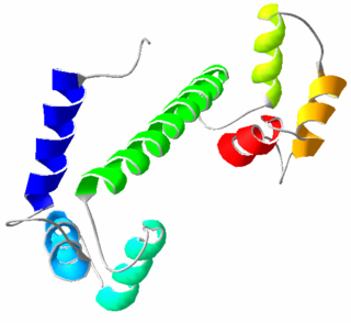
Calmodulin (CaM) (an abbreviation for calcium-modulated protein) is a multifunctional intermediate calcium-binding messenger protein expressed in all eukaryotic cells. It is an intracellular target of the secondary messenger Ca2+, and the binding of Ca2+ is required for the activation of calmodulin. Once bound to Ca2+, calmodulin acts as part of a calcium signal transduction pathway by modifying its interactions with various target proteins such as kinases or phosphatases.

<span class="mw-page-title-main">Calbindin</span> Protein

Calbindins are three different calcium-binding proteins: calbindin, calretinin and S100G. They were originally described as vitamin D-dependent calcium-binding proteins in the intestine and kidney of chicks and mammals. They are now classified in different subfamilies as they differ in the number of Ca2+ binding EF hands.

<span class="mw-page-title-main">TRPV6</span> Protein-coding gene in the species Homo sapiens

TRPV6 is a membrane calcium (Ca2+) channel protein which is particularly involved in the first step in Ca2+absorption in the intestine.

Calcium-binding proteins are proteins that participate in calcium cell signaling pathways by binding to Ca2+, the calcium ion that plays an important role in many cellular processes. Calcium-binding proteins have specific domains that bind to calcium and are known to be heterogeneous.

p16 Mammalian protein found in Homo sapiens

p16, is a protein that slows cell division by slowing the progression of the cell cycle from the G1 phase to the S phase, thereby acting as a tumor suppressor. It is encoded by the CDKN2A gene. A deletion in this gene can result in insufficient or non-functional p16, accelerating the cell cycle and resulting in many types of cancer.

Plasma membrane Ca<sup>2+</sup> ATPase Transport protein

The plasma membrane Ca2+ ATPase (PMCA) is a transport protein in the plasma membrane of cells that functions as a calcium pump to remove calcium (Ca2+) from the cell. PMCA function is vital for regulating the amount of Ca2+ within all eukaryotic cells. There is a very large transmembrane electrochemical gradient of Ca2+ driving the entry of the ion into cells, yet it is very important that they maintain low concentrations of Ca2+ for proper cell signalling. Thus, it is necessary for cells to employ ion pumps to remove the Ca2+. The PMCA and the sodium calcium exchanger (NCX) are together the main regulators of intracellular Ca2+ concentrations. Since it transports Ca2+ into the extracellular space, the PMCA is also an important regulator of the calcium concentration in the extracellular space.

<span class="mw-page-title-main">Parvalbumin</span>

Parvalbumin (PV) is a calcium-binding protein with low molecular weight. In humans, it is encoded by the PVALB gene. It is not a member of the albumin family; it is named for its size and its ability to coagulate.

<span class="mw-page-title-main">Fatty acid-binding protein</span>

The fatty-acid-binding proteins (FABPs) are a family of transport proteins for fatty acids and other lipophilic substances such as eicosanoids and retinoids. These proteins are thought to facilitate the transfer of fatty acids between extra- and intracellular membranes. Some family members are also believed to transport lipophilic molecules from outer cell membrane to certain intracellular receptors such as PPAR. The FABPs are intracellular carriers that “solubilize” the endocannabinoid anandamide (AEA), transporting AEA to the breakdown by FAAH, and compounds that bind to FABPs block AEA breakdown, raising its level. The cannabinoids are also discovered to bind human FABPs that function as intracellular carriers, as THC and CBD inhibit the cellular uptake and catabolism of AEA by targeting FABPs. Competition for FABPs may in part or wholly explain the increased circulating levels of endocannabinoids reported after consumption of cannabinoids. Levels of fatty-acid-binding protein have been shown to decline with ageing in the mouse brain, possibly contributing to age-associated decline in synaptic activity.

<span class="mw-page-title-main">TRPV2</span> Protein-coding gene in the species Homo sapiens

Transient receptor potential cation channel subfamily V member 2 is a protein that in humans is encoded by the TRPV2 gene. TRPV2 is a nonspecific cation channel that is a part of the TRP channel family. This channel allows the cell to communicate with its extracellular environment through the transfer of ions, and responds to noxious temperatures greater than 52 °C. It has a structure similar to that of potassium channels, and has similar functions throughout multiple species; recent research has also shown multiple interactions in the human body.

<span class="mw-page-title-main">S100A2</span> Protein-coding gene in the species Homo sapiens

S100 calcium-binding protein A2 (S100A2) is a protein that in humans is encoded by the S100A2 gene and it is located on chromosome 1q21 with other S100 proteins.

<span class="mw-page-title-main">Inositol-trisphosphate 3-kinase</span> Class of enzymes

Inositol (1,4,5) trisphosphate 3-kinase (EC 2.7.1.127), abbreviated here as ITP3K, is an enzyme that facilitates a phospho-group transfer from adenosine triphosphate to 1D-myo-inositol 1,4,5-trisphosphate. This enzyme belongs to the family of transferases, specifically those transferring phosphorus-containing groups (phosphotransferases) with an alcohol group as acceptor. The systematic name of this enzyme class is ATP:1D-myo-inositol-1,4,5-trisphosphate 3-phosphotransferase. ITP3K catalyzes the transfer of the gamma-phosphate from ATP to the 3-position of inositol 1,4,5-trisphosphate to form inositol 1,3,4,5-tetrakisphosphate. ITP3K is highly specific for the 1,4,5-isomer of IP3, and it exclusively phosphorylates the 3-OH position, producing Ins(1,3,4,5)P4, also known as inositol tetrakisphosphate or IP4.

<span class="mw-page-title-main">ATP2B4</span> Protein-coding gene in the species Homo sapiens

Plasma membrane calcium-transporting ATPase 4 is an enzyme that in humans is encoded by the ATP2B4 gene.

<span class="mw-page-title-main">WNT3A</span> Protein-coding gene in the species Homo sapiens

Protein Wnt-3a is a protein that in humans is encoded by the WNT3A gene.

<span class="mw-page-title-main">FIS1</span> Protein-coding gene in the species Homo sapiens

Mitochondrial fission 1 protein (FIS1) is a protein that in humans is encoded by the FIS1 gene on chromosome 7. This protein is a component of a mitochondrial complex, the ARCosome, that promotes mitochondrial fission. Its role in mitochondrial fission thus implicates it in the regulation of mitochondrial morphology, the cell cycle, and apoptosis. By extension, the protein is involved in associated diseases, including neurodegenerative diseases and cancers.

<span class="mw-page-title-main">CPNE1</span> Protein-coding gene in humans

Copine-1 is a protein that in humans is encoded by the CPNE1 gene.

<span class="mw-page-title-main">Cellular senescence</span> Phenomenon characterized by the cessation of cell division

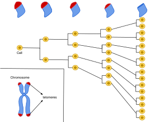
Cellular senescence is a phenomenon characterized by the cessation of cell division. In their experiments during the early 1960s, Leonard Hayflick and Paul Moorhead found that normal human fetal fibroblasts in culture reach a maximum of approximately 50 cell population doublings before becoming senescent. This process is known as "replicative senescence", or the Hayflick limit. Hayflick's discovery of mortal cells paved the path for the discovery and understanding of cellular aging molecular pathways. Cellular senescence can be initiated by a wide variety of stress inducing factors. These stress factors include both environmental and internal damaging events, abnormal cellular growth, oxidative stress, autophagy factors, among many other things.

<span class="mw-page-title-main">SEC16B</span> Protein-coding gene in the species Homo sapiens

Protein transport protein Sec16B also known as regucalcin gene promoter region-related protein p117 (RGPR-p117) and leucine zipper transcription regulator 2 (LZTR2) is a protein that in humans is encoded by the SEC16B gene.

<span class="mw-page-title-main">FOXA2</span> Mammalian protein found in Homo sapiens

Forkhead box protein A2 (FOXA2), also known as hepatocyte nuclear factor 3-beta (HNF-3B), is a transcription factor that plays an important role during development, in mature tissues and, when dysregulated or mutated, also in cancer.

miR-137

In molecular biology, miR-137 is a short non-coding RNA molecule that functions to regulate the expression levels of other genes by various mechanisms. miR-137 is located on human chromosome 1p22 and has been implicated to act as a tumor suppressor in several cancer types including colorectal cancer, squamous cell carcinoma and melanoma via cell cycle control.

<span class="mw-page-title-main">HINT2</span> Protein-coding gene in the species Homo sapiens

Histidine triad nucleotide binding protein 2 (HINT2) is a mitochondrial protein that in humans is encoded by the HINT2 gene on chromosome 9. This protein is an AMP-lysine hydrolase and phosphoamidase and may contribute to tumor suppression.

References

  1. 1 2 3 GRCh38: Ensembl release 89: ENSG00000130988 - Ensembl, May 2017
  2. 1 2 3 GRCm38: Ensembl release 89: ENSMUSG00000023070 - Ensembl, May 2017
  3. "Human PubMed Reference:". National Center for Biotechnology Information, U.S. National Library of Medicine.
  4. "Mouse PubMed Reference:". National Center for Biotechnology Information, U.S. National Library of Medicine.
  5. Shimokawa N, Matsuda Y, Yamaguchi M (October 1995). "Genomic cloning and chromosomal assignment of rat regucalcin gene". Molecular and Cellular Biochemistry. 151 (2): 157–63. doi:10.1007/BF01322338. PMID   8569761. S2CID   20648596.
  6. Fujita T, Mandel JL, Shirasawa T, Hino O, Shirai T, Maruyama N (September 1995). "Isolation of cDNA clone encoding human homologue of senescence marker protein-30 (SMP30) and its location on the X chromosome". Biochimica et Biophysica Acta (BBA) - Gene Structure and Expression. 1263 (3): 249–52. doi:10.1016/0167-4781(95)00120-6. PMID   7548213.
  7. 1 2 "Entrez Gene: RGN regucalcin (senescence marker protein-30)".
  8. Yamaguchi M, Isogai M, Kato S, Mori S (June 1991). "Immunohistochemical demonstration of calcium-binding protein regucalcin in the tissues of rats: the protein localizes in liver and brain". Chemical & Pharmaceutical Bulletin. 39 (6): 1601–3. doi: 10.1248/cpb.39.1601 . PMID   1934180.
  9. Shimokawa N, Yamaguchi M (June 1992). "Calcium administration stimulates the expression of calcium-binding protein regucalcin mRNA in rat liver". FEBS Letters. 305 (2): 151–4. doi: 10.1016/0014-5793(92)80884-J . PMID   1618342. S2CID   24974683.
  10. Yamaguchi M, Isogai M (May 1993). "Tissue concentration of calcium-binding protein regucalcin in rats by enzyme-linked immunoadsorbent assay". Molecular and Cellular Biochemistry. 122 (1): 65–8. doi:10.1007/BF00925738. PMID   8350865. S2CID   22140722.
  11. 1 2 Misawa H, Yamaguchi M (August 2000). "The gene of Ca2+-binding protein regucalcin is highly conserved in vertebrate species". International Journal of Molecular Medicine. 6 (2): 191–6. doi:10.3892/ijmm.6.2.191. PMID   10891565.
  12. Yamaguchi M, Yamamoto T (June 1978). "Purification of calcium binding substance from soluble fraction of normal rat liver". Chemical & Pharmaceutical Bulletin. 26 (6): 1915–8. doi: 10.1248/cpb.26.1915 . PMID   699201.
  13. 1 2 Yamaguchi M, Sugii K (February 1981). "Properties of calcium-binding protein isolated from the soluble fraction of normal rat liver". Chemical & Pharmaceutical Bulletin. 29 (2): 567–70. doi: 10.1248/cpb.29.567 . PMID   7273253.
  14. Yamaguchi M, Mori S (January 1988). "Effect of Ca2+ and Zn2+ on 5'-nucleotidase activity in rat liver plasma membranes: Hepatic calcium-binding protein (regucalcin) reverses the Ca2+ effect". Chemical & Pharmaceutical Bulletin. 36 (1): 321–325. doi: 10.1248/cpb.36.321 . PMID   2837338.
  15. 1 2 Yamaguchi M (1992). "A novel Ca2+-binding protein regucalcin and calcium inhibition. Regulatory role in liver cell function". Calcium Inhibition. Boca Raton: CRC Press. pp. 19–41.
  16. Fujita T, Uchida K, Maruyama N (April 1992). "Purification of senescence marker protein-30 (SMP30) and its androgen-independent decrease with age in the rat liver". Biochimica et Biophysica Acta (BBA) - General Subjects. 1116 (2): 122–8. doi:10.1016/0304-4165(92)90108-7. PMID   1581340.
  17. Fujita T, Shirasawa T, Uchida K, Maruyama N (October 1992). "Isolation of cDNA clone encoding rat senescence marker protein-30 (SMP30) and its tissue distribution". Biochimica et Biophysica Acta (BBA) - Gene Structure and Expression. 1132 (3): 297–305. doi:10.1016/0167-4781(92)90164-u. PMID   1420310.
  18. 1 2 Shimokawa N, Yamaguchi M (August 1993). "Molecular cloning and sequencing of the cDNA coding for a calcium-binding protein regucalcin from rat liver". FEBS Letters. 327 (3): 251–5. doi: 10.1016/0014-5793(93)80998-a . PMID   8348951. S2CID   1303220.
  19. Yamaguchi M, Makino R, Shimokawa N (December 1996). "The 5' end sequences and exon organization in rat regucalcin gene". Molecular and Cellular Biochemistry. 165 (2): 145–50. doi:10.1007/bf00229476. PMID   8979263. S2CID   10508949.
  20. Yamaguchi M (January 2011). "The transcriptional regulation of regucalcin gene expression". Molecular and Cellular Biochemistry. 346 (1–2): 147–71. doi:10.1007/s11010-010-0601-8. PMID   20936536. S2CID   22077914.
  21. Yamaguchi M (March 2005). "Role of regucalcin in maintaining cell homeostasis and function (review)". International Journal of Molecular Medicine. 15 (3): 371–89. doi:10.3892/ijmm.15.3.371. PMID   15702226.
  22. Yamaguchi M (August 2010). "Regucalcin and metabolic disorders: osteoporosis and hyperlipidemia are induced in regucalcin transgenic rats". Molecular and Cellular Biochemistry. 341 (1–2): 119–33. doi:10.1007/s11010-010-0443-4. PMID   20349117. S2CID   12577305.
  23. 1 2 Yamaguchi M (2017). The Role of Regucalcin in Cell Homeostasis and Disorder. New York: Nova Science Publishers. pp. 1–288. ISBN   978-3-319-39855-6.
  24. Yamaguchi M, ed. (2019). Regucalcin: Metabolic Regulation and Disease. New York: Nova Science Publishers. pp. 1–176. ISBN   978-1536161724.
  25. Yamaguchi M (August 2015). "Involvement of regucalcin as a suppressor protein in human carcinogenesis: Insight into the gene therapy". Journal of Cancer Research and Clinical Oncology. 141 (8): 133–1341. doi:10.1007/s00432-014-1831-z. PMID   25230901. S2CID   25371567.
  26. Yamaguchi M, Osuka S, Weitzmann MN, El-Rayes BF, Shoji M, Murata T (May 2016). "Prolonged survival in pancreatic cancer patients with increased regucalcin gene expression: Overexpression of regucalcin suppresses the proliferation in human pancreatic cancer MIA PaCa-2 cells in vitro". International Journal of Oncology. 48 (5): 1955–1964. doi: 10.3892/ijo.2016.3409 . PMID   26935290.
  27. Cardosoa AL, Fernandes A, Aguilar-Pimentelc JA, de Angelisd MH, Guedes JR, Britob MA, Ortolano S, Pani G, Athanasopoulou S, Gonos ES, Schosserer M, Grillari J, Peterson P, Tuna BG, Dogan S, Meyer A, van Os R, Trendelenburg AU (July 2018). "Towards frailty biomarkers: Candidates from genes and pathways regulated in aging and age-related diseases". Journal of Cancer Research and Clinical Oncology. 47: 214–277. doi: 10.1016/j.arr.2018.07.004 . hdl: 10807/130553 . PMID   30071357.
  28. Nitschkowski D, Marwitz S, Kotanidou SA, Reck M, Kugler C, Rabe KF, Ammerpohl O, Goldmann T (November 2019). "Live and let die: epigenetic modifications of Survivin and Regucalcin in non-small cell lung cancer tissues contribute to malignancy". Clinical Epigenetics. 11 (1): 157. doi: 10.1186/s13148-019-0770-6 . PMC   6852724 . PMID   31718698.

Further reading