Renin–angiotensin system

Last updated

Anatomical diagram of RAS Renin-angiotensin system in man shadow.svg
Anatomical diagram of RAS

The renin-angiotensin system (RAS), or renin-angiotensin-aldosterone system (RAAS), is a hormone system that regulates blood pressure, fluid, and electrolyte balance, and systemic vascular resistance. [2] [3]

Contents

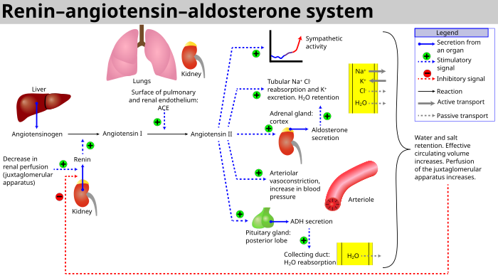
When renal blood flow is reduced, juxtaglomerular cells in the kidneys convert the precursor prorenin (already present in the blood) into renin and secrete it directly into the circulation. Plasma renin then carries out the conversion of angiotensinogen, released by the liver, to a decapeptide called angiotensin I. [4] Angiotensin I is subsequently converted to angiotensin II (an octapeptide) by the angiotensin-converting enzyme (ACE) found on the surface of vascular endothelial cells, predominantly those of the lungs. [5] Angiotensin II has a short life of about 1 to 2 minutes. Then, it is rapidly degraded into a heptapeptide called angiotensin III by angiotensinases which are present in red blood cells and vascular beds in many tissues.

Angiotensin III increases blood pressure and stimulates aldosterone secretion from the adrenal cortex; it has 100% adrenocortical stimulating activity and 40% vasopressor activity of angiotensin II.

Angiotensin IV also has adrenocortical and vasopressor activities.

Angiotensin II is a potent vasoconstrictive peptide that causes blood vessels to narrow, resulting in increased blood pressure. [6] Angiotensin II also stimulates the secretion of the hormone aldosterone [6] from the adrenal cortex. Aldosterone causes the renal tubules to increase the reabsorption of sodium which in consequence causes the reabsorption of water into the blood, while at the same time causing the excretion of potassium (to maintain electrolyte balance). This increases the volume of extracellular fluid in the body, which also increases blood pressure.

If the RAS is abnormally active, blood pressure will be too high. There are several types of drugs which includes ACE inhibitors, angiotensin II receptor blockers (ARBs), and renin inhibitors that interrupt different steps in this system to improve blood pressure. These drugs are one of the primary ways to control high blood pressure, heart failure, kidney failure, and harmful effects of diabetes. [7] [8]

Activation

RAAS schematic Renin-angiotensin-aldosterone system.svg
RAAS schematic

The system can be activated when there is a loss of blood volume or a drop in blood pressure (such as in hemorrhage or dehydration). This loss of pressure is interpreted by baroreceptors in the carotid sinus. It can also be activated by a decrease in the filtrate sodium chloride (NaCl) concentration or a decreased filtrate flow rate that will stimulate the macula densa to signal the juxtaglomerular cells to release renin.[ citation needed ]

  1. If the perfusion of the juxtaglomerular apparatus in the kidney's macula densa decreases, then the juxtaglomerular cells (granular cells, modified pericytes in the glomerular capillary) release the enzyme renin.
  2. Renin cleaves a decapeptide from angiotensinogen, a globular protein. The decapeptide is known as angiotensin I.
  3. Angiotensin I is then converted to an octapeptide, angiotensin II by angiotensin-converting enzyme (ACE), [9] which is thought to be found mainly in endothelial cells of the capillaries throughout the body, within the lungs and the epithelial cells of the kidneys. One study in 1992 found ACE in all blood vessel endothelial cells. [10]
  4. Angiotensin II is the major bioactive product of the renin–angiotensin system, binding to receptors on intraglomerular mesangial cells, causing these cells to contract along with the blood vessels surrounding them; and to receptors on the zona glomerulosa cells, causing the release of aldosterone from the zona glomerulosa in the adrenal cortex. Angiotensin II acts as an endocrine, autocrine/paracrine, and intracrine hormone.

Cardiovascular effects

Renal hormone regulation schematic RenalHormoneRegulation.png
Renal hormone regulation schematic

Angiotensin I may have some minor activity, but angiotensin II is the major bio-active product. Angiotensin II has a variety of effects on the body:[ citation needed ]

These effects directly act together to increase blood pressure and are opposed by atrial natriuretic peptide (ANP).

Local renin–angiotensin systems

Locally expressed renin–angiotensin systems have been found in a number of tissues, including the kidneys, adrenal glands, the heart, vasculature and nervous system, and have a variety of functions, including local cardiovascular regulation, in association or independently of the systemic renin–angiotensin system, as well as non-cardiovascular functions. [9] [11] [12] Outside the kidneys, renin is predominantly picked up from the circulation but may be secreted locally in some tissues; its precursor prorenin is highly expressed in tissues and more than half of circulating prorenin is of extrarenal origin, but its physiological role besides serving as precursor to renin is still unclear. [13] Outside the liver, angiotensinogen is picked up from the circulation or expressed locally in some tissues; with renin they form angiotensin I, and locally expressed angiotensin-converting enzyme, chymase or other enzymes can transform it into angiotensin II. [13] [14] [15] This process can be intracellular or interstitial. [9]

In the adrenal glands, it is likely involved in the paracrine regulation of aldosterone secretion; in the heart and vasculature, it may be involved in remodeling or vascular tone; and in the brain, where it is largely independent of the circulatory RAS, it may be involved in local blood pressure regulation. [9] [12] [16] In addition, both the central and peripheral nervous systems can use angiotensin for sympathetic neurotransmission. [17] Other places of expression include the reproductive system, the skin and digestive organs. Medications aimed at the systemic system may affect the expression of those local systems, beneficially or adversely. [9]

Fetal renin–angiotensin system

In the fetus, the renin–angiotensin system is predominantly a sodium-losing system,[ citation needed ] as angiotensin II has little or no effect on aldosterone levels. Renin levels are high in the fetus, while angiotensin II levels are significantly lower; this is due to the limited pulmonary blood flow, preventing ACE (found predominantly in the pulmonary circulation) from having its maximum effect.[ citation needed ]

Clinical significance

Flowchart showing the clinical effects of RAAS activity and the sites of action of ACE inhibitors and angiotensin receptor blockers. Renin angiotensin aldosterone system.png
Flowchart showing the clinical effects of RAAS activity and the sites of action of ACE inhibitors and angiotensin receptor blockers.

See also

Related Research Articles

<span class="mw-page-title-main">ACE inhibitor</span> Class of medications used primarily to treat high blood pressure

Angiotensin-converting-enzyme inhibitors are a class of medication used primarily for the treatment of high blood pressure and heart failure. This class of medicine works by causing relaxation of blood vessels as well as a decrease in blood volume, which leads to lower blood pressure and decreased oxygen demand from the heart.

Azotemia is a medical condition characterized by abnormally high levels of nitrogen-containing compounds in the blood. It is largely related to insufficient or dysfunctional filtering of blood by the kidneys. It can lead to uremia and acute kidney injury if not controlled.

<span class="mw-page-title-main">Nephron</span> Microscopic structural and functional unit of the kidney

The nephron is the minute or microscopic structural and functional unit of the kidney. It is composed of a renal corpuscle and a renal tubule. The renal corpuscle consists of a tuft of capillaries called a glomerulus and a cup-shaped structure called Bowman's capsule. The renal tubule extends from the capsule. The capsule and tubule are connected and are composed of epithelial cells with a lumen. A healthy adult has 1 to 1.5 million nephrons in each kidney. Blood is filtered as it passes through three layers: the endothelial cells of the capillary wall, its basement membrane, and between the foot processes of the podocytes of the lining of the capsule. The tubule has adjacent peritubular capillaries that run between the descending and ascending portions of the tubule. As the fluid from the capsule flows down into the tubule, it is processed by the epithelial cells lining the tubule: water is reabsorbed and substances are exchanged ; first with the interstitial fluid outside the tubules, and then into the plasma in the adjacent peritubular capillaries through the endothelial cells lining that capillary. This process regulates the volume of body fluid as well as levels of many body substances. At the end of the tubule, the remaining fluid—urine—exits: it is composed of water, metabolic waste, and toxins.

<span class="mw-page-title-main">Renin</span> Aspartic protease protein and enzyme

Renin, also known as an angiotensinogenase, is an aspartic protease protein and enzyme secreted by the kidneys that participates in the body's renin-angiotensin-aldosterone system (RAAS)—also known as the renin-angiotensin-aldosterone axis—that increases the volume of extracellular fluid and causes arterial vasoconstriction. Thus, it increases the body's mean arterial blood pressure.

<span class="mw-page-title-main">Angiotensin</span> Group of peptide hormones in mammals

Angiotensin is a peptide hormone that causes vasoconstriction and an increase in blood pressure. It is part of the renin–angiotensin system, which regulates blood pressure. Angiotensin also stimulates the release of aldosterone from the adrenal cortex to promote sodium retention by the kidneys.

<span class="mw-page-title-main">Aldosterone</span> Mineralocorticoid steroid hormone

Aldosterone is the main mineralocorticoid steroid hormone produced by the zona glomerulosa of the adrenal cortex in the adrenal gland. It is essential for sodium conservation in the kidney, salivary glands, sweat glands, and colon. It plays a central role in the homeostatic regulation of blood pressure, plasma sodium (Na+), and potassium (K+) levels. It does so primarily by acting on the mineralocorticoid receptors in the distal tubules and collecting ducts of the nephron. It influences the reabsorption of sodium and excretion of potassium (from and into the tubular fluids, respectively) of the kidney, thereby indirectly influencing water retention or loss, blood pressure, and blood volume. When dysregulated, aldosterone is pathogenic and contributes to the development and progression of cardiovascular and kidney disease. Aldosterone has exactly the opposite function of the atrial natriuretic hormone secreted by the heart.

<span class="mw-page-title-main">Atrial natriuretic peptide</span> Cardiac hormone which increases renal sodium excretion

Atrial Natriuretic Peptide (ANP) or atrial natriuretic factor (ANF) is a natriuretic peptide hormone secreted from the cardiac atria that in humans is encoded by the NPPA gene. Natriuretic peptides are a family of hormone/paracrine factors that are structurally related. The main function of ANP is causing a reduction in expanded extracellular fluid (ECF) volume by increasing renal sodium excretion. ANP is synthesized and secreted by cardiac muscle cells in the walls of the atria in the heart. These cells contain volume receptors which respond to increased stretching of the atrial wall due to increased atrial blood volume.

<span class="mw-page-title-main">Juxtaglomerular apparatus</span> Structure that regulates function of each nephron

The juxtaglomerular apparatus is a structure in the kidney that regulates the function of each nephron, the functional units of the kidney. The juxtaglomerular apparatus is named because it is next to (juxta-) the glomerulus.

<span class="mw-page-title-main">Renal physiology</span> Study of the physiology of the kidney

Renal physiology is the study of the physiology of the kidney. This encompasses all functions of the kidney, including maintenance of acid-base balance; regulation of fluid balance; regulation of sodium, potassium, and other electrolytes; clearance of toxins; absorption of glucose, amino acids, and other small molecules; regulation of blood pressure; production of various hormones, such as erythropoietin; and activation of vitamin D.

<span class="mw-page-title-main">Angiotensin-converting enzyme</span> Mammalian protein found in humans

Angiotensin-converting enzyme, or ACE, is a central component of the renin–angiotensin system (RAS), which controls blood pressure by regulating the volume of fluids in the body. It converts the hormone angiotensin I to the active vasoconstrictor angiotensin II. Therefore, ACE indirectly increases blood pressure by causing blood vessels to constrict. ACE inhibitors are widely used as pharmaceutical drugs for treatment of cardiovascular diseases.

<span class="mw-page-title-main">Macula densa</span> A region of juxtaglomerular apparatus in nephron of kidney

In the kidney, the macula densa is an area of closely packed specialized cells lining the wall of the distal tubule where it touches the glomerulus. Specifically, the macula densa is found in the terminal portion of the distal straight tubule, after which the distal convoluted tubule begins.

An osmoreceptor is a sensory receptor primarily found in the hypothalamus of most homeothermic organisms that detects changes in osmotic pressure. Osmoreceptors can be found in several structures, including two of the circumventricular organs – the vascular organ of the lamina terminalis, and the subfornical organ. They contribute to osmoregulation, controlling fluid balance in the body. Osmoreceptors are also found in the kidneys where they also modulate osmolality.

<span class="mw-page-title-main">Juxtaglomerular cell</span> Cell in kidneys that produces & secretes renin

Juxtaglomerular cells, also known as juxtaglomerular granular cells are cells in the kidney that synthesize, store, and secrete the enzyme renin. They are specialized smooth muscle cells mainly in the walls of the afferent arterioles that deliver blood to the glomerulus. In synthesizing renin, they play a critical role in the renin–angiotensin system and thus in autoregulation of the kidney.

<span class="mw-page-title-main">Zona glomerulosa</span> Part of the adrenal gland

The zona glomerulosa of the adrenal gland is the most superficial layer of the adrenal cortex, lying directly beneath the renal capsule. Its cells are ovoid and arranged in clusters or arches.

<span class="mw-page-title-main">Bartter syndrome</span> Medical condition

Bartter syndrome (BS) is a rare inherited disease characterised by a defect in the thick ascending limb of the loop of Henle, which results in low potassium levels (hypokalemia), increased blood pH (alkalosis), and normal to low blood pressure. There are two types of Bartter syndrome: neonatal and classic. A closely associated disorder, Gitelman syndrome, is milder than both subtypes of Bartter syndrome.

<span class="mw-page-title-main">Afferent arterioles</span> Blood vessels supplying nephrons of kidneys

The afferent arterioles are a group of blood vessels that supply the nephrons in many excretory systems. They play an important role in the regulation of blood pressure as a part of the tubuloglomerular feedback mechanism.

<span class="mw-page-title-main">Reabsorption</span> Part of the function of the kidney

In renal physiology, reabsorption or tubular reabsorption is the process by which the nephron removes water and solutes from the tubular fluid (pre-urine) and returns them to the circulating blood. It is called reabsorption (and not absorption) because these substances have already been absorbed once (particularly in the intestines) and the body is reclaiming them from a postglomerular fluid stream that is on its way to becoming urine (that is, they will soon be lost to the urine unless they are reabsorbed from the tubule into the peritubular capillaries. This happens as a result of sodium transport from the lumen into the blood by the Na+/K+ATPase in the basolateral membrane of the epithelial cells. Thus, the glomerular filtrate becomes more concentrated, which is one of the steps in forming urine. Nephrons are divided into five segments, with different segments responsible for reabsorbing different substances. Reabsorption allows many useful solutes (primarily glucose and amino acids), salts and water that have passed through Bowman's capsule, to return to the circulation. These solutes are reabsorbed isotonically, in that the osmotic potential of the fluid leaving the proximal convoluted tubule is the same as that of the initial glomerular filtrate. However, glucose, amino acids, inorganic phosphate, and some other solutes are reabsorbed via secondary active transport through cotransport channels driven by the sodium gradient.

In the physiology of the kidney, tubuloglomerular feedback (TGF) is a feedback system inside the kidneys. Within each nephron, information from the renal tubules is signaled to the glomerulus. Tubuloglomerular feedback is one of several mechanisms the kidney uses to regulate glomerular filtration rate (GFR). It involves the concept of purinergic signaling, in which an increased distal tubular sodium chloride concentration causes a basolateral release of adenosine from the macula densa cells. This initiates a cascade of events that ultimately brings GFR to an appropriate level.

<span class="mw-page-title-main">Aliskiren</span> Medication

Aliskiren is the first in a class of drugs called direct renin inhibitors. It is used for essential (primary) hypertension. While used for high blood pressure, other better studied medications are typically recommended due to concerns of higher side effects and less evidence of benefit.

Glucocorticoid remediable aldosteronism also describable as aldosterone synthase hyperactivity, is an autosomal dominant disorder in which the increase in aldosterone secretion produced by ACTH is no longer transient.

References

  1. Boron WF, Boulpaep EL, eds. (2003). "Integration of Salt and Water Balance (pp. 866–867); The Adrenal Gland (p. 1059)". Medical Physiology: A Cellular And Molecular Approach. Elsevier/Saunders. ISBN   978-1-4160-2328-9. OCLC   1127823558. Archived from the original on 9 April 2022. Retrieved 8 April 2022.
  2. Lappin S, Fountain JH (5 May 2019). "Physiology, Renin-Angiotensin System". NCBI. NIH. PMID   29261862. Archived from the original on 29 April 2019. Retrieved 9 May 2019.
  3. Nakagawa P, Gomez J, Grobe JL, Sigmund CD (January 2020). "The Renin-Angiotensin System in the Central Nervous System and Its Role in Blood Pressure Regulation". Current Hypertension Reports. 22 (1): 7. doi:10.1007/s11906-019-1011-2. PMC   7101821 . PMID   31925571.
  4. Kumar A, Fausto A (2010). "11". Pathologic Basis of Disease (8th ed.). Saunders Elsevier. p. 493. ISBN   978-1-4160-3121-5. OCLC   758251143. Archived from the original on 9 April 2022. Retrieved 8 April 2022.
  5. Golan D, Tashjian A, Armstrong E, Armstrong A (15 December 2011). Principles of Pharmacology – The Pathophysiologic Basis of Drug Therapy. Lippincott Williams & Wolters. p. 335. ISBN   978-1-60831-270-2. OCLC   1058067942. Archived from the original on 9 April 2022. Retrieved 8 April 2022.
  6. 1 2 3 Yee AH, Burns JD, Wijdicks EF (April 2010). "Cerebral salt wasting: pathophysiology, diagnosis, and treatment". Neurosurg Clin N Am. 21 (2): 339–352. doi:10.1016/j.nec.2009.10.011. PMID   20380974.
  7. Bakris GL (November 2022). "High Blood Pressure: Heart and Blood Vessel Disorders". Merck Manual Home Edition. Archived from the original on 5 November 2010. Retrieved 6 June 2008.
  8. Solomon SD, Anavekar N (2005). "A Brief Overview of Inhibition of the Renin–Angiotensin System: Emphasis on Blockade of the Angiotensin II Type-1 Receptor". Medscape Cardiology. 9 (2). Archived from the original on 15 December 2019. Retrieved 6 June 2008.
  9. 1 2 3 4 5 Paul M, Poyan Mehr A, Kreutz R (July 2006). "Physiology of local renin–angiotensin systems". Physiol. Rev. 86 (3): 747–803. doi:10.1152/physrev.00036.2005. PMID   16816138.
  10. Rogerson FM, Chai SY, Schlawe I, Murray WK, Marley PD, Mendelsohn FA (July 1992). "Presence of angiotensin converting enzyme in the adventitia of large blood vessels". J. Hypertens. 10 (7): 615–620. doi:10.1097/00004872-199207000-00003. PMID   1321187. S2CID   25785488.
  11. Kobori H, Nangaku M, Navar LG, Nishiyama A (September 2007). "The intrarenal renin-angiotensin system: from physiology to the pathobiology of hypertension and kidney disease". Pharmacological Reviews. 59 (3): 251–287. doi:10.1124/pr.59.3.3. PMC   2034302 . PMID   17878513.
  12. 1 2 Ehrhart-Bornstein M, Hinson JP, Bornstein SR, Scherbaum WA, Vinson GP (April 1998). "Intraadrenal interactions in the regulation of adrenocortical steroidogenesis". Endocrine Reviews. 19 (2): 101–143. doi: 10.1210/edrv.19.2.0326 . PMID   9570034.[ permanent dead link ]
  13. 1 2 Nguyen G (March 2011). "Renin, (pro)renin and receptor: an update". Clinical Science. 120 (5): 169–178. doi:10.1042/CS20100432. PMID   21087212.
  14. Kumar R, Singh VP, Baker KM (March 2008). "The intracellular renin-angiotensin system: implications in cardiovascular remodeling". Current Opinion in Nephrology and Hypertension. 17 (2): 168–173. doi:10.1097/MNH.0b013e3282f521a8. PMID   18277150. S2CID   39068591.
  15. Kumar R, Singh VP, Baker KM (April 2009). "The intracellular renin-angiotensin system in the heart". Current Hypertension Reports. 11 (2): 104–110. doi:10.1007/s11906-009-0020-y. PMID   19278599. S2CID   46657557.
  16. McKinley MJ, Albiston AL, Allen AM, Mathai ML, May CN, McAllen RM, et al. (June 2003). "The brain renin-angiotensin system: location and physiological roles". The International Journal of Biochemistry & Cell Biology. 35 (6): 901–918. doi:10.1016/S1357-2725(02)00306-0. PMID   12676175.
  17. Patil J, Heiniger E, Schaffner T, Mühlemann O, Imboden H (April 2008). "Angiotensinergic neurons in sympathetic coeliac ganglia innervating rat and human mesenteric resistance blood vessels". Regulatory Peptides. 147 (1–3): 82–87. doi:10.1016/j.regpep.2008.01.006. PMID   18308407. S2CID   23123825.
  18. Odaka C, Mizuochi T (September 2000). "Angiotensin-converting enzyme inhibitor captopril prevents activation-induced apoptosis by interfering with T cell activation signals". Clinical and Experimental Immunology. 121 (3): 515–522. doi: 10.1046/j.1365-2249.2000.01323.x . PMC   1905724 . PMID   10971519.
  19. Mehta A (January 2011). "Direct Renin Inhibitors as Antihypertensive Drugs". Pharmaxchange. Archived from the original on 7 December 2010.
  20. Gradman A, Schmieder R, Lins R, Nussberger J, Chiangs Y, Bedigian M (2005). "Aliskiren, a novel orally effective renin inhibitor, provides dose-dependent antihypertensive efficacy and placebo-like tolerability in hypertensive patients". Circulation. 111 (8): 1012–1018. doi: 10.1161/01.CIR.0000156466.02908.ED . PMID   15723979.
  21. Richter WF, Whitby BR, Chou RC (1996). "Distribution of remikiren, a potent orally active inhibitor of human renin, in laboratory animals". Xenobiotica. 26 (3): 243–254. doi:10.3109/00498259609046705. PMID   8730917.
  22. Tissot AC, Maurer P, Nussberger J, Sabat R, Pfister T, Ignatenko S, et al. (March 2008). "Effect of immunisation against angiotensin II with CYT006-AngQb on ambulatory blood pressure: a double-blind, randomised, placebo-controlled phase IIa study". Lancet. 371 (9615): 821–827. doi:10.1016/S0140-6736(08)60381-5. PMID   18328929. S2CID   15175992.
  23. Brown MJ (October 2009). "Success and failure of vaccines against renin-angiotensin system components". Nature Reviews. Cardiology. 6 (10): 639–647. doi:10.1038/nrcardio.2009.156. PMID   19707182. S2CID   15949.

Further reading