Cytochrome P450 aromatic O-demethylase

Last updated
Aromatic O-demethylase, cytochrome P450 subunit
PDB 5NCB.png
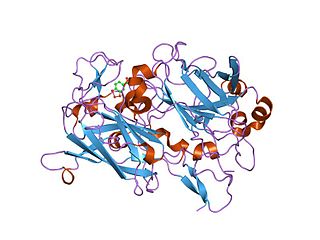
Crystal structure of gcoA (cartoon diagram) in complex with heme (green spheres) and guaiacol (magenta) based on PDB: 5NCB . [1]
Identifiers
Organism Amycolatopsis sp.
SymbolgcoA
PDB 5NCB
UniProt P0DPQ7
Other data
EC number 1.14.14.-
Search for
Structures Swiss-model
Domains InterPro
Aromatic O-demethylase, reductase subunit
PDB 5OGX.png
Crystal structure of gcoB (cartoon diagram) complexed with FAD (magenta spheres) and an iron–sulfur cluster (orange/yellow) based on PDB: 5OGX . [1]
Identifiers
Organism Amycolatopsis sp.
SymbolgcoB
PDB 5OGX
UniProt P0DPQ8
Other data
EC number 1.6.2.-
Search for
Structures Swiss-model
Domains InterPro

Cytochrome P450 aromatic O-demethylase is a bacterial enzyme that catalyzes the demethylation of lignin and various lignols. The net reaction follows the following stoichiometry, illustrated with a generic methoxy arene: [1]

Contents

ArOCH3 + O2 + 2 e + 2 H+ → ArOH + CH2O + H2O

The enzyme is notable for its promiscuity, affecting the O-demethylation of a range of substrates, including lignin.

It is a heterodimeric protein derived from the products of two genes. The component proteins are a cytochrome P450 enzyme (encoded by the gcoA gene from the family CYP255A) and a three-domain reductase (encoded by the gcoB gene) complexed with three cofactors (2Fe-2S, FAD, and NADH). [1]

A proposed structure of lignin highlighting the pervasiveness of the O-methyl groups. Lignin structure.svg
A proposed structure of lignin highlighting the pervasiveness of the O-methyl groups.

Mechanism

GcoA and GcoB form a dimer complex in solution. GcoA process the substrate while GcoB provides the electrons to support the mixed function oxidase. As with other P450's, monooxygenation of the substrate proceeds concomitantly with reduction of half an equivalent of O2 to water. An oxygen rebound mechanism can be assumed. GcoA positions the aromatic ring within the hydrophobic active site cavity where the heme is located. [2] [3]

GcoA and GcoB interacting Cytochrome P450 aromatic O-demethylase GcoA GcoB.png
GcoA and GcoB interacting

Structure

GcoA has a typical P450 structure: a thiolate-ligated heme next to a buried active site. GcoB is however unusual. Cytochrome P450s normally are complemented by either a cytochrome P450 reductase [4] or a ferredoxin and ferredoxin reductase;  its electrons are carried by NAD+ or NADP+. GcoB however has a single polypeptide. This polypeptide has an N-terminal ferredoxin with both an NAD(P)+ and also an FAD binding region.

GcoB: FAD relative to FES GcoB FAD FES.png
GcoB: FAD relative to FES

CcoA and GcoB are closely interlinked, acting as an heterodimer in solution. The surface of GcoB has an acidic patch that must interact with the matching basic region in GcoA. It is assumed that the part of GcoB interacting with GcoA is at the intersection between the FAD binding domain and ferredoxin domain. To achieve this GcoB would have to go through some structural change, which would represent a new class of P450 systems (family N). [5] [6] [7]

Potential applications

Cytochrome P450 aromatic O-demethylase assists in the partial O-demethylation of lignin. The resulting 1,2-diols are well suited for oxidative degradation via intra- and extra-diol dioxygenases. Thus O-demethylated lignins are potentially susceptible to partial depolymerization. [8]  With fewer crosslinks, the modified ligand is potentially more useful than the precursor., [9] ranging from fuels [10] [11]

Related Research Articles

Demethylation is the chemical process resulting in the removal of a methyl group (CH3) from a molecule. A common way of demethylation is the replacement of a methyl group by a hydrogen atom, resulting in a net loss of one carbon and two hydrogen atoms.

<span class="mw-page-title-main">Cytochrome P450</span> Class of enzymes

Cytochromes P450 are a superfamily of enzymes containing heme as a cofactor that mostly, but not exclusively, function as monooxygenases. In mammals, these proteins oxidize steroids, fatty acids, and xenobiotics, and are important for the clearance of various compounds, as well as for hormone synthesis and breakdown. In 1963, Estabrook, Cooper, and Rosenthal described the role of CYP as a catalyst in steroid hormone synthesis and drug metabolism. In plants, these proteins are important for the biosynthesis of defensive compounds, fatty acids, and hormones.

Ferredoxins are iron–sulfur proteins that mediate electron transfer in a range of metabolic reactions. The term "ferredoxin" was coined by D.C. Wharton of the DuPont Co. and applied to the "iron protein" first purified in 1962 by Mortenson, Valentine, and Carnahan from the anaerobic bacterium Clostridium pasteurianum.

<span class="mw-page-title-main">Flavin adenine dinucleotide</span> Redox-active coenzyme

In biochemistry, flavin adenine dinucleotide (FAD) is a redox-active coenzyme associated with various proteins, which is involved with several enzymatic reactions in metabolism. A flavoprotein is a protein that contains a flavin group, which may be in the form of FAD or flavin mononucleotide (FMN). Many flavoproteins are known: components of the succinate dehydrogenase complex, α-ketoglutarate dehydrogenase, and a component of the pyruvate dehydrogenase complex.

<span class="mw-page-title-main">Rieske protein</span> Protein family with an iron–sulfur center transferring electrons

Rieske proteins are iron–sulfur protein (ISP) components of cytochrome bc1 complexes and cytochrome b6f complexes and are responsible for electron transfer in some biological systems. John S. Rieske and co-workers first discovered the protein and in 1964 isolated an acetylated form of the bovine mitochondrial protein. In 1979 Trumpower's lab isolated the "oxidation factor" from bovine mitochondria and showed it was a reconstitutively-active form of the Rieske iron-sulfur protein
It is a unique [2Fe-2S] cluster in that one of the two Fe atoms is coordinated by two histidine residues rather than two cysteine residues. They have since been found in plants, animals, and bacteria with widely ranging electron reduction potentials from -150 to +400 mV.

Aromatic-ring-hydroxylating dioxygenases (ARHD) incorporate two atoms of dioxygen (O2) into their substrates in the dihydroxylation reaction. The product is (substituted) cis-1,2-dihydroxycyclohexadiene, which is subsequently converted to (substituted) benzene glycol by a cis-diol dehydrogenase.

Any enzyme system that includes cytochrome P450 protein or domain can be called a P450-containing system.

<span class="mw-page-title-main">Cholesterol side-chain cleavage enzyme</span> Mammalian protein found in Homo sapiens

Cholesterol side-chain cleavage enzyme is commonly referred to as P450scc, where "scc" is an acronym for side-chain cleavage. P450scc is a mitochondrial enzyme that catalyzes conversion of cholesterol to pregnenolone. This is the first reaction in the process of steroidogenesis in all mammalian tissues that specialize in the production of various steroid hormones.

<span class="mw-page-title-main">Cytochrome P450 reductase</span> Mammalian protein found in Homo sapiens

Cytochrome P450 reductase is a membrane-bound enzyme required for electron transfer from NADPH to cytochrome P450 and other heme proteins including heme oxygenase in the endoplasmic reticulum of the eukaryotic cell.

<span class="mw-page-title-main">Lanosterol 14 alpha-demethylase</span> Protein-coding gene in the species Homo sapiens

Lanosterol 14α-demethylase (CYP51A1) is the animal version of a cytochrome P450 enzyme that is involved in the conversion of lanosterol to 4,4-dimethylcholesta-8(9),14,24-trien-3β-ol. The cytochrome P450 isoenzymes are a conserved group of proteins that serve as key players in the metabolism of organic substances and the biosynthesis of important steroids, lipids, and vitamins in eukaryotes. As a member of this family, lanosterol 14α-demethylase is responsible for an essential step in the biosynthesis of sterols. In particular, this protein catalyzes the removal of the C-14α-methyl group from lanosterol. This demethylation step is regarded as the initial checkpoint in the transformation of lanosterol to other sterols that are widely used within the cell.

<span class="mw-page-title-main">Nitric oxide dioxygenase</span>

Nitric oxide dioxygenase (EC 1.14.12.17) is an enzyme that catalyzes the conversion of nitric oxide (NO) to nitrate (NO
3
) . The net reaction for the reaction catalyzed by nitric oxide dioxygenase is shown below:

In enzymology, a ferredoxin-NADP+ reductase (EC 1.18.1.2) abbreviated FNR, is an enzyme that catalyzes the chemical reaction

<span class="mw-page-title-main">NADPH—hemoprotein reductase</span> Enzyme

In enzymology, a NADPH—hemoprotein reductase is an enzyme that catalyzes the chemical reaction

<span class="mw-page-title-main">Adrenal ferredoxin</span> Mammalian protein found in Homo sapiens

Adrenal ferredoxin is a protein that in humans is encoded by the FDX1 gene. In addition to the expressed gene at this chromosomal locus (11q22), there are pseudogenes located on chromosomes 20 and 21.

<span class="mw-page-title-main">Adrenodoxin reductase</span> Protein-coding gene in the species Homo sapiens

Adrenodoxin reductase, was first isolated from bovine adrenal cortex where it functions as the first enzyme in the mitochondrial P450 systems that catalyze essential steps in steroid hormone biosynthesis. Examination of complete genome sequences revealed that adrenodoxin reductase gene is present in most metazoans and prokaryotes.
<span class="mw-page-title-main">Dioxygenase</span> Class of enzymes

Dioxygenases are oxidoreductase enzymes. Aerobic life, from simple single-celled bacteria species to complex eukaryotic organisms, has evolved to depend on the oxidizing power of dioxygen in various metabolic pathways. From energetic adenosine triphosphate (ATP) generation to xenobiotic degradation, the use of dioxygen as a biological oxidant is widespread and varied in the exact mechanism of its use. Enzymes employ many different schemes to use dioxygen, and this largely depends on the substrate and reaction at hand.

Flavoprotein pyridine nucleotide cytochrome reductases catalyse the interchange of reducing equivalents between one-electron carriers and the two-electron-carrying nicotinamide dinucleotides. The enzymes include ferredoxin-NADP+ reductases, plant and fungal NAD(P)H:nitrate reductases, cytochrome b5 reductases, cytochrome P450 reductases, sulphite reductases, nitric oxide synthases, phthalate dioxygenase reductase, and various other flavoproteins.

Oxidoreductase NAD-binding domain is an evolutionary conserved protein domain present in a variety of proteins that include, bacterial flavohemoprotein, mammalian NADH-cytochrome b5 reductase, eukaryotic NADPH-cytochrome P450 reductase, nitrate reductase from plants, nitric-oxide synthase, bacterial vanillate demethylase and others.

1,8-Cineole 2-endo-monooxygenase (EC 1.14.14.133, Formerly EC 1.14.13.156, P450cin, CYP176A, CYP176A1) is an enzyme with systematic name 1,8-cineole,NADPH:oxygen oxidoreductase (2-endo-hydroxylating). This enzyme catalyses the following chemical reaction

Adrenodoxin-NADP+ reductase (EC 1.18.1.6, adrenodoxin reductase, nicotinamide adenine dinucleotide phosphate-adrenodoxin reductase, ADR, NADPH:adrenal ferredoxin oxidoreductase) is an enzyme with systematic name adrendoxin:NADP+ oxidoreductase. This enzyme catalyses the following chemical reaction

References

  1. 1 2 3 4 Mallinson SJ, Machovina MM, Silveira RL, Garcia-Borràs M, Gallup N, Johnson CW, et al. (June 2018). "A promiscuous cytochrome P450 aromatic O-demethylase for lignin bioconversion". Nature Communications. 9 (1): 2487. Bibcode:2018NatCo...9.2487M. doi:10.1038/s41467-018-04878-2. PMC   6021390 . PMID   29950589.
  2. Vaillancourt FH, Bolin JT, Eltis LD (2006). "The ins and outs of ring-cleaving dioxygenases". Critical Reviews in Biochemistry and Molecular Biology. 41 (4): 241–67. doi:10.1080/10409230600817422. PMID   16849108. S2CID   24145324.
  3. Huang WC, Ellis J, Moody PC, Raven EL, Roberts GC (September 2013). "Redox-linked domain movements in the catalytic cycle of cytochrome p450 reductase". Structure. 21 (9): 1581–9. doi:10.1016/j.str.2013.06.022. PMC   3763376 . PMID   23911089.
  4. Wang M, Roberts DL, Paschke R, Shea TM, Masters BS, Kim JJ (August 1997). "Three-dimensional structure of NADPH-cytochrome P450 reductase: prototype for FMN- and FAD-containing enzymes". Proceedings of the National Academy of Sciences of the United States of America. 94 (16): 8411–6. Bibcode:1997PNAS...94.8411W. doi: 10.1073/pnas.94.16.8411 . PMC   22938 . PMID   9237990.
  5. Sevrioukova IF, Li H, Zhang H, Peterson JA, Poulos TL (March 1999). "Structure of a cytochrome P450-redox partner electron-transfer complex". Proceedings of the National Academy of Sciences of the United States of America. 96 (5): 1863–8. Bibcode:1999PNAS...96.1863S. doi: 10.1073/pnas.96.5.1863 . PMC   26702 . PMID   10051560.
  6. Tripathi S, Li H, Poulos TL (June 2013). "Structural basis for effector control and redox partner recognition in cytochrome P450". Science. 340 (6137): 1227–30. Bibcode:2013Sci...340.1227T. doi:10.1126/science.1235797. PMID   23744947. S2CID   23421892.
  7. Hasemann CA, Kurumbail RG, Boddupalli SS, Peterson JA, Deisenhofer J (January 1995). "Structure and function of cytochromes P450: a comparative analysis of three crystal structures". Structure. 3 (1): 41–62. doi: 10.1016/s0969-2126(01)00134-4 . PMID   7743131.
  8. Bugg TD, Rahmanpour R (December 2015). "Enzymatic conversion of lignin into renewable chemicals". Current Opinion in Chemical Biology. 29: 10–7. doi:10.1016/j.cbpa.2015.06.009. PMID   26121945.
  9. Beckham GT, Johnson CW, Karp EM, Salvachúa D, Vardon DR (December 2016). "Opportunities and challenges in biological lignin valorization". Current Opinion in Biotechnology. 42: 40–53. doi: 10.1016/j.copbio.2016.02.030 . PMID   26974563.
  10. Vardon DR, Franden MA, Johnson CW, Karp EM, Guarnieri MT, Linger JG, et al. (2015). "Adipic acid production from lignin". Energy & Environmental Science. 8 (2): 617–628. doi:10.1039/c4ee03230f. ISSN   1754-5692.
  11. Lin L, Cheng Y, Pu Y, Sun S, Li X, Jin M, et al. (2016). "Systems biology-guided biodesign of consolidated lignin conversion". Green Chemistry. 18 (20): 5536–5547. doi:10.1039/c6gc01131d. ISSN   1463-9262. OSTI   1326560.