21-Hydroxylase

Last updated

Steroid 21-hydroxylase
Full Structure of 21-Hydroxylase.png
Identifiers
EC no. 1.14.14.16
CAS no. 9029-68-9
Alt. names"Cytochrome P450, family 21, subfamily A, polypeptide 2", CYP21A2, CYP21, CYP21B, [1] P45021A2, cytochrome P450c21, [2] [3] [4] steroid 21-monooxygenase, [5] 21-hydroxylase, 21α-hydroxylase, [6] [7] 21β-hydroxylase [8] [9]
Databases
IntEnz IntEnz view
BRENDA BRENDA entry
ExPASy NiceZyme view
KEGG KEGG entry
MetaCyc metabolic pathway
PRIAM profile
PDB structures RCSB PDB PDBe PDBsum
Gene Ontology AmiGO / QuickGO
Search
PMC articles
PubMed articles
NCBI proteins

Steroid 21-hydroxylase is a protein that in humans is encoded by the CYP21A2 gene. The protein is an enzyme that hydroxylates steroids at the C21 position on the molecule. [10] [11] Naming conventions for enzymes are based on the substrate acted upon and the chemical process performed. Biochemically, this enzyme is involved in the biosynthesis of the adrenal gland hormones aldosterone and cortisol, which are important in blood pressure regulation, sodium homeostasis and blood sugar control. The enzyme converts progesterone and 17α-hydroxyprogesterone into 11-deoxycorticosterone and 11-deoxycortisol, respectively, [12] [13] within metabolic pathways which in humans ultimately lead to aldosterone and cortisol creation—deficiency in the enzyme may cause congenital adrenal hyperplasia.

Contents

Steroid 21-hydroxylase is a member of the cytochrome P450 family of monooxygenase enzymes that use an iron-containing heme cofactor to oxidize substrates.

In humans, the enzyme is localized in endoplasmic reticulum membranes of cells in adrenal cortex, [14] [15] and is encoded by the CYP21A2 gene which is located near the CYP21A1P pseudogene that has high degree of sequence similarity. This similarity makes it difficult to analyze the gene at the molecular level, and sometimes leads to loss-of-function mutations of the gene due to intergenic exchange of DNA.

Gene

CYP21A2
21-hydroxylase subunit.png
Available structures
PDB Ortholog search: PDBe RCSB
Identifiers
Aliases CYP21A2 , CA21H, CAH1, CPS1, CYP21, CYP21B, P450c21B, cytochrome P450 family 21 subfamily A member 2
External IDs OMIM: 613815 MGI: 88591 HomoloGene: 68063 GeneCards: CYP21A2
EC number 1.14.14.16,1.14.14.16
Orthologs
SpeciesHumanMouse
Entrez
Ensembl
UniProt
RefSeq (mRNA)

NM_000500
NM_001128590

NM_009995

RefSeq (protein)

NP_034125

Location (UCSC) Chr 6: 32.04 – 32.04 Mb Chr 17: 35.02 – 35.02 Mb
PubMed search [18] [19]
Wikidata
View/Edit Human View/Edit Mouse
Evolution of the CYP21A locus in humans and mice. CYP21Aevolution.png
Evolution of the CYP21A locus in humans and mice.

Steroid 21-hydroxylase in humans is encoded by the CYP21A2 gene that may be accompanied by one or several copies of the nonfunctional pseudogene CYP21A1P, [20] [21] this pseudogene shares 98% of the exonic informational identity with the actual functional gene. [22] [23]

Pseudogenes are common in genomes, and they originate as artifacts during the duplication process. Though often thought of as "junk DNA", research has shown that retaining these faulty copies can have a beneficial role, often providing regulation of their parent genes. [24]

In the mouse genome, the Cyp21a2 is a pseudogene and the Cyp21a1 is a functional gene. [25] In the chicken and quail, there is only a single Cyp21 gene, which locus is located between complement component C4 and TNX gene, along with Cenpa. [26]

CYP21A2 in humans is located in chromosome 6, in the major histocompatibility complex III (MHC class III) [27] close to the Complement component 4 genes C4A and C4B , the Tenascin X gene TNXB and STK19 . [28] MHC class III is the most gene-dense region of the human genome, containing many genes that have, as of 2023 - unknown functions or structures. [29] [27]

Inside the MHC class III, CYP21A2 is located within the RCCX cluster (an abbreviation composed of the names of the genes RP (a former name for STK19 serine/threonine kinase 19), [30] [31] C4 , CYP21 and TNX ), [32] which is the most complex gene cluster in the human genome. [33] The number of RCCX segments varies between one and four in a chromosome, [30] with the prevalence of approximately 15% for monomodular, 75% for bimodular (STK19-C4A-CYP21A1P-TNXA-STK19B-C4B-CYP21A2-TNXB), [31] [34] and 10% for trimodular in Europeans. [35] The quadrimodular structure of the RCCX unit is very rare. [36] [30] [35] In a monomodular structure, all of the genes are functional i.e. protein-coding, but if a module count is two or more, there is only one copy of each functional gene rest being non-coding pseudogenes with the exception of the C4 gene which always has active copies. [30] [35]

Due to the high degree of homology between the CYP21A2 gene and the CYP21A1P pseudogene and the complexity of the RCCX locus, it is difficult to perform molecular diagnostics for CYP21A2. The pseudogene can have single-nucleotide polymorphisms (SNP) that are identical or similar to those in the functional gene, making it difficult to distinguish between them. The pseudogene can also recombine with the functional gene, creating hybrid genes that have features of both. This can result in false-positive or false-negative results when testing for SNPs in the CYP21A2. [37]

The whole genome sequencing technology relies on breaking the DNA into small fragments, sequencing them, and then assembling them back together based on their overlaps. However, because of the high homology and variability of the CYP21A2 and its pseudogene, the fragments cannot be mapped unambiguously to either copy of the gene. This can lead to errors or gaps in the assembly, or missing some variants that are present in the gene. [38] [37]

Polymerase chain reaction (PCR) molecular diagnostics uses selective primers to amplify specific segments of the DNA sequence that are relevant for diagnosing or detecting a certain disease or condition. If the primers are not designed carefully, they may bind to both the CYP21A2 and the CYP21A1P pseudogene, or to different segments of the RCCX cluster, resulting in false-positive or false-negative results. Therefore, PCR for the CYP21A2 requires the use of locus-specific primers that can distinguish between the gene and the pseudogene, and between different RCCX modules. Moreover, PCR may not be able to detect complex variants such as large gene conversions, deletions, or duplications, which are frequent in the case of the CYP21A2. [39] [40] [38]

Southern blotting, a method used for detecting and quantifying a specific DNA sequence in DNA samples, also has limitations in analyzing CYP21A2. This method is time-consuming and requires a large amount of good-quality DNA, which makes it less applicable in routine diagnostic settings. This method comes with a radioactive biohazard, which poses safety concerns and makes it labor-intensive. Southern blotting is unable to detect the junction sites of chimeras. The CYP21A2 gene is prone to mismatch and rearrangement, producing different types of complex variations that include copy number variants, large gene conversions, small insertions/deletions, and single-nucleotide (SNP) variants. Southern blotting is not capable of detecting all these types of variants simultaneously. Besides that, southern blotting requires genetic analysis of the parents, which is not always feasible or practical. [38] [41]

Therefore, to analyze the CYP21A2 gene accurately, a more specialized and sensitive method is needed, such as targeted long-read sequencing, which can sequence longer DNA fragments and capture more information about the gene structure and variation. However, this method is not widely available or affordable for clinical use. [42] [43] [44]

Protein

Steroid 21-hydroxylase, is a member of the cytochrome P450 family of monooxygenase enzymes, the protein has 494 amino acid residues with a molecular weight of 55,000. This enzyme is at most 28% homologous to other P-450 enzymes that have been studied. [45]

Structurally, the protein contains an evolutionarily conserved core of four α-helix bundles (the importance of such genetic conservation is in demonstrating the functional importance of this aspect of this protein's structure). In addition, it has two additional alpha helices, two sets of β-sheets, and a heme cofactor binding loop. [46] Each subunit in the human enzyme consists of a total of 13 α-helices and 9 β-strands that folds into a triangular prism-like tertiary structure. [12]

The iron(III) heme group that defines the active site resides in the center of each subunit. The human enzyme binds one substrate at a time. [12] In contrast, the well-characterized bovine enzyme can bind two substrates. [47] The human and bovine enzyme share 80% amino acid sequence identity, but are structurally different, particularly in loop regions, and also evident in secondary structure elements. [12]

Species

Variations of the steroid 21-hydroxylase can be found in all vertebrates. [48]

Cyp21 first emerged in chordates before the speciation between basal chordates and vertebrates. [49] The sea lamprey, an early jawless fish species that originated over 500 million years ago, provides valuable insights into the evolution and emergence of Cyp21. Sea lampreys lack the 11β-hydroxylase enzyme responsible for converting 11-deoxycortisol to cortisol as observed in mammals. Instead, they rely on 11-deoxycortisol, a product of a reaction catalyzed by CYP21, as their primary glucocorticoid hormone with mineralocorticoid properties. This suggests the presence of a complex and highly specific corticosteroid signaling pathway that emerged at least half a billion years ago during early vertebrate evolution. [50]

In vertebrates, such as fish, amphibians, reptiles, birds, and mammals, Cyp21 participates in the biosynthesis of glucocorticoids and mineralocorticoids, therefore, Cyp21 is essential for the regulation of stress response, electrolyte balance and blood pressure, immune system, and metabolism in vertebrates. [51]

Cyp21 is relatively conserved among mammals, and shows some variations in its structure, expression, and regulation. [51] Rhesus macaques and orangutans possess two copies of Cyp21, while chimpanzees have three, still, a pseudogene (CYP21A1P) is only present in humans among primates. [52]

Tissue and subcellular distribution

Steroid 21-hydroxylase is localized in microsomes of endoplasmic reticulum membranes within adrenal cortex. [10] It is one of three microsomal steroidogenic cytochrome P450 enzymes, the others being steroid 17-hydroxylase and aromatase. [53]

Unlike other enzymes of the cytochrome P450 superfamily of enzymes that are expressed in multiple tissues, with most abundant expression in the liver, in adult humans steroid 21-hydroxylase, along with steroid 11β-hydroxylase and aldosterone synthase, is almost exclusively expressed in the adrenal gland. [54] [55]

As of 2023, the main subcellular location for the encoded protein in human cells is not known, and is pending cell analysis. [56]

Function

Steroid numbering, C21 is in the side chain of C17 Trimethyl steroid-nomenclature.svg
Steroid numbering, C21 is in the side chain of C17

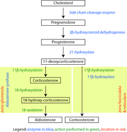
The enzyme, steroid 21-hydroxylase hydroxylates steroids at the C21 position. [13] Steroids are a group of naturally occurring and synthetically produced organic compounds, steroids all share a four ring primary structure. The enzyme catalyzes the chemical reaction in which the hydroxyl group (-OH) is added at the C21 position of the steroid biomolecule. This location is on a side chain of the D ring.

The enzyme is a member of the cytochrome P450 superfamily of monooxygenase enzymes. The cytochrome P450 enzymes catalyze many reactions involved in drug metabolism and synthesis of cholesterol, steroids and other lipids.

Steroid 21-hydroxylase is essential for the biosynthesis of cortisol and aldosterone. [57] [58]

Mechanism

Progesterone oxidation by 21-hydroxylase.svg
17a-hydroxyprogesterone oxidation by 21-hydroxylase.svg
Reaction scheme showing hydroxylation of progesterone (top) and 17a-hydroxyprogesterone (bottom)
Human steroidogenesis, showing both reactions of 21-Hydroxylase at center top. Steroidogenesis.svg
Human steroidogenesis, showing both reactions of 21-Hydroxylase at center top.
Corticosteroid biosynthetic pathway in the rat. Corticosteroid-biosynthetic-pathway-rat.png
Corticosteroid biosynthetic pathway in the rat.

Steroid 21-hydroxylase is a cytochrome P450 enzyme that is notable for its substrate specificity and relatively high catalytic efficiency. [48]

Like other cytochrome P450 enzymes, steroid 21-hydroxylase participates in the cytochrome P450 catalytic cycle and engages in one-electron transfer with NADPH-P450 reductase. Steroid 21-hydroxylase is highly specific for hydroxylation of progesterone and 17-hydroxyprogesterone. This is in marked contrast to the evolutionarily and functionally related P450 enzyme 17-hydroxylase, which has a broad range of substrates. [59]

The chemical reaction in which steroid 21-hydroxylase catalyzes the addition of hydroxyl (-OH) to the C21 position of progesterone, 17α-hydroxyprogesterone and 21-desoxycortisone [60] was first described in 1952. [61]

Studies of the human enzyme expressed in yeast initially classified 17-hydroxyprogesterone as the preferred substrate for steroid 21-hydroxylase, [59] [62] [63] however, later analysis of the purified human enzyme found a lower KM and greater catalytic efficiency for progesterone over 17-hydroxyprogesterone. [12]

The catalytic efficiency of steroid 21-hydroxylase for conversion of progesterone in humans is approximately 1.3 x 10⁷ M−1s−1 at 37 °C. This makes it the most catalytically efficient P450 enzyme of those reported to date, and catalytically more efficient than the closely related bovine steroid 21-hydroxylase enzyme. [14] C-H bond breaking to create a primary carbon radical is thought to be the rate-limiting step in the hydroxylation. [12]

Clinical significance

Congenital adrenal hyperplasia

Genetic variants in the CYP21A2 gene cause a disturbance in the development of the enzyme, leading to congenital adrenal hyperplasia (CAH) due to 21-hydroxylase deficiency. Gene conversion events involving the functional gene and the pseudogene account for many cases of steroid 21-hydroxylase deficiency. [64] CAH is an autosomal recessive disorder. There are multiple forms of CAH, defined as classical and nonclassical forms based on the amount of enzyme function still present in the patient.

The classical forms occur in approximately 1 in 10000 to 1 in 20000 births globally, [58] [65] and includes both the salt-wasting (excessive excretion of sodium via the urine causing hyponatremia and dehydration) and simple-virilizing forms. Complete loss of enzymatic activity causes the salt-wasting form. Variations in the structure of steroid 21-hydroxylase are related to the clinical severity of congenital adrenal hyperplasia. Cortisol and aldosterone deficits are associated with life-threatening sodium loss, as the steroids play roles in regulating sodium homeostasis. Simple-virilizing CAH patients (~1-2% enzyme function) [58] maintain adequate sodium homeostasis, but exhibit other symptoms shared by the salt-wasting form, including accelerated growth in childhood and ambiguous genitalia in female neonates.

The nonclassical form is the mildest condition, retaining about 20% to 50% of enzyme function. [58] This form is associated with mild and clinically silent cortisol impairment, [65] but an excess of androgens post-puberty. [66]

Non-classic congenital adrenal hyperplasia

Non-classical congenital adrenal hyperplasia caused by 21-hydroxylase deficiency (NCCAH) is a milder and late-onset congenital adrenal hyperplasia. Its prevalence rate in different ethnic groups varies from 1 in 1000 to 1 in 50. [58] Some people affected by the condition have no relevant signs and symptoms, while others experience symptoms of hyperandrogenism. [58] [65] [66]

Women with NCCAH usually have normal female genitalia at birth. In later life, the signs and symptoms of the condition may include acne, hirsutism, male-pattern baldness, irregular menstruation, and infertility. [58] [65] [25]

Fewer studies have been published about males with NCCAH comparing to those about females, because males are generally asymptomatic. [25] [58] Males, however, may present with acne [67] [68] and early balding. [69] [70]

While symptoms are usually diagnosed after puberty, children may present with premature adrenarche. [71]

Research on other conditions

There is ongoing research on how Genetic variants in the CYP21A2 gene may lead to pathogenic conditions. A variant of this gene has been reported to cause autosomal dominant posterior polar cataract, suggesting that steroid 21-hydroxylase may be involved in the extra-adrenal biosynthesis of aldosterone and cortisol in the lens of the eye. [72]

History

In the 1950s and 1960s, steroidogenic pathways that included cholesterol conversion to progesterone through a complex pathway involving multiple steps were identified, and, among them, a pathway for cortisol synthesis showing specific enzymatic steps that included hydroxylation reactions at position 21 (21-hydroxylation) mediated by cytochrome P450 enzymes. [73] Cytochrome P450 enzymes were then described, and steroid 21-hydroxylation was associated with cytochrome P450. [74] [73]

In the 1980s and 1990s, partial-length bovine Cyp21 cDNA clones were identified as related to human CYP21A2. [75] [73] Researchers discovered mutations in the CYP21A2 gene associated with congenital adrenal hyperplasia (CAH). [73]

From the 1990s onward, specific mutations were correlated with different forms/severity levels of CAH. Genotype/phenotype correlations were investigated for improved diagnostic accuracy. [73]

See also

Related Research Articles

<span class="mw-page-title-main">Adrenal gland</span> Endocrine gland

The adrenal glands are endocrine glands that produce a variety of hormones including adrenaline and the steroids aldosterone and cortisol. They are found above the kidneys. Each gland has an outer cortex which produces steroid hormones and an inner medulla. The adrenal cortex itself is divided into three main zones: the zona glomerulosa, the zona fasciculata and the zona reticularis.

<span class="mw-page-title-main">Congenital adrenal hyperplasia</span> Medical condition

Congenital adrenal hyperplasia (CAH) is a group of autosomal recessive disorders characterized by impaired cortisol synthesis. It results from the deficiency of one of the five enzymes required for the synthesis of cortisol in the adrenal cortex. Most of these disorders involve excessive or deficient production of hormones such as glucocorticoids, mineralocorticoids, or sex steroids, and can alter development of primary or secondary sex characteristics in some affected infants, children, or adults. It is one of the most common autosomal recessive disorders in humans.

<span class="mw-page-title-main">Lipoid congenital adrenal hyperplasia</span> Medical condition

Lipoid congenital adrenal hyperplasia is an endocrine disorder that is an uncommon and potentially lethal form of congenital adrenal hyperplasia (CAH). It arises from defects in the earliest stages of steroid hormone synthesis: the transport of cholesterol into the mitochondria and the conversion of cholesterol to pregnenolone—the first step in the synthesis of all steroid hormones. Lipoid CAH causes mineralocorticoid deficiency in affected infants and children. Male infants are severely undervirilized causing their external genitalia to look feminine. The adrenals are large and filled with lipid globules derived from cholesterol.

<span class="mw-page-title-main">Congenital adrenal hyperplasia due to 11β-hydroxylase deficiency</span> Medical condition

Congenital adrenal hyperplasia due to 11β-hydroxylase deficiency is a form of congenital adrenal hyperplasia (CAH) which produces a higher than normal amount of androgen, resulting from a defect in the gene encoding the enzyme steroid 11β-hydroxylase (11β-OH) which mediates the final step of cortisol synthesis in the adrenal. 11β-OH CAH results in hypertension due to excessive mineralocorticoid effects. It also causes excessive androgen production both before and after birth and can virilize a genetically female fetus or a child of either sex.

Congenital adrenal hyperplasia due to 17α-hydroxylase deficiency is an uncommon form of congenital adrenal hyperplasia resulting from a defect in the gene CYP17A1, which encodes for the enzyme 17α-hydroxylase. It causes decreased synthesis of cortisol and sex steroids, with resulting increase in mineralocorticoid production. Thus, common symptoms include mild hypocortisolism, ambiguous genitalia in genetic males or failure of the ovaries to function at puberty in genetic females, and hypokalemic hypertension (respectively). However, partial (incomplete) deficiency is notable for having inconsistent symptoms between patients, and affected genetic (XX) females may be wholly asymptomatic except for infertility.

<span class="mw-page-title-main">Congenital adrenal hyperplasia due to 21-hydroxylase deficiency</span> Medical condition

Congenital adrenal hyperplasia due to 21-hydroxylase deficiency (CAH) is a genetic disorder characterized by impaired production of cortisol in the adrenal glands.

<span class="mw-page-title-main">Aldosterone synthase</span> Protein-coding gene in the species Homo sapiens

Aldosterone synthase, also called steroid 18-hydroxylase, corticosterone 18-monooxygenase or P450C18, is a steroid hydroxylase cytochrome P450 enzyme involved in the biosynthesis of the mineralocorticoid aldosterone and other steroids. The enzyme catalyzes sequential hydroxylations of the steroid angular methyl group at C18 after initial 11β-hydroxylation. It is encoded by the CYP11B2 gene in humans.

<span class="mw-page-title-main">CYP17A1</span> Mammalian protein found in Homo sapiens

Cytochrome P450 17A1 is an enzyme of the hydroxylase type that in humans is encoded by the CYP17A1 gene on chromosome 10. It is ubiquitously expressed in many tissues and cell types, including the zona reticularis and zona fasciculata of the adrenal cortex as well as gonadal tissues. It has both 17α-hydroxylase and 17,20-lyase activities, and is a key enzyme in the steroidogenic pathway that produces progestins, mineralocorticoids, glucocorticoids, androgens, and estrogens. More specifically, the enzyme acts upon pregnenolone and progesterone to add a hydroxyl (-OH) group at carbon 17 position (C17) of the steroid D ring, or acts upon 17α-hydroxyprogesterone and 17α-hydroxypregnenolone to split the side-chain off the steroid nucleus.

3β-Hydroxysteroid dehydrogenase/Δ5-4 isomerase (3β-HSD) is an enzyme that catalyzes the biosynthesis of the steroid progesterone from pregnenolone, 17α-hydroxyprogesterone from 17α-hydroxypregnenolone, and androstenedione from dehydroepiandrosterone (DHEA) in the adrenal gland. It is the only enzyme in the adrenal pathway of corticosteroid synthesis that is not a member of the cytochrome P450 family. It is also present in other steroid-producing tissues, including the ovary, testis and placenta. In humans, there are two 3β-HSD isozymes encoded by the HSD3B1 and HSD3B2 genes.

<span class="mw-page-title-main">Cholesterol side-chain cleavage enzyme</span> Mammalian protein found in Homo sapiens

Cholesterol side-chain cleavage enzyme is commonly referred to as P450scc, where "scc" is an acronym for side-chain cleavage. P450scc is a mitochondrial enzyme that catalyzes conversion of cholesterol to pregnenolone. This is the first reaction in the process of steroidogenesis in all mammalian tissues that specialize in the production of various steroid hormones.

<span class="mw-page-title-main">Steroid 11β-hydroxylase</span> Protein found in mammals

Steroid 11β-hydroxylase, also known as steroid 11β-monooxygenase, is a steroid hydroxylase found in the zona glomerulosa and zona fasciculata of the adrenal cortex. Named officially the cytochrome P450 11B1, mitochondrial, it is a protein that in humans is encoded by the CYP11B1 gene. The enzyme is involved in the biosynthesis of adrenal corticosteroids by catalyzing the addition of hydroxyl groups during oxidation reactions.

<span class="mw-page-title-main">Cytochrome P450 reductase</span> Mammalian protein found in Homo sapiens

Cytochrome P450 reductase is a membrane-bound enzyme required for electron transfer from NADPH to cytochrome P450 and other heme proteins including heme oxygenase in the endoplasmic reticulum of the eukaryotic cell.

<span class="mw-page-title-main">Inborn errors of steroid metabolism</span> Medical condition

An inborn error of steroid metabolism is an inborn error of metabolism due to defects in steroid metabolism.

<span class="mw-page-title-main">21-Deoxycortisol</span> Chemical compound

21-Deoxycortisol, also known as 11β,17α-dihydroxyprogesterone or as 11β,17α-dihydroxypregn-4-ene-3,20-dione, is a naturally occurring, endogenous steroid related to cortisol (11β,17α,21-trihydroxyprogesterone) which is formed as a metabolite from 17α-hydroxyprogesterone via 11β-hydroxylase.

<span class="mw-page-title-main">11β-Hydroxyprogesterone</span> Chemical compound

11β-Hydroxyprogesterone (11β-OHP), also known as 21-deoxycorticosterone, as well as 11β-hydroxypregn-4-ene-3,20-dione, is a naturally occurring, endogenous steroid and derivative of progesterone. It is a potent mineralocorticoid. Syntheses of 11β-OHP from progesterone is catalyzed by the steroid 11β-hydroxylase (CYP11B1) enzyme, and, to a lesser extent, by the aldosterone synthase enzyme (CYP11B2).

Walter L. Miller is an American endocrinologist and professor emeritus of pediatrics at the University of California, San Francisco (UCSF). Miller is expert in the field of human steroid biosynthesis and disorders of steroid metabolism. Over the past 40 years Miller's group at UCSF has described molecular basis of several metabolic disorders including, congenital adrenal hyperplasia, pseudo vitamin D dependent rickets, severe, recessive form of Ehlers-Danlos syndrome, 17,20 lyase deficiency caused by CYP17A1 defects, P450scc deficiency caused by CYP11A1 defects, P450 oxidoreductase deficiency.

Cytochrome P450 oxidoreductase deficiency (PORD) is a rare disease and inborn error of metabolism caused by deficiency of cytochrome P450 oxidoreductase (POR). POR is a 2-flavin protein that is responsible for the transfer of electrons from NADPH to all 50 microsomal cytochrome P450 (CYP450) enzymes. This includes the steroidogenic enzymes CYP17A1 (17α-hydroxylase/17,20-lyase), CYP19A1 (aromatase), and CYP21A2 (21-hydroxylase); CYP26B1 ; and the hepatic drug-metabolizing CYP450 enzymes, among many other CYP450 enzymes. Virilization of female infants in PORD may also be caused by alternative biosynthesis of 5α-dihydrotestosterone via the so-called "androgen backdoor pathway". The ABS component of severe forms of PORD is probably caused by CYP26B1 deficiency, which results in retinoic acid excess and defects during skeletal embryogenesis. All forms of PORD in humans are likely partial, as POR knockout in mice results in death during prenatal development.

Late onset congenital adrenal hyperplasia (LOCAH), also known as nonclassic congenital adrenal hyperplasia, is a milder form of congenital adrenal hyperplasia (CAH), a group of autosomal recessive disorders characterized by impaired cortisol synthesis that leads to variable degrees of postnatal androgen excess.

The androgen backdoor pathway is a collective name for all metabolic pathways where physiologically significant androgens are synthesized from 21-carbon steroids (pregnanes) by their 5α-reduction, bypassing testosterone and/or androstenedione.

RCCX is a complex, multiallelic, and tandem copy number variation (CNV) human DNA locus on chromosome 6p21.3, a cluster located in the major histocompatibility complex (MHC) class III region. CNVs are segments of DNA that vary in copy number compared to a reference genome and play a significant role in human phenotypic variation and disease development. The RCCX cluster consists of one or more modules each having a series of genes close to each other: serine/threonine kinase 19 (STK19), complement 4 (C4), steroid 21-hydroxylase (CYP21), and tenascin-X (TNX).

References

  1. "UniProt". www.uniprot.org.
  2. Marino S, Perez Garrido N, Ramírez P, Pujana M, Dratler G, Belgorosky A, Marino R (2020). "Molecular analysis of the CYP21A2 gene in dried blood spot samples". Medicina. 80 (3): 197–202. PMID   32442933.
  3. Kaewkot A, Boonkaewwan C, Noosud J, Kayan A (November 2017). "Expression level of the cytochrome P450c21 (CYP21) protein correlating to drip loss in pigs". Animal Science Journal. 88 (11): 1855–1859. doi:10.1111/asj.12863. PMID   28677294.
  4. Mizrachi D, Wang Z, Sharma KK, Gupta MK, Xu K, Dwyer CR, Auchus RJ (May 2011). "Why human cytochrome P450c21 is a progesterone 21-hydroxylase". Biochemistry. 50 (19): 3968–74. doi:10.1021/bi102078e. PMC   3165045 . PMID   21446712.
  5. "Information on EC 1.14.14.16 - steroid 21-monooxygenase - BRENDA Enzyme Database". www.brenda-enzymes.org.
  6. Mukangwa M, Takizawa K, Aoki Y, Hamano S, Tetsuka M (February 2020). "Expression of genes encoding mineralocorticoid biosynthetic enzymes and the mineralocorticoid receptor, and levels of mineralocorticoids in the bovine follicle and corpus luteum". The Journal of Reproduction and Development. 66 (1): 75–81. doi:10.1262/jrd.2019-127. PMC   7040213 . PMID   31839646.
  7. Sarafoglou K, Lorentz CP, Otten N, Oetting WS, Grebe SK (July 2012). "Molecular testing in congenital adrenal hyperplasia due to 21α-hydroxylase deficiency in the era of newborn screening". Clinical Genetics. 82 (1): 64–70. doi:10.1111/j.1399-0004.2011.01694.x. PMID   21534945. S2CID   7197547.
  8. Bergamaschi R, Livieri C, Uggetti C, Candeloro E, Egitto MG, Pichiecchio A, Cosi V, Bastianello S (March 2006). "Brain white matter impairment in congenital adrenal hyperplasia". Archives of Neurology . 63 (3): 413–6. doi:10.1001/archneur.63.3.413. PMID   16540460.
  9. Marcol W, Kalina-Faska B, Wackermann-Ramos A, Koehler B (2000). "Congenital adrenal hyperplasia conditioned by 21beta-hydroxylase deficiency - clinical considerations". Endokrynologia, Diabetologia I Choroby Przemiany Materii Wieku Rozwojowego (in Polish). 6 (1): 67–9. PMID   14640134.
  10. 1 2 PD-icon.svg This article incorporates public domain material from "NCBI: CYP21A2 cytochrome P450 family 21 subfamily A member 2". Reference Sequence collection . National Center for Biotechnology Information. Retrieved 30 November 2020. This gene encodes a member of the cytochrome P450 superfamily of enzymes. The cytochrome P450 proteins are monooxygenases that catalyze many reactions involved in drug metabolism and synthesis of cholesterol, steroids, and other lipids. This protein localizes to the endoplasmic reticulum and hydroxylates steroids at the 21 position. Its activity is required for the synthesis of steroid hormones including cortisol and aldosterone. Mutations in this gene cause congenital adrenal hyperplasia. A related pseudogene is located near this gene; gene conversion events involving the functional gene and the pseudogene are thought to account for many cases of steroid 21-hydroxylase deficiency. Two transcript variants encoding different isoforms have been found for this gene.
  11. Ryan KJ, Engel LL (March 1957). "Hydroxylation of steroids at carbon 21" (PDF). The Journal of Biological Chemistry. 225 (1): 103–14. doi: 10.1016/S0021-9258(18)64913-0 . PMID   13416221.
  12. 1 2 3 4 5 6 Pallan PS, Wang C, Lei L, Yoshimoto FK, Auchus RJ, Waterman MR, Guengerich FP, Egli M (May 2015). "Human Cytochrome P450 21A2, the Major Steroid 21-Hydroxylase: structure of the enzyme progesterone substrate complex and rate-limiting c-h bond cleavage". The Journal of Biological Chemistry. 290 (21): 13128–43. doi: 10.1074/jbc.M115.646307 . PMC   4505568 . PMID   25855791.
  13. 1 2 Neunzig J, Milhim M, Schiffer L, Khatri Y, Zapp J, Sánchez-Guijo A, et al. (March 2017). "The steroid metabolite 16(β)-OH-androstenedione generated by CYP21A2 serves as a substrate for CYP19A1". The Journal of Steroid Biochemistry and Molecular Biology. 167: 182–191. doi:10.1016/j.jsbmb.2017.01.002. PMID   28065637. S2CID   36860068.
  14. 1 2 Guengerich FP, Waterman MR, Egli M (August 2016). "Recent Structural Insights into Cytochrome P450 Function". Trends in Pharmacological Sciences. 37 (8): 625–40. doi:10.1016/j.tips.2016.05.006. PMC   4961565 . PMID   27267697.
  15. Sushko TA, Gilep AA, Usanov SA (June 2012). "Mechanism of intermolecular interactions of microsomal cytochrome P450s CYP17 and CYP21 involved in steroid hormone biosynthesis". Biochemistry. Biokhimiia . 77 (6): 585–92. doi:10.1134/S0006297912060041. PMID   22817457. S2CID   18927484.
  16. 1 2 3 ENSG00000231852, ENSG00000206338, ENSG00000233151, ENSG00000232414, ENSG00000235134 GRCh38: Ensembl release 89: ENSG00000198457, ENSG00000231852, ENSG00000206338, ENSG00000233151, ENSG00000232414, ENSG00000235134 - Ensembl, May 2017
  17. 1 2 3 GRCm38: Ensembl release 89: ENSMUSG00000024365 - Ensembl, May 2017
  18. "Human PubMed Reference:". National Center for Biotechnology Information, U.S. National Library of Medicine.
  19. "Mouse PubMed Reference:". National Center for Biotechnology Information, U.S. National Library of Medicine.
  20. PD-icon.svg This article incorporates public domain material from "CYP21A2 cytochrome P450 family 21 subfamily A member 2 [ Homo sapiens (human) ]". Reference Sequence collection . National Center for Biotechnology Information.
  21. Baumgartner-Parzer S, Witsch-Baumgartner M, Hoeppner W (October 2020). "EMQN best practice guidelines for molecular genetic testing and reporting of 21-hydroxylase deficiency". European Journal of Human Genetics. 28 (10): 1341–1367. doi: 10.1038/s41431-020-0653-5 . PMC   7609334 . PMID   32616876. S2CID   220295067.
  22. Higashi Y, Yoshioka H, Yamane M, Gotoh O, Fujii-Kuriyama Y (May 1986). "Complete nucleotide sequence of two steroid 21-hydroxylase genes tandemly arranged in human chromosome: a pseudogene and a genuine gene". Proceedings of the National Academy of Sciences of the United States of America. 83 (9): 2841–5. Bibcode:1986PNAS...83.2841H. doi: 10.1073/pnas.83.9.2841 . PMC   323402 . PMID   3486422.
  23. Concolino P, Rizza R, Costella A, Carrozza C, Zuppi C, Capoluongo E (June 2017). "CYP21A2 intronic variants causing 21-hydroxylase deficiency". Metabolism: Clinical and Experimental. 71: 46–51. doi:10.1016/j.metabol.2017.03.003. PMID   28521877.
  24. Witek J, Mohiuddin SS (2023). Biochemistry, Pseudogenes. Treasure Island (FL): StatPearls Publishing. PMID   31751022. NCBI   NBK549832.
  25. 1 2 3 Parker KL, Chaplin DD, Wong M, Seidman JG, Smith JA, Schimmer BP (December 1985). "Expression of murine 21-hydroxylase in mouse adrenal glands and in transfected Y1 adrenocortical tumor cells". Proceedings of the National Academy of Sciences of the United States of America. 82 (23): 7860–4. Bibcode:1985PNAS...82.7860P. doi: 10.1073/pnas.82.23.7860 . PMC   390869 . PMID   2999780.
  26. Shiina T, Shimizu S, Hosomichi K, Kohara S, Watanabe S, Hanzawa K, et al. (June 2004). "Comparative genomic analysis of two avian (quail and chicken) MHC regions". Journal of Immunology. 172 (11): 6751–63. doi: 10.4049/jimmunol.172.11.6751 . PMID   15153492.
  27. 1 2 Yu CY (1999). "Molecular genetics of the human MHC complement gene cluster". Experimental and Clinical Immunogenetics. 15 (4): 213–230. doi:10.1159/000019075. PMID   10072631. S2CID   25061446.
  28. White PC, Grossberger D, Onufer BJ, Chaplin DD, New MI, Dupont B, Strominger JL (February 1985). "Two genes encoding steroid 21-hydroxylase are located near the genes encoding the fourth component of complement in man". Proceedings of the National Academy of Sciences of the United States of America. 82 (4): 1089–1093. Bibcode:1985PNAS...82.1089W. doi: 10.1073/pnas.82.4.1089 . PMC   397199 . PMID   2983330.
  29. Xie T, Rowen L, Aguado B, Ahearn ME, Madan A, Qin S, et al. (December 2003). "Analysis of the gene-dense major histocompatibility complex class III region and its comparison to mouse". Genome Research. 13 (12): 2621–2636. doi:10.1101/gr.1736803. PMC   403804 . PMID   14656967.
  30. 1 2 3 4 Bánlaki Z, Doleschall M, Rajczy K, Fust G, Szilágyi A (October 2012). "Fine-tuned characterization of RCCX copy number variants and their relationship with extended MHC haplotypes". Genes and Immunity. 13 (7): 530–535. doi: 10.1038/gene.2012.29 . PMID   22785613. S2CID   36582994.
  31. 1 2 Carrozza C, Foca L, De Paolis E, Concolino P (2021). "Genes and Pseudogenes: Complexity of the RCCX Locus and Disease". Frontiers in Endocrinology. 12: 709758. doi: 10.3389/fendo.2021.709758 . PMC   8362596 . PMID   34394006.
  32. Sweeten TL, Odell DW, Odell JD, Torres AR (January 2008). "C4B null alleles are not associated with genetic polymorphisms in the adjacent gene CYP21A2 in autism". BMC Medical Genetics. 9: 1. doi: 10.1186/1471-2350-9-1 . PMC   2265260 . PMID   18179706.
  33. Milner CM, Campbell RD (August 2001). "Genetic organization of the human MHC class III region". Frontiers in Bioscience. 6: D914–D926. doi: 10.2741/milner . PMID   11487476.
  34. Kim JH, Kim GH, Yoo HW, Choi JH (June 2023). "Molecular basis and genetic testing strategies for diagnosing 21-hydroxylase deficiency, including CAH-X syndrome". Annals of Pediatric Endocrinology & Metabolism. 28 (2): 77–86. doi:10.6065/apem.2346108.054. PMC   10329939 . PMID   37401054.
  35. 1 2 3 Bánlaki Z, Szabó JA, Szilágyi Á, Patócs A, Prohászka Z, Füst G, Doleschall M (2013). "Intraspecific evolution of human RCCX copy number variation traced by haplotypes of the CYP21A2 gene". Genome Biol Evol. 5 (1): 98–112. doi:10.1093/gbe/evs121. PMC   3595039 . PMID   23241443.
  36. Tsai LP, Lee HH (September 2012). "Analysis of CYP21A1P and the duplicated CYP21A2 genes". Gene. 506 (1): 261–262. doi:10.1016/j.gene.2012.06.045. PMID   22771554.
  37. 1 2 Arriba M, Ezquieta B (2022). "Molecular Diagnosis of Steroid 21-Hydroxylase Deficiency: A Practical Approach". Front Endocrinol. 13: 834549. doi: 10.3389/fendo.2022.834549 . PMC   9001848 . PMID   35422767.
  38. 1 2 3 Karaoğlan M, Nacarkahya G, Aytaç EH, Keskin M (November 2021). "Challenges of CYP21A2 genotyping in children with 21-hydroxylase deficiency: determination of genotype-phenotype correlation using next generation sequencing in Southeastern Anatolia". J Endocrinol Invest. 44 (11): 2395–2405. doi:10.1007/s40618-021-01546-z. PMID   33677812. S2CID   232133292.
  39. Xu Z, Chen W, Merke DP, McDonnell NB (November 2013). "Comprehensive mutation analysis of the CYP21A2 gene: an efficient multistep approach to the molecular diagnosis of congenital adrenal hyperplasia". J Mol Diagn. 15 (6): 745–53. doi:10.1016/j.jmoldx.2013.06.001. PMC   5803549 . PMID   24071710.
  40. "Polymerase Chain Reaction (PCR) Fact Sheet". Genome.gov.
  41. "Southern blotting — Knowledge Hub". GeNotes.
  42. Guo X, Zhang Y, Yu Y, Zhang L, Ullah K, Ji M, Jin B, Shu J (2023). "Corrigendum: Getting pregnant with congenital adrenal hyperplasia: assisted reproduction and pregnancy complications. A systematic review and meta-analysis". Front Endocrinol. 14: 1269711. doi: 10.3389/fendo.2023.1269711 . PMC   10575760 . PMID   37842302.
  43. Adachi E, Nakagawa R, Tsuji-Hosokawa A, Gau M, Kirino S, Yogi A, Nakatani H, Takasawa K, Yamaguchi T, Kosho T, Murakami M, Tajima T, Hasegawa T, Yamada T, Morio T, Ohara O, Kashimada K (October 2023). "A MinION-Based Long-Read Sequencing Application with One-Step PCR for the Genetic Diagnosis of 21-Hydroxylase Deficiency". J Clin Endocrinol Metab. doi: 10.1210/clinem/dgad577 . PMID   37804107. S2CID   263742489.
  44. Zhang X, Gao Y, Lu L, Cao Y, Zhang W, Sun B, Wu X, Tong A, Chen S, Wang X, Mao J, Nie M (2023). "Targeted long-read sequencing for comprehensive detection of CYP21A2 mutations in patients with 21-hydroxylase deficiency". Journal of Endocrinological Investigation. doi:10.1007/s40618-023-02197-y. PMID   37815751. S2CID   263800944.
  45. "Steroid 21-hydroxylase | DrugBank Online". go.drugbank.com. Retrieved 27 November 2023.
  46. Werck-Reichhart D, Feyereisen R (2000). "Cytochromes P450: a success story". Genome Biology. 1 (6): REVIEWS3003. doi: 10.1186/gb-2000-1-6-reviews3003 . PMC   138896 . PMID   11178272.
  47. Zhao B, Lei L, Kagawa N, Sundaramoorthy M, Banerjee S, Nagy LD, Guengerich FP, Waterman MR (March 2012). "Three-dimensional structure of steroid 21-hydroxylase (cytochrome P450 21A2) with two substrates reveals locations of disease-associated variants". The Journal of Biological Chemistry. 287 (13): 10613–22. doi: 10.1074/jbc.M111.323501 . PMC   3323056 . PMID   22262854.
  48. 1 2 Graham SE, Peterson JA (2002). "Sequence alignments, variabilities, and vagaries". Cytochrome P450 Part C. Methods in Enzymology. Vol. 357. Academic Press. pp. 15–28. doi:10.1016/s0076-6879(02)57661-8. ISBN   9780121822606. PMID   12424893.
  49. Baker ME, Nelson DR, Studer RA (July 2015). "Origin of the response to adrenal and sex steroids: Roles of promiscuity and co-evolution of enzymes and steroid receptors". J Steroid Biochem Mol Biol. 151: 12–24. doi:10.1016/j.jsbmb.2014.10.020. PMID   25445914. S2CID   21649057.
  50. Barany A, Shaughnessy CA, McCormick SD (March 2021). "Corticosteroid control of Na+/K+-ATPase in the intestine of the sea lamprey (Petromyzon marinus)". General and Comparative Endocrinology. 307: 113756. doi:10.1016/j.ygcen.2021.113756. PMID   33741310. S2CID   232296805.
  51. 1 2 Cameron PU, Tabarias HA, Pulendran B, Robinson W, Dawkins RL (1990). "Conservation of the central MHC genome: PFGE mapping and RFLP analysis of complement, HSP70, and TNF genes in the goat". Immunogenetics. 31 (4): 253–64. doi:10.1007/BF00204897. PMID   1970334. S2CID   22716959.
  52. Kawashima A, Satta Y (2014). "Substrate-dependent evolution of cytochrome P450: rapid turnover of the detoxification-type and conservation of the biosynthesis-type". PLOS ONE. 9 (6): e100059. Bibcode:2014PLoSO...9j0059K. doi: 10.1371/journal.pone.0100059 . PMC   4076195 . PMID   24977709.
  53. Auchus RJ, Miller WL (2015). "P450 Enzymes in Steroid Processing". Cytochrome P450: Structure, Mechanism, and Biochemistry (Fourth ed.). Springer International Publishing. pp. 851–879. doi:10.1007/978-3-319-12108-6_12. ISBN   978-3-319-12107-9.
  54. Korzekwa K (2014). "Enzyme Kinetics of Oxidative Metabolism: Cytochromes P450". Enzyme Kinetics in Drug Metabolism. Methods in Molecular Biology (Clifton, N.J.). Vol. 1113. Humana Press. pp. 149–166. doi:10.1007/978-1-62703-758-7_8. ISBN   978-1-62703-757-0. ISSN   1940-6029. PMID   24523112.
  55. "Tissue expression of CYP21A2". The Human Protein Atlas.
  56. "CYP21A2 Subcellular RNA expression". The Human Protein Atlas.
  57. Araújo RS, Mendonca BB, Barbosa AS, Lin CJ, Marcondes JA, Billerbeck AE, Bachega TA (October 2007). "Microconversion between CYP21A2 and CYP21A1P promoter regions causes the nonclassical form of 21-hydroxylase deficiency". The Journal of Clinical Endocrinology and Metabolism. 92 (10): 4028–34. doi: 10.1210/jc.2006-2163 . PMID   17666484.
  58. 1 2 3 4 5 6 7 8 Speiser PW, Arlt W, Auchus RJ, Baskin LS, Conway GS, Merke DP, et al. (November 2018). "Congenital Adrenal Hyperplasia Due to Steroid 21-Hydroxylase Deficiency: An Endocrine Society Clinical Practice Guideline". The Journal of Clinical Endocrinology and Metabolism. 103 (11): 4043–4088. doi:10.1210/jc.2018-01865. PMC   6456929 . PMID   30272171.
  59. 1 2 Auchus RJ, Sampath Kumar A, Andrew Boswell C, Gupta MK, Bruce K, Rath NP, Covey DF (January 2003). "The enantiomer of progesterone (ent-progesterone) is a competitive inhibitor of human cytochromes P450c17 and P450c21". Archives of Biochemistry and Biophysics. 409 (1): 134–44. doi:10.1016/s0003-9861(02)00491-5. PMID   12464252.
  60. Rosenfeld G, Ungar F, Dorfman RI, Pincus G (1955). "Irradiation and adrenal steroidogenesis: steroid transformations by irradiated isolated perfused calf adrenals". Endocrinology. 56 (1): 24–9. doi:10.1210/endo-56-1-24. PMID   13220521.
  61. Dorfman RI, Hayano M (March 1952). "The action of adrenal homogenates on progesterone, 17-hydroxyprogesterone and 21-desoxycortisone". Archives of Biochemistry and Biophysics. 36 (1): 237–9. doi:10.1016/0003-9861(52)90397-4. PMID   14934270.
  62. Lorence MC, Trant JM, Mason JI, Bhasker CR, Fujii-Kuriyama Y, Estabrook RW, Waterman MR (August 1989). "Expression of a full-length cDNA encoding bovine adrenal cytochrome P450C21". Archives of Biochemistry and Biophysics. 273 (1): 79–88. doi:10.1016/0003-9861(89)90164-1. PMID   2502949.
  63. Wu DA, Hu MC, Chung BC (April 1991). "Expression and functional study of wild-type and mutant human cytochrome P450c21 in Saccharomyces cerevisiae". DNA and Cell Biology. 10 (3): 201–9. doi:10.1089/dna.1991.10.201. PMID   1707279.
  64. "NCBI: CYP21A2 cytochrome P450 family 21 subfamily A member 2". National Center for Biotechnology Information. Retrieved 30 November 2020. This gene encodes a member of the cytochrome P450 superfamily of enzymes. The cytochrome P450 proteins are monooxygenases that catalyze many reactions involved in drug metabolism and synthesis of cholesterol, steroids, and other lipids. This protein localizes to the endoplasmic reticulum and hydroxylates steroids at the 21 position. Its activity is required for the synthesis of steroid hormones including cortisol and aldosterone. Mutations in this gene cause congenital adrenal hyperplasia. A related pseudogene is located near this gene; gene conversion events involving the functional gene and the pseudogene are thought to account for many cases of steroid 21-hydroxylase deficiency. Two transcript variants encoding different isoforms have been found for this gene.PD-icon.svg This article incorporates text from this source, which is in the public domain .
  65. 1 2 3 4 Merke DP, Auchus RJ (September 2020). "Congenital Adrenal Hyperplasia Due to 21-Hydroxylase Deficiency". The New England Journal of Medicine. 383 (13): 1248–1261. doi:10.1056/NEJMra1909786. PMID   32966723. S2CID   221884108.
  66. 1 2 Miller WL, Auchus RJ (February 2011). "The molecular biology, biochemistry, and physiology of human steroidogenesis and its disorders". Endocrine Reviews. 32 (1): 81–151. doi:10.1210/er.2010-0013. PMC   3365799 . PMID   21051590.
  67. Sharquie KE, Noaimi AA, Saleh BO, Anbar ZN (December 2009). "The frequency of 21-alpha hydroxylase enzyme deficiency and related sex hormones in Iraqi healthy male subjects versus patients with acne vulgaris". Saudi Medical Journal. 30 (12): 1547–50. PMID   19936418.
  68. Falhammar H, Nordenström A (September 2015). "Nonclassic congenital adrenal hyperplasia due to 21-hydroxylase deficiency: clinical presentation, diagnosis, treatment, and outcome". Endocrine. 50 (1): 32–50. doi:10.1007/s12020-015-0656-0. PMID   26082286. S2CID   23469344.
  69. New MI (November 2006). "Extensive clinical experience: nonclassical 21-hydroxylase deficiency". The Journal of Clinical Endocrinology and Metabolism. 91 (11): 4205–14. doi: 10.1210/jc.2006-1645 . PMID   16912124. Loss of scalp hair in females and males is embarrassing, requiring treatment with 5α-reductase inhibitors
  70. Feingold KR, Anawalt B, Boyce A, Chrousos G, de Herder WW, Dungan K, Grossman A, Hershman JM, Hofland J, Kaltsas G, Koch C, Kopp P, Korbonits M, McLachlan R, Morley JE, New M, Purnell J, Singer F, Stratakis CA, Trence DL, Wilson DP, Yau M, Gujral J, New MI (April 2019). Congenital Adrenal Hyperplasia: Diagnosis and Emergency Treatment. MDText.com. PMID   25905311.
  71. Witchel SF, Azziz R (2010). "Nonclassic congenital adrenal hyperplasia". International Journal of Pediatric Endocrinology. 2010: 625105. doi: 10.1155/2010/625105 . PMC   2910408 . PMID   20671993.
  72. Berry V, Pontikos N, Ionides A, Kalitzeos A, Quinlan RA, Michaelides M (April 2022). "Pathogenic variants in the CYP21A2 gene cause isolated autosomal dominant congenital posterior polar cataracts". Ophthalmic Genet. 43 (2): 218–223. doi: 10.1080/13816810.2021.1998556 . PMID   34748434. S2CID   243861798.
  73. 1 2 3 4 5 Miller WL, White PC (January 2023). "History of Adrenal Research: From Ancient Anatomy to Contemporary Molecular Biology". Endocr Rev. 44 (1): 70–116. doi:10.1210/endrev/bnac019. PMC   9835964 . PMID   35947694.
  74. Yamazaki H, ed. (2014). Fifty Years of Cytochrome P450 Research. doi:10.1007/978-4-431-54992-5. ISBN   978-4-431-54991-8. S2CID   27237884.
  75. Miller WL (2020). "Tenascin-X-Discovery and Early Research". Front Immunol. 11: 612497. doi: 10.3389/fimmu.2020.612497 . PMC   7829301 . PMID   33505400.

This article incorporates text from the United States National Library of Medicine, which is in the public domain.