Posterior cingulate

Last updated
Posterior cingulate cortex

Gray727.svg

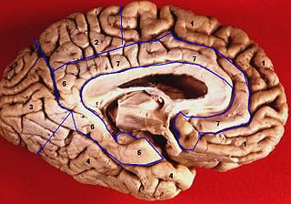
Medial surface of left cerebral hemisphere.

Gray727-Brodman.png

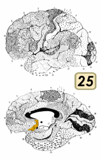
Medial surface. (Areas 23 and 31 at center right. The image is reversed from image above.)
Details
Part of Cingulate gyrus
Identifiers
Latin Cortex cingularis posterior
NeuroNames 162
NeuroLex ID birnlex_950
FMA 61924

Anatomical terms of neuroanatomy

Sagittal MRI slice with highlighting indicating location of the posterior cingulate. MRI posterior cingulate.png
Sagittal MRI slice with highlighting indicating location of the posterior cingulate.

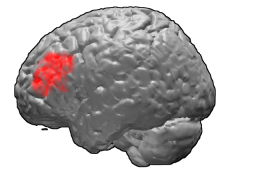
The posterior cingulate cortex (PCC) is the caudal part of the cingulate cortex, located posterior to the anterior cingulate cortex. This is the upper part of the "limbic lobe". The cingulate cortex is made up of an area around the midline of the brain. Surrounding areas include the retrosplenial cortex and the precuneus.

Cingulate cortex

The cingulate cortex is a part of the brain situated in the medial aspect of the cerebral cortex. The cingulate cortex includes the entire cingulate gyrus, which lies immediately above the corpus callosum, and the continuation of this in the cingulate sulcus. The cingulate cortex is usually considered part of the limbic lobe.

Anterior cingulate cortex brain region

The anterior cingulate cortex (ACC) is the frontal part of the cingulate cortex that resembles a "collar" surrounding the frontal part of the corpus callosum. It consists of Brodmann areas 24, 32, and 33.

Limbic lobe area of the brain

The limbic lobe is an arc-shaped region of cortex on the medial surface of each cerebral hemisphere of the mammalian brain, consisting of parts of the frontal, parietal and temporal lobes. The term is ambiguous, with some authors including the paraterminal gyrus, the subcallosal area, the cingulate gyrus, the parahippocampal gyrus, the dentate gyrus, the hippocampus and the subiculum; while the Terminologia Anatomica includes the cingulate sulcus, the cingulate gyrus, the isthmus of cingulate gyrus, the fasciolar gyrus, the parahippocampal gyrus, the parahippocampal sulcus, the dentate gyrus, the fimbrodentate sulcus, the fimbria of hippocampus, the collateral sulcus, and the rhinal sulcus, and omits the hippocampus.

Contents

Cytoarchitectonically the posterior cingulate cortex is associated with Brodmann areas 23 and 31.

Brodmann area

A Brodmann area is a region of the cerebral cortex, in the human or other primate brain, defined by its cytoarchitecture, or histological structure and organization of cells.

Brodmann area 23

Brodmann area 23 (BA23) is a region in the brain corresponding to some portion of the posterior cingulate cortex. It lies between Brodmann area 30 and Brodmann area 31 and is located on the medial wall of the cingulate gyrus between the callosal sulcus and the cingulate sulcus.

Brodmann area 31

Brodmann area 31, also known as dorsal posterior cingulate area 31, is a subdivision of the cytoarchitecturally defined cingulate region of the cerebral cortex. In the human it occupies portions of the posterior cingulate gyrus and medial aspect of the parietal lobe. Approximate boundaries are the cingulate sulcus dorsally and the parieto-occipital sulcus caudally. It partially surrounds the subparietal sulcus, the ventral continuation of the cingulate sulcus in the parietal lobe. Cytoarchitecturally it is bounded rostrally by the ventral anterior cingulate area 24, ventrally by the ventral posterior cingulate area 23, dorsally by the gigantopyramidal area 4 and preparietal area 5 and caudally by the superior parietal area 7 (H) (Brodmann-1909).

The posterior cingulate cortex forms a central node in the default mode network of the brain. It has been shown to communicate with various brain networks simultaneously and is involved in various functions. [1] Along with the precuneus, the posterior cingulate cortex has been implicated as a neural substrate for human awareness in numerous studies of both the anesthesized and vegetative (coma) state. Imaging studies indicate a prominent role for the posterior cingulate cortex in pain and episodic memory retrieval. [2] Increased size of posterior ventral cingulate cortex is related to declines in working memory performance. [3] The posterior cingulate cortex has been strongly implicated as a key part of several intrinsic control networks. [4] [5]

Default mode network large scale brain network, active when one is not focused on the outside world and the brain is at wakeful rest (e.g. daydreaming), and also while thinking about others, thinking about oneself, remembering the past, and planning for the future

In neuroscience, the default mode network (DMN), also default network, or default state network, is a large scale brain network of interacting brain regions known to have activity highly correlated with each other and distinct from other networks in the brain.

Anatomy

Location and boundaries

The posterior cingulate cortex is the backmost part of the cingulate cortex, lying behind the anterior cingulate cortex. The PCC forms a part of the posteromedial cortex, along with the retrosplenial cortex (Brodmann areas 29 and 30) and precuneus (located posterior and superior to the PCC). The PCC, together with the retrosplenial cortex, forms the retrosplenial gyrus. The posterior cingulate cortex is bordered by the following brain regions: the marginal ramus of the cingulate sulcus (superiorly), the corpus callosum (inferiorly), the parieto-occipital sulcus (posteriorly), and Brodmann area 24 (anteriorly). [4]

Retrosplenial cortex Part of the brains cerebral cortex

The retrosplenial cortex (RSC) is a cortical area in the brain, located posteriorly and comprising Brodmann areas 29 and 30 .The region's name refers to its anatomical location immediately behind the splenium of the corpus callosum in primates, although in rodents it is located more towards the brain surface and is relatively larger. Its function is currently not well understood, but its location close to visual areas and also to the hippocampal spatial/memory system suggest it may have a role in mediating between perceptual and memory functions.

Brodmann area 29

Brodmann area 29, also known as granular retrolimbic area 29 or granular retrosplenial cortex, is a cytoarchitecturally defined portion of the retrosplenial region of the cerebral cortex. In the human it is a narrow band located in the isthmus of cingulate gyrus. Cytoarchitecturally it is bounded internally by the ectosplenial area 26 and externally by the agranular retrolimbic area 30 (Brodmann-1909).

Brodmann area 30

Brodmann area 30, also known as agranular retrolimbic area 30, is a subdivision of the cytoarchitecturally defined retrosplenial region of the cerebral cortex. In the human it is located in the isthmus of cingulate gyrus. Cytoarchitecturally it is bounded internally by the granular retrolimbic area 29, dorsally by the ventral posterior cingulate area 23 and ventrolaterally by the ectorhinal area 36 (Brodmann-1909).

Cytoarchitectural organization

The posterior cingulate cortex is considered a paralimbic cortical structure, consisting of Brodmann areas 23 and 31. As part of the paralimbic cortex, it has fewer than six layers, placing its cell architecture in between the six-layered neocortex and the more primitive allocortex of core limbic structures. It has also been associated with the hippocampocentric subdivision of the paralimbic zone. The cytoarchitecture of the PCC is not entirely uniform, instead it contains distinct anterior and dorsal subregions, which are increasingly understood as distinct in function as well as cytoarchitectural structure. [4]

Paralimbic cortex

The paralimbic cortex is an area of three-layered cortex that includes the following regions: the piriform cortex, entorhinal cortex, the parahippocampal cortex on the medial surface of the temporal lobe, and the cingulate cortex just above the corpus callosum.

Neocortex Mammalian structure involved in higher-order brain functions

The neocortex, also called the neopallium and isocortex, is the part of the mammalian brain involved in higher-order brain functions such as sensory perception, cognition, generation of motor commands, spatial reasoning and language.

The allocortex is one of the two types of cerebral cortex, the other being the neocortex. It is characterized by having just three or four cell layers, in contrast with the six layers of the neocortex, and takes up a much smaller area than the neocortex. There are three subtypes of allocortex: the paleocortex, the archicortex, and the periallocortex – a transitional zone between the neocortex and the allocortex.

Structural connections

Nonhuman structure

In non-human primates the following structural connections of the posterior cingulate cortex are well documented: [4]

As is true in other areas of the posteromedial cortex, the posterior cingulate cortex has no apparent connections to primary sensory or motor areas. Thus, it is unlikely to be involved in low-level sensory or motor processing. [4]

Human structure

While many of the connections in non-human primates may be present in humans, they are less well documented. Studies have shown strong reciprocal connections to medial temporal lobe memory structures, such as the entorhinal cortex and the parahippocampal gyrus, which is known to be involved in associative learning and episodic memory. [6] The human posterior cingulate cortex is also connected to areas involved in emotion and social behavior, attention (the lateral intraparietal cortex and precuneus), and learning and motivation (the anterior and lateral thalamic nucleus, caudate nucleus, orbitofrontal cortex, and anterior cingulate cortex). [5] [7]

Function

The posterior cingulate cortex is highly connected and one of the most metabolically active regions in the brain, but there is no consensus as to its cognitive role. [4] [5] Cerebral blood flow and metabolic rate in the posterior cingulate cortex are approximately 40% higher than average across the brain. The posterior cingulate cortex also has high functional connectivity, signifying extensive intrinsic connectivity networks (networks of brain regions involved in a range of tasks that share common spatio-temporal patterns of activity). [4]

Emotion and memory

The posterior cingulate cortex has been linked by lesion studies to spatial memory, configural learning, and maintenance of discriminative avoidance learning. [6] More recently the PCC was shown to display intense activity when autobiographical memories (such as those concerning friends and family) are recalled successfully. In a study involving autobiographical recollection, the caudal part of the left posterior cingulate cortex was the only brain structure highly active in all subjects. [6] Furthermore, the PCC does not show this same activation during attempted but unsuccessful retrieval, implying an important role in successful memory retrieval (see below: Alzheimer’s Disease). [6]

The posterior cingulate cortex has also been firmly linked to emotional salience. [6] [7] Thus, it has been hypothesized that the emotional importance of autobiographical memories may contribute to the strength and consistency of activity in the posterior cingulate cortex upon successful recollection of these memories. [6] The posterior cingulate cortex is significantly bilaterally activated by emotional stimuli, independent of valence (positive or negative). This is in contrast to other structures in the limbic system, such as the amygdala, which responded disproportionately to negative stimuli, or the left frontal pole, which activated only in response to positive stimuli. These results support the hypothesis that the posterior cingulate cortex mediates interactions between emotion and memory.

Intrinsic control networks

The posterior cingulate cortex exhibits connectivity with a wide range of intrinsic control networks. Its most widely known role is as a central node in the default mode network (DMN). The default mode network (and the PCC) is highly reactive and quickly deactivates during tasks with externally directed, or presently centered, attention (such as working memory or meditation). [4] [8] Conversely, the DMN is active when attention is internally directed (during episodic memory retrieval, planning, and daydreaming). A failure of the DMN to deactivate at proper times is associated with poor cognitive function, thereby indicating its importance in attention. [4]

In addition to the default mode network, the posterior cingulate cortex is also involved in the dorsal attention network (a top-down control of visual attention and eye movement) and the frontoparietal control network (involved in executive motor control). [4] Furthermore, fMRI studies have shown that the posterior cingulate cortex activates during visual tasks when some form of monetary incentive is involved, essentially functioning as a neural interface between motivation-related areas and top-down control of visual attention. [9] [10]

The relationship between these networks in and within the PCC is not clearly understood. When activity increases in the dorsal attention network and the frontoparietal control network, it must simultaneously decrease in the DMN in a closely correlated way. This anti-correlated pattern is indicative of the various differences and importance of subregions in the posterior cingulate cortex. [4]

Considering the PCC's relation with the DMN, since suppressed posterior cingulate activity favors low cognitive introspection and higher external attention while increased activity indicates memory retrieval and planning, it has been hypothesized that this brain region is heavily involved in noticing internal and external changes and facilitating novel behavior or thought in response. High activity, then, would indicate continued operation with the current cognitive set, while lower activity would indicate exploration, flexibility, and renewed learning. [5]

An alternative hypothesis is focused more on the difference between the dorsal and ventral subregions and takes into consideration their functional separation. In this model, the PCC is hypothesized to take a chief regulatory role in focusing internal and external attention. Mounting evidence that the posterior cingulate cortex is involved in both integrating memories of experiences and initiating a signal to change behavioral strategies supports this hypothesis. Under this model, the PCC plays a crucial role in controlling state of arousal, the breadth of focus, and the internal or external focus of attention. This hypothesis emphasizes the PCC as a dynamic network, rather than a fixed and unchanging structure. [4]

While both of the hypotheses are the result of scientific studies, the role of the PCC is still not well understood and there remains much work to be done to investigate the extent of their veracity. [4] [5]

Meditation

From neuroimaging and subjective descriptions, the PCC has been found to be activated during self-related thinking and deactivated during meditation. [11] Using generative topographic mapping, it was further found that undistracted, effortless mind wandering corresponded with PCC deactivation, whereas distracted and controlled awareness corresponded with PCC activation. [11] These results track closely with findings about the PCC's role in the DMN.

Disorders

Structural and functional abnormalities in the PCC result in a range of neurological and psychiatric disorders. The PCC likely integrates and mediates information in the brain. Therefore, the functional abnormalities of the PCC might be an accumulation of remote and widespread damage in the brain. [4]

Alzheimer's disease

The PCC is commonly affected by neurodegenerative disease. [12] In fact, reduced metabolism in the PCC is an early sign of Alzheimer's disease, and is frequently present before a clinical diagnosis. [4] The reduced metabolism in the PCC is typically one part in a diffuse pattern of metabolic dysfunction in the brain that includes medial Temporal lobe structures and the anterior thalamus, and these metabolic abnormalities may be due to damage in isolated but connected regions. [4] For instance, Meguro et al. (1999) show that experimental damage of the rhinal cortex results in hypometabolism of the PCC. [13] In Alzheimer's disease, the metabolic abnormality is linked to Amyloid deposition and brain Atrophy with a spatial distribution that resembles the nodes of the Default mode network (DMN). [4] In early Alzheimer's, the functional connectivity within the DMN is reduced and affects the connection between the PCC and the hippocampus, and these altered patterns can reflect ApoE genetic status (a risk factor associated with the disease). [4] It has been found that neurodegenerative diseases spread 'prion-like' through the brain. [4] For example, when the proteins amyloid-b and TDP-43 are in their abnormal form, they spread trans-synaptically and are associated with Neurodegeneration. [4] This transmission of abnormal protein would be constrained by the organization of white matter connections and could potentially explain the spatial distribution of Alzheimer's pathology within the DMN. [4] In Alzheimer’s disease, the topology of white matter connectivity helps in predicting atrophic patterns, [14] which could explain why the PCC is affected in the early stages of the disease. [4]

Autism spectrum disorder

Autism spectrum disorders (ASDs) are associated with metabolic and functional abnormalities of the PCC. Individuals with ASDs show reduction in metabolism, exhibit abnormal functional responses and demonstrate reductions in functional connectivity. [4] One study showed these reductions are prominent in the PCC.[ non-primary source needed ] [15] Studies have shown that the abnormalities in cingulate responses during interpersonal interaction correlate with the severity of autistic symptoms, and the failure to show task dependent deactivation in the PCC correlates with overall social function. [4] Finally, post-mortem studies show that the PCCs of patients with ASDs have cytoarchitectonic abnormalities, as well as reduced levels of GABA A receptors, and benzodiazepine binding sites. [4]

Attention deficit hyperactivity disorder

It has been suggested that ADHD is a disorder of the DMN, where neural systems are disrupted by uncontrolled activity that leads to attentional lapses. [16] Nakao et al. (2011) performed a meta-analysis of structural MRI studies and found that patients with ADHD exhibit an increased left PCC, [17] which could mean that developmental abnormalities affect the PCC. In fact, PCC function is abnormal in ADHD. [4] Within the DMN, functional connectivity is reduced and the resting state activity is a way to diagnose children with ADHD. [4] Treatment for ADHD, includes psychostimulant medication that directly affects PCC activity. [4] Other studies looking at medications for abnormalities in the PCC found that the PCC may only respond to stimulant treatments and that the effectiveness of medication can be dependent on motivation levels. [4] Furthermore, ADHD has been associated with the gene SNAP25. In healthy children, SNAP25 polymorphism is linked to working memory capacity, altered PCC structure, and task-dependent PCC deactivation patterns on working memory task. [18]

Depression

Abnormal PCC functional connectivity has been linked to major depression. However, the conclusions of the studies vary. One found increased PCC functional connectivity, [19] while another showed that untreated patients had decreased functional connectivity from the PCC to the caudate. [20] Other studies have looked at interactions between the PCC and the sub-genual cingulate region (Brodmann area 25), a region of the brain that potentially causes depression. [4] The anterior node of the DMN is formed, in part, by the highly connected PCC and Brodmann area 25. The two regions are metabolically overactive in treatment resistant major depression. [21] The link between the activity in the PCC and Brodmann area 25 correlates with rumination, a feature of depression. [22] This link between the two regions could influence medication responses in patients. Already, it has been found that both regions show alterations in metabolism after antidepressant treatment. Furthermore, patients who undergo Deep brain stimulation, had increased glucose metabolism and cerebral flow in the PCC, while also altering Brodmann area 25. [4]

Schizophrenia

Abnormal activity in the PCC has been linked to schizophrenia, a mental disorder with common symptoms such as hallucinations, delusions, disorganized thinking, and a lack of emotional intelligence. What is common between symptoms is that they have to do with an inability to distinguish between internal and external events; they lack insight. Two PET studies on patients with schizophrenia showed abnormal metabolism in the PCC. One study found that glucose metabolism was decreased in schizophrenics, [23] while another found abnormal glucose metabolism that was highly correlated in the pulvinar and the PCC. [24] In the latter study, thalamic interactions with the frontal lobes were reduced, which could mean that schizophrenia affects thalamocortical connections. Further abnormalities in the PCC, abnormal NMDA, cannabinoid, and DABAergic receptor binding have been found with post-mortem autoradiography of schizophrenics. [25] Abnormalities in the structure and white matter connections of the PCC have also been found in schizophrenic patients. Schizophrenics with a poor outcome often have reduced PCC volume. [24] It has further been found that white matter abnormalities in the cingulum bundle, a structure that connects the PCC to other limbic structures. [26] In functional MRI studies, there is more evidence of abnormal PCC function. There can be increases and decreases in the functional connectivity. [27] There are also abnormal PCC responses during task performance. [28] All of the previously mentioned network abnormalities may be the reason for psychotic symptoms. Recently it was found that the psychedelic drug psilocybin induces an altered state of consciousness and is related to abnormal metabolism and functional connectivity of the PCC, as well as a reduction in the strength of anti-correlations between the DMN and the FPCN. [29] These networks contribute to internal and external cognition, so abnormalities in these networks might be responsible for the psychosis in schizophrenia.

Traumatic brain injury

After traumatic brain injury (TBI), abnormalities have been shown in the PCC. Often, head injuries produce widespread axonal injury that disconnect brain regions and lead to cognitive impairment. This is also related to reduced metabolism within the PCC. [30] Studies have looked at performance on simple choice reaction time tasks after TBIs. [31] This study, in particular, found that the pattern of functional connectivity from the PCC to the rest of the DMN could predict TBI impairments even before symptoms had manifested. They also found that greater damage to the cingulum bundle, that connects the PCC to the anterior DMN, was linked to impairments of sustained attention. In a subsequent study, it was found that TBIs are related to a difficulty in switching from automatic to controlled responses. [32] Within selected tasks, patients with TBIs showed impaired motor inhibition that was associated with failure to rapidly reactive the PCC. Collectively, this suggests that the failure to control the PCC/DMN activity can lead to attentional lapses in TBI patients.

Anxiety disorders

There is accumulating evidence for PCC dysfunction underlying many childhood/adolescent-onset mental disorders. [33] Further, anxiety disorder patients show an association between increased extinction–related PCC activity and greater symptom severity. [34] PCC dysfunction may also play a role in anxiety disorders during adolescence. [35]

See also

Related Research Articles

Precuneus part of the brain

The precuneus is the portion of the superior parietal lobule on the medial surface of each brain hemisphere. It is located in front of the cuneus. The precuneus is bounded in front by the marginal branch of the cingulate sulcus, at the rear by the parietooccipital sulcus, and underneath by the subparietal sulcus. It is involved with episodic memory, visuospatial processing, reflections upon self, and aspects of consciousness.

Brodmann area 9

Brodmann area 9, or BA9, is part of the frontal cortex in the brain of humans and other primates. It contributes to the dorsolateral and medial prefrontal cortex.

Brodmann area 46

Brodmann area 46, or BA46, is part of the frontal cortex in the human brain. It is between BA10 and BA45.

Claustrum

The claustrum is a thin, bilateral structure which connects to cortical and subcortical regions of the brain. It is located between the insula laterally and the putamen medially, separated by the extreme and external capsules respectively. The blood supply to the claustrum is fulfilled via the middle cerebral artery. It is considered to be the most densely connected structure in the brain allowing for integration of various cortical inputs into one experience rather than singular events. The claustrum is difficult to study given the limited number of individuals with claustral lesions and the poor resolution of neuroimaging.

Insular cortex

In each hemisphere of the mammalian brain the insular cortex is a portion of the cerebral cortex folded deep within the lateral sulcus.

Brodmann area 25

Brodmann area 25 (BA25) is the subgenual area, area subgenualis or subgenual cingulatea area in the cerebral cortex of the brain and delineated based on its cytoarchitectonic characteristics.

Orbitofrontal cortex prefrontal cortex region

The orbitofrontal cortex (OFC) is a prefrontal cortex region in the frontal lobes of the brain which is involved in the cognitive process of decision-making. In non-human primates it consists of the association cortex areas Brodmann area 11, 12 and 13; in humans it consists of Brodmann area 10, 11 and 47.

Superior longitudinal fasciculus

The superior longitudinal fasciculus (SLF) is an association fiber tract that is composed of three separate components. It is present in both hemispheres and can be found lateral to the centrum ovale and connects the frontal, occipital, parietal, and temporal lobes. These bundles of axon tracts pass from the frontal lobe through the operculum to the posterior end of the lateral sulcus where they either radiate to and synapse on neurons in the occipital lobe or turn downward and forward around the putamen and then radiate to and synapse on neurons in anterior portions of the temporal lobe.

The task-positive network (TPN) is a network of areas in the human brain that typically responds with activation increases to attention-demanding tasks in functional imaging studies. The task-positive network encompasses regions of the dorsal attention system, but in addition includes dorsolateral and ventrolateral prefrontal regions, the insular cortex, and the SMA/pre-SMA. Notably, the nodes of this network are also correlated during rest. The task-positive network is anti-correlated with the default mode network.

Brain activity and meditation

Meditation and its effect on brain activity and the central nervous system became a focus of collaborative research in neuroscience, psychology and neurobiology during the latter half of the 20th century. Research on meditation sought to define and characterize various practices. Meditation’s effect on the brain can be broken up into two categories: state changes and trait changes, respectively alterations in brain activities during the act of meditating and changes that are the outcome of long-term practice.

Resting state fMRI

Resting state fMRI is a method of functional magnetic resonance imaging (fMRI) that is used in brain mapping to evaluate regional interactions that occur in a resting or task-negative state, when an explicit task is not being performed. A number of resting-state conditions are identified in the brain, one of which is the default mode network. These resting brain state conditions are observed through changes in blood flow in the brain which creates what is referred to as a blood-oxygen-level dependent (BOLD) signal that can be measured using fMRI. Because brain activity is intrinsic, present even in the absence of an externally prompted task, any brain region will have spontaneous fluctuations in BOLD signal. The resting state approach is useful to explore the brain's functional organization and to examine if it is altered in neurological or mental disorders. Resting-state functional connectivity research has revealed a number of networks which are consistently found in healthy subjects, different stages of consciousness and across species, and represent specific patterns of synchronous activity.

The dorsal nexus is an area within the dorsal medial prefrontal cortex that serves as an intersection point for multiple brain networks. Research suggests it plays a role in the maintenance and manipulation of information, as well as supporting the control of cognitive functions such as behavior, memory, and conflict resolution. Abnormally increased connectivity between these networks through the Dorsal Nexus has been associated with certain types of depression. The activity generated by this abnormally high level of connectivity during a depressive state can be identified through Magnetic resonance imaging (MRI) and Positron emission tomography (PET).

Fronto-cerebellar dissociation

Fronto-cerebellar dissociation is the disconnection and independent function of frontal and cerebellar regions of the brain. It is characterized by inhibited communication between the two regions, and is notably observed in cases of ADHD, schizophrenia, alcohol abuse, and heroin abuse. The frontal and cerebellar regions make distinctive contributions to cognitive performance, with the left-frontal activations being responsible for selecting a response to a stimulus, while the right-cerebellar activation is responsible for the search for a given response to a stimulus. Left-frontal activation increases when there are many appropriate responses to a stimulus, and right-cerebellar activation increases when there is a single appropriate response to a stimulus. A person with dissociated frontal and cerebellar regions may have difficulties with selecting a response to a stimuli, or difficulties with response initiation. Fronto-cerebellar dissociation can often result in either the frontal lobe or the cerebellum becoming more active in place of the less active region as a compensatory effect.

Salience network large scale brain network of the human brain, composed of the anterior insula and dorsal anterior cingulate cortex, which is involved in detecting and filtering salient stimuli, as well as in recruiting relevant functional networks

The salience network (SN) is a large scale brain network of the human brain that is primarily composed of the anterior insula (AI) and dorsal anterior cingulate cortex (dACC). It is involved in detecting and filtering salient stimuli, as well as in recruiting relevant functional networks. Together with its interconnected brain networks, the SN contributes to a variety of complex functions, including communication, social behavior, and self-awareness through the integration of sensory, emotional, and cognitive information.

Social cognitive neuroscience is the scientific study of the biological processes underpinning social cognition. Specifically, it uses the tools of neuroscience to study "the mental mechanisms that create, frame, regulate, and respond to our experience of the social world". Social cognitive neuroscience uses the epistemological foundations of cognitive neuroscience, and is closely related to social neuroscience. Social cognitive neuroscience employs human neuroimaging, typically using functional magnetic resonance imaging (fMRI). Human brain stimulation techniques such as transcranial magnetic stimulation and transcranial direct-current stimulation are also used. In nonhuman animals, direct electrophysiological recordings and electrical stimulation of single cells and neuronal populations are utilized for investigating lower-level social cognitive processes.

References

  1. R Leech; R Braga; DJ Sharp (2013). "Echoes of the brain within the posterior cingulate cortex". The Journal of Neuroscience. 32 (1): 215–222. doi:10.1523/JNEUROSCI.3689-11.2012. PMID   22219283.
  2. Nielsen FA, Balslev D, Hansen LK (2005). "Mining the posterior cingulate: segregation between memory and pain components". NeuroImage. 27 (3): 520–532. doi:10.1016/j.neuroimage.2005.04.034. PMID   15946864.
  3. Kozlovskiy SA, Vartanov AV, Nikonova EY, Pyasik MM, Velichkovsky BM (2012). "The Cingulate Cortex and Human Memory Processes". Psychology in Russia: State of the Art. 5: 231–243. doi:10.11621/pir.2012.0014.
  4. 1 2 3 4 5 6 7 8 9 10 11 12 13 14 15 16 17 18 19 20 21 22 23 24 25 26 27 28 29 30 31 Leech R, Sharp DJ (July 2013). "The role of the posterior cingulate cortex in cognition and disease". Brain. 137 (Pt 1): 12–32. doi:10.1093/brain/awt162. PMC   3891440 Lock-green.svg. PMID   23869106.
  5. 1 2 3 4 5 Pearson, John M.; Heilbronner, Sarah R.; Barack, David L.; Hayden, Benjamin Y.; Platt, Michael L. (April 2011). "Posterior cingulate cortex: adapting behavior to a changing world". Trends in Cognitive Sciences. 15 (4): 143–151. doi:10.1016/j.tics.2011.02.002. PMC   3070780 Lock-green.svg. PMID   21420893.
  6. 1 2 3 4 5 6 Maddock, R. J.; A. S. Garrett; M. H. Buonocore (2001). "Remembering Familiar People: The Posterior Cingulate Cortex and Autobiographical Memory Retrieval". Neuroscience. 104 (3): 667–676. doi:10.1016/s0306-4522(01)00108-7.
  7. 1 2 Maddock, Richard J.; Garrett, Amy S.; Buonocore, Michael H. (January 2003). "Posterior cingulate cortex activation by emotional words: fMRI evidence from a valence decision task". Human Brain Mapping. 18 (1): 30–41. doi:10.1002/hbm.10075.
  8. Brewer, Judson A.; Garrison, Kathleen A.; Whitfield-Gabrieli, Susan (2013). "What about the "Self" is Processed in the Posterior Cingulate Cortex?". Frontiers in Human Neuroscience. 7. doi:10.3389/fnhum.2013.00647.
  9. Small, Dana A.; Gitelman, Darren; Simmons, Katharine; Bloise, Suzanne; Parrish, Todd; Mesulam, Marsel M. (2005). "Monetary Incentives Enhance Processing in Brain Regions Mediating Top-down Control of Attention". Cerebral Cortex. 15: 1855–1865. doi:10.1093/cercor/bhi063.
  10. Engelmann, Jan B.; Damaraju, Eswar; Padmala, Srikanth; Pessoa, Luiz (2009). "Combined Effects of Attention and Motivation on Visual Task Performance: Transient and Sustained Motivational Effects". Frontiers in Human Neuroscience. 3. doi:10.3389/neuro.09.004.2009.
  11. 1 2 Garrison KA, Santoyo JF, Davis JH, Thornhill TA, Kerr CE, Brewer JA (2013). "Effortless awareness: using real time neurofeedback to investigate correlates of posterior cingulate cortex activity in meditators' self-report". Front Hum Neurosci. 7: 440. doi:10.3389/fnhum.2013.00440. PMC   3734786 Lock-green.svg. PMID   23964222.
  12. Buckner, RL; Andrews-Hanna JR; Schacter DL (2008). "The brain's default network: anatomy, function, and relevance to disease". Ann N Y Acad Sci. 1124: 1–38. doi:10.1196/annals.1440.011. PMID   18400922.
  13. Meguro, K. (1999). "Neocortical and hippocampal glucose hypometabolism following neurotoxic lesions of the entorhinal and perirhinal cortices in the non-human primate as shown by PET: Implications for Alzheimer's disease". Brain. 122 (8): 1519–1531. doi:10.1093/brain/122.8.1519. ISSN   1460-2156.
  14. Raj, A; Kuceyeski A; Weiner M (2012). "A network diffusion model of disease progression in dementia". Neuron. 73: 1204–15. doi:10.1016/j.neuron.2011.12.040.
  15. Cherkassky, VL; Kana, RK; Keller, TA; Just, MA (Nov 6, 2006). "Functional connectivity in a baseline resting-state network in autism". NeuroReport. 17 (16): 1687–90. doi:10.1097/01.wnr.0000239956.45448.4c. PMID   17047454.
  16. Sonuga-Barke, EJ; Castellanos, FX (2007). "Spontaneous attentional fluctuations in impaired states and pathological conditions: a neurobiological hypothesis". Neuroscience and Biobehavioral Reviews. 31 (7): 977–86. doi:10.1016/j.neubiorev.2007.02.005. PMID   17445893.
  17. Nakao, T; Radua, J; Rubia, K; Mataix-Cols, D (November 2011). "Gray matter volume abnormalities in ADHD: voxel-based meta-analysis exploring the effects of age and stimulant medication". The American Journal of Psychiatry. 168 (11): 1154–63. doi:10.1176/appi.ajp.2011.11020281. PMID   21865529.
  18. Latasch, L; Christ, R (March 1988). "[Problems in anesthesia of drug addicts]". Der Anaesthesist. 37 (3): 123–39. PMID   3289412.
  19. Zhou, Y; Yu, C; Zheng, H; Liu, Y; Song, M; Qin, W; Li, K; Jiang, T (March 2010). "Increased neural resources recruitment in the intrinsic organization in major depression". Journal of Affective Disorders. 121 (3): 220–30. doi:10.1016/j.jad.2009.05.029. PMID   19541369.
  20. Bluhm, R; Williamson, P; Lanius, R; Théberge, J; Densmore, M; Bartha, R; Neufeld, R; Osuch, E (December 2009). "Resting state default-mode network connectivity in early depression using a seed region-of-interest analysis: decreased connectivity with caudate nucleus". Psychiatry and clinical neurosciences. 63 (6): 754–61. doi:10.1111/j.1440-1819.2009.02030.x. PMID   20021629.
  21. Mayberg, HS; Liotti, M; Brannan, SK; McGinnis, S; Mahurin, RK; Jerabek, PA; Silva, JA; Tekell, JL; Martin, CC; Lancaster, JL; Fox, PT (May 1999). "Reciprocal limbic-cortical function and negative mood: converging PET findings in depression and normal sadness". The American Journal of Psychiatry. 156 (5): 675–82. doi:10.1176/ajp.156.5.675. PMID   10327898.
  22. Berman, MG; Peltier, S; Nee, DE; Kross, E; Deldin, PJ; Jonides, J (October 2011). "Depression, rumination and the default network". Social cognitive and affective neuroscience. 6 (5): 548–55. doi:10.1093/scan/nsq080. PMC   3190207 Lock-green.svg. PMID   20855296.
  23. Haznedar, MM; Buchsbaum, MS; Hazlett, EA; Shihabuddin, L; New, A; Siever, LJ (Dec 1, 2004). "Cingulate gyrus volume and metabolism in the schizophrenia spectrum". Schizophrenia Research. 71 (2–3): 249–62. doi:10.1016/j.schres.2004.02.025. PMID   15474896.
  24. 1 2 Mitelman, SA; Byne, W; Kemether, EM; Hazlett, EA; Buchsbaum, MS (September 2005). "Metabolic disconnection between the mediodorsal nucleus of the thalamus and cortical Brodmann's areas of the left hemisphere in schizophrenia". The American Journal of Psychiatry. 162 (9): 1733–5. doi:10.1176/appi.ajp.162.9.1733. PMID   16135634.
  25. Newell, KA; Zavitsanou, K; Huang, XF (Aug 22, 2005). "Ionotropic glutamate receptor binding in the posterior cingulate cortex in schizophrenia patients". NeuroReport. 16 (12): 1363–7. doi:10.1097/01.wnr.0000174056.11403.71. PMID   16056140.
  26. Kubicki, M; McCarley, RW; Nestor, PG; Huh, T; Kikinis, R; Shenton, ME; Wible, CG (December 2003). "An fMRI study of semantic processing in men with schizophrenia". NeuroImage. 20 (4): 1923–33. doi:10.1016/s1053-8119(03)00383-5. PMC   2806220 Lock-green.svg. PMID   14683698.
  27. Liang, M; Zhou, Y; Jiang, T; Liu, Z; Tian, L; Liu, H; Hao, Y (Feb 6, 2006). "Widespread functional disconnectivity in schizophrenia with resting-state functional magnetic resonance imaging". NeuroReport. 17 (2): 209–13. doi:10.1097/01.wnr.0000198434.06518.b8. PMID   16407773.
  28. Whitfield-Gabrieli, S; Thermenos, HW; Milanovic, S; Tsuang, MT; Faraone, SV; McCarley, RW; Shenton, ME; Green, AI; Nieto-Castanon, A; LaViolette, P; Wojcik, J; Gabrieli, JD; Seidman, LJ (Jan 27, 2009). "Hyperactivity and hyperconnectivity of the default network in schizophrenia and in first-degree relatives of persons with schizophrenia". Proceedings of the National Academy of Sciences of the United States of America. 106 (4): 1279–84. doi:10.1073/pnas.0809141106. PMC   2633557 Lock-green.svg. PMID   19164577.
  29. Carhart-Harris RL, Erritzoe D, Williams T, Stone JM, Reed LJ, Colasanti A, Tyacke RJ, Leech R, Malizia AL, Murphy K, Hobden P, Evans J, Feilding A, Wise RG, Nutt DJ (2012). "Neural correlates of the psychedelic state as determined by fMRI studies with psilocybin". Proceedings of the National Academy of Sciences of the United States of America. 109 (6): 2138–43. doi:10.1073/pnas.1119598109. PMC   3277566 Lock-green.svg. PMID   22308440.
  30. Nakashima, T; Nakayama, N; Miwa, K; Okumura, A; Soeda, A; Iwama, T (February 2007). "Focal brain glucose hypometabolism in patients with neuropsychologic deficits after diffuse axonal injury". AJNR. American journal of neuroradiology. 28 (2): 236–42. PMID   17296986.
  31. Bonnelle V, Leech R, Kinnunen KM, Ham TE, Beckmann CF, De Boissezon X, Greenwood RJ, Sharp DJ (2011). "Default mode network connectivity predicts sustained attention deficits after traumatic brain injury". J. Neurosci. 31 (38): 13442–51. doi:10.1523/JNEUROSCI.1163-11.2011. PMID   21940437.
  32. Bonnelle, V; Ham, TE; Leech, R; Kinnunen, KM; Mehta, MA; Greenwood, RJ; Sharp, DJ (Mar 20, 2012). "Salience network integrity predicts default mode network function after traumatic brain injury". Proceedings of the National Academy of Sciences of the United States of America. 109 (12): 4690–5. doi:10.1073/pnas.1113455109. PMC   3311356 Lock-green.svg. PMID   22393019.
  33. Leech, Robert; Sharp, David J. (2014-01-01). "The role of the posterior cingulate cortex in cognition and disease". Brain. 137 (1): 12–32. doi:10.1093/brain/awt162. ISSN   0006-8950.
  34. Milad, Mohammed R.; Furtak, Sharon C.; Greenberg, Jennifer L.; Keshaviah, Aparna; Im, Jooyeon J.; Falkenstein, Martha J.; Jenike, Michael; Rauch, Scott L.; Wilhelm, Sabine (2013-06-01). "Deficits in Conditioned Fear Extinction in Obsessive-Compulsive Disorder and Neurobiological Changes in the Fear Circuit". JAMA Psychiatry. 70 (6): 608–618. doi:10.1001/jamapsychiatry.2013.914. ISSN   2168-622X.
  35. Ganella, Despina E.; Drummond, Katherine D.; Ganella, Eleni P.; Whittle, Sarah; Kim, Jee Hyun (2018). "Extinction of Conditioned Fear in Adolescents and Adults: A Human fMRI Study". Frontiers in Human Neuroscience. 11. doi:10.3389/fnhum.2017.00647. ISSN   1662-5161.

For details regarding MRI definitions of the cingulate cortex based on the Desikan-Killiany Brain atlas, see: