Bovine pancreatic ribonuclease

Last updated
Pancreatic ribonuclease
RNase A.png
Structure of RNase A
Identifiers
EC no. 3.1.27.5
CAS no. 9001-99-4
Databases
IntEnz IntEnz view
BRENDA BRENDA entry
ExPASy NiceZyme view
KEGG KEGG entry
MetaCyc metabolic pathway
PRIAM profile
PDB structures RCSB PDB PDBe PDBsum
Gene Ontology AmiGO / QuickGO
Search
PMC articles
PubMed articles
NCBI proteins
A vial of RNAse A for use in molecular biology. RNAseA.jpg
A vial of RNAse A for use in molecular biology.

Bovine pancreatic ribonuclease, also often referred to as bovine pancreatic ribonuclease A or simply RNase A, is a pancreatic ribonuclease enzyme that cleaves single-stranded RNA. Bovine pancreatic ribonuclease is one of the classic model systems of protein science. [1] Two Nobel Prizes in Chemistry have been awarded in recognition of work on bovine pancreatic ribonuclease: in 1972, the Prize was awarded to Christian Anfinsen for his work on protein folding and to Stanford Moore and William Stein for their work on the relationship between the protein's structure and its chemical mechanism; [2] in 1984, the Prize was awarded to Robert Bruce Merrifield for development of chemical synthesis of proteins. [3]

Contents

History

Bovine pancreatic ribonuclease became a common model system in the study of proteins largely because it was extremely stable and could be purified in large quantities. In the 1940s Armour and Company purified a kilogram of protein - a very large quantity, particularly by the protein purification standards of the time - and offered samples at low cost to interested scientists. [4] The ability to have a single lot of purified enzyme made it a predominant model system for protein studies. It remains commonly referred to as ribonuclease A or RNase A as the most prominent member of its protein family, known variously as pancreatic ribonuclease, ribonuclease A, or ribonuclease I.

Christian Anfinsen's studies of the oxidative folding process of bovine pancreatic ribonuclease laid the groundwork for understanding the relationship between amino acid sequence and a protein's folded three-dimensional structure and solidified the thermodynamic hypothesis of protein folding, according to which the folded form of a protein represents its free energy minimum. [4] [5]

RNase A was the first enzyme for which a correct catalytic mechanism was proposed, even before its structure was known. [6] RNase A was the first protein for showing the effects of non-native isomers of peptide bonds preceding proline residues in protein folding. [7]

Bovine pancreatic ribonuclease was also the model protein used to work out many spectroscopic methods for assaying protein structure, including absorbance, circular dichroism, Raman, electron paramagnetic resonance (EPR) and nuclear magnetic resonance (NMR) spectroscopy. It was the first model protein for the development of chemical methods for the study of proteins, such as chemical modification of exposed side chains, antigenic recognition, and limited proteolysis of disordered segments. Ribonuclease S, which is RNase A that has been treated with the protease subtilisin, was the third protein to have its crystallographic structure solved, in 1967. [8]

Structure and properties

Labeled ribbon diagram dalla ribonuclease A pancreatica bovina (PDB accession code 7RSA). The backbone ribbon is colored from blue (N-terminus) to red (C-terminus). The side chains of the four disulfide-bonded cysteines are shown in yellow, with their sulfur atoms highlighted as small spheres. Residues important for catalysis are shown in magenta. Ribonuclease A 7rsa.png
Labeled ribbon diagram dalla ribonuclease A pancreatica bovina (PDB accession code 7RSA). The backbone ribbon is colored from blue (N-terminus) to red (C-terminus). The side chains of the four disulfide-bonded cysteines are shown in yellow, with their sulfur atoms highlighted as small spheres. Residues important for catalysis are shown in magenta.

RNase A is a relatively small protein (124 residues, ~13.7 kDa). It can be characterized as a two-layer protein with a deep cleft for binding the RNA substrate. The first layer is composed of three alpha helices (residues 3-13, 24-34 and 50-60) from the N-terminal half of the protein. The second layer consist of three β-hairpins (residues 61-74, 79-104 and 105-124 from the C-terminal half) arranged in two β-sheets. The hairpins 61-74 and 105-124 form a four-stranded, antiparallel β-sheet that lies on helix 3 (residues 50-60). The longest β-hairpin 79-104 mates with a short β-strand (residues 42-45) to form a three-stranded, antiparallel β-sheet that lies on helix 2 (residues 24-34).

RNase A has four disulfide bonds in its native state: Cys26-Cys84, Cys58-110, Cys40-95 and Cys65-72. The first two (26-84 and 58-110) are essential for conformational folding; each joins an alpha helix of the first layer to a beta sheet of the second layer, forming a small hydrophobic core in its vicinity. The latter two disulfide bonds (40-95 and 65-72) are less essential for folding; either one can be reduced (but not both) without affecting the native structure under physiological conditions. These disulfide bonds connect loop segments and are relatively exposed to solvent. The 65-72 disulfide bond has an extraordinarily high propensity to form, significantly more than would be expected from its loop entropy, both as a peptide and in the full-length protein. This suggests that the 61-74 β-hairpin has a high propensity to fold conformationally.

RNase A is a basic protein (pI = 9.63); its many positive charges are consistent with its binding to RNA (a poly-anion). More generally, RNase A is unusually polar or, rather, unusually lacking in hydrophobic groups, especially aliphatic ones. This may account for its need of four disulfide bonds to stabilize its structure. The low hydrophobic content may also serve to reduce the physical repulsion between highly charged groups (its own and those of its substrate RNA) and regions of low dielectric constant (the nonpolar residues).

The N-terminal α-helix of RNase A (residues 3-13) is connected to the rest of RNase A by a flexible linker (residues 16-23). As shown by F. M. Richards, this linker may be cleaved by subtilisin between residues 20 and 21 without causing the N-terminal helix to dissociate from the rest of RNase A. The peptide-protein complex is called "RNase S", the peptide (residues 1-20) is called the "S-peptide" and the remainder (residues 21-124) is called the "S-protein". The dissociation constant of the S-peptide for the S-protein is roughly 30 pM; this tight binding can be exploited for protein purification by attaching the S-peptide to the protein of interest and passing a mixture over an affinity column with bound S-protein. [A smaller C-peptide (residues 1-13) also works.] The RNase S model system has also been used for studying protein folding by coupling folding and association. The S-peptide was the first peptide from a native protein shown to have (flickering) secondary structure in isolation (by Klee and Brown in 1967).

RNase A cleaves specifically after pyrimidine nucleotides. [9] Cleavage takes place in two steps: first, the 3’,5’-phosphodiester bond is cleaved to generate a 2’,3’-cyclic phosphodiester intermediate; second, the cyclic phosphodiester is hydrolyzed to a 3’-monophosphate. [10] It can be inhibited by ribonuclease inhibitor protein, by heavy metal ions, and by uridine-vanadate complexes. [10]

Enzymatic mechanism

The positive charges of RNase A lie mainly in a deep cleft between two lobes. The RNA substrate lies in this cleft and is cleaved by two catalytic histidine residues, His12 and His119, to form a 2',3'-cyclic phosphate intermediate that is stabilized by nearby Lys41.

Enzyme regulation

This protein may use the morpheein model of allosteric regulation. [11]

See also

Related Research Articles

<span class="mw-page-title-main">Beta sheet</span> Protein structural motif

The beta sheet is a common motif of the regular protein secondary structure. Beta sheets consist of beta strands (β-strands) connected laterally by at least two or three backbone hydrogen bonds, forming a generally twisted, pleated sheet. A β-strand is a stretch of polypeptide chain typically 3 to 10 amino acids long with backbone in an extended conformation. The supramolecular association of β-sheets has been implicated in the formation of the fibrils and protein aggregates observed in amyloidosis, Alzheimer's disease and other proteinopathies.

<span class="mw-page-title-main">Proinsulin</span> Precursor protein in humans

Proinsulin is the prohormone precursor to insulin made in the beta cells of the Pancreatic Islets, specialized regions of the pancreas. In humans, proinsulin is encoded by the INS gene. The pancreatic islets only secrete between 1% and 3% of proinsulin intact. However, because proinsulin has a longer half life than insulin, it can account for anywhere from 5–30% of the insulin-like structures circulating in the blood. There are higher concentrations of proinsulin after meals and lower levels when a person is fasting. Additionally, while proinsulin and insulin have structural differences, proinsulin does demonstrate some affinity for the insulin receptor. Due to the relative similarities in structure, proinsulin can produce between 5% and 10% of the metabolic activity similarly induced by insulin.

<span class="mw-page-title-main">Ribonuclease</span> Class of enzyme that catalyzes the degradation of RNA

Ribonuclease is a type of nuclease that catalyzes the degradation of RNA into smaller components. Ribonucleases can be divided into endoribonucleases and exoribonucleases, and comprise several sub-classes within the EC 2.7 and 3.1 classes of enzymes.

<span class="mw-page-title-main">Protein disulfide-isomerase</span> Class of enzymes

Protein disulfide isomerase, or PDI, is an enzyme in the endoplasmic reticulum (ER) in eukaryotes and the periplasm of bacteria that catalyzes the formation and breakage of disulfide bonds between cysteine residues within proteins as they fold. This allows proteins to quickly find the correct arrangement of disulfide bonds in their fully folded state, and therefore the enzyme acts to catalyze protein folding.

<span class="mw-page-title-main">Ribonuclease H</span> Enzyme family

Ribonuclease H is a family of non-sequence-specific endonuclease enzymes that catalyze the cleavage of RNA in an RNA/DNA substrate via a hydrolytic mechanism. Members of the RNase H family can be found in nearly all organisms, from bacteria to archaea to eukaryotes.

<span class="mw-page-title-main">Ranpirnase</span> Enzyme from the Northern Leopard Frog

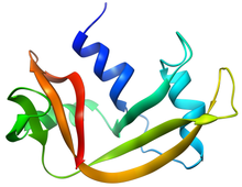
Ranpirnase is a ribonuclease enzyme found in the oocytes of the Northern Leopard Frog. Ranpirnase is a member of the pancreatic ribonuclease protein superfamily and degrades RNA substrates with a sequence preference for uracil and guanine nucleotides. Along with amphinase, another leopard frog ribonuclease, Ranpirnase has been studied as a potential cancer and antiviral treatment due to its unusual mechanism of cytotoxicity tested against transformed cells and antiviral activity.

<span class="mw-page-title-main">Enteropeptidase</span> Class of enzymes

Enteropeptidase is an enzyme produced by cells of the duodenum and is involved in digestion in humans and other animals. Enteropeptidase converts trypsinogen into its active form trypsin, resulting in the subsequent activation of pancreatic digestive enzymes. Absence of enteropeptidase results in intestinal digestion impairment.

<span class="mw-page-title-main">Angiogenin</span> Protein-coding gene in the species Homo sapiens

Angiogenin (ANG) also known as ribonuclease 5 is a small 123 amino acid protein that in humans is encoded by the ANG gene. Angiogenin is a potent stimulator of new blood vessels through the process of angiogenesis. Ang hydrolyzes cellular RNA, resulting in modulated levels of protein synthesis and interacts with DNA causing a promoter-like increase in the expression of rRNA. Ang is associated with cancer and neurological disease through angiogenesis and through activating gene expression that suppresses apoptosis.

<span class="mw-page-title-main">Ribonuclease P</span> Class of enzymes

Ribonuclease P is a type of ribonuclease which cleaves RNA. RNase P is unique from other RNases in that it is a ribozyme – a ribonucleic acid that acts as a catalyst in the same way that a protein-based enzyme would. Its function is to cleave off an extra, or precursor, sequence of RNA on tRNA molecules. Further, RNase P is one of two known multiple turnover ribozymes in nature, the discovery of which earned Sidney Altman and Thomas Cech the Nobel Prize in Chemistry in 1989: in the 1970s, Altman discovered the existence of precursor tRNA with flanking sequences and was the first to characterize RNase P and its activity in processing of the 5' leader sequence of precursor tRNA. Recent findings also reveal that RNase P has a new function. It has been shown that human nuclear RNase P is required for the normal and efficient transcription of various small noncoding RNAs, such as tRNA, 5S rRNA, SRP RNA and U6 snRNA genes, which are transcribed by RNA polymerase III, one of three major nuclear RNA polymerases in human cells.

<span class="mw-page-title-main">Ribonuclease inhibitor</span>

Ribonuclease inhibitor (RI) is a large (~450 residues, ~49 kDa), acidic (pI ~4.7), leucine-rich repeat protein that forms extremely tight complexes with certain ribonucleases. It is a major cellular protein, comprising ~0.1% of all cellular protein by weight, and appears to play an important role in regulating the lifetime of RNA.

<span class="mw-page-title-main">Beta hairpin</span>

The beta hairpin is a simple protein structural motif involving two beta strands that look like a hairpin. The motif consists of two strands that are adjacent in primary structure, oriented in an antiparallel direction, and linked by a short loop of two to five amino acids. Beta hairpins can occur in isolation or as part of a series of hydrogen bonded strands that collectively comprise a beta sheet.

<span class="mw-page-title-main">Prolyl isomerase</span> Enzyme

Prolyl isomerase is an enzyme found in both prokaryotes and eukaryotes that interconverts the cis and trans isomers of peptide bonds with the amino acid proline. Proline has an unusually conformationally restrained peptide bond due to its cyclic structure with its side chain bonded to its secondary amine nitrogen. Most amino acids have a strong energetic preference for the trans peptide bond conformation due to steric hindrance, but proline's unusual structure stabilizes the cis form so that both isomers are populated under biologically relevant conditions. Proteins with prolyl isomerase activity include cyclophilin, FKBPs, and parvulin, although larger proteins can also contain prolyl isomerase domains.

Ribonuclease T<sub>1</sub> Class of enzymes

Ribonuclease T1 (EC 4.6.1.24, guanyloribonuclease, Aspergillus oryzae ribonuclease, RNase N1, RNase N2, ribonuclease N3, ribonuclease U1, ribonuclease F1, ribonuclease Ch, ribonuclease PP1, ribonuclease SA, RNase F1, ribonuclease C2, binase, RNase Sa, guanyl-specific RNase, RNase G, RNase T1, ribonuclease guaninenucleotido-2'-transferase (cyclizing), ribonuclease N3, ribonuclease N1) is a fungal endonuclease that cleaves single-stranded RNA after guanine residues, i.e., on their 3' end; the most commonly studied form of this enzyme is the version found in the mold Aspergillus oryzae. Owing to its specificity for guanine, RNase T1 is often used to digest denatured RNA prior to sequencing. Similar to other ribonucleases such as barnase and RNase A, ribonuclease T1 has been popular for folding studies.

<span class="mw-page-title-main">Pancreatic ribonuclease family</span> Class of enzymes

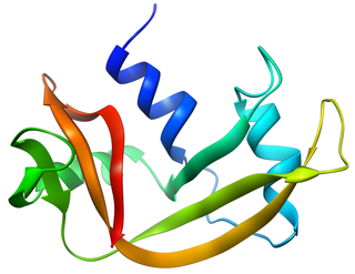
Pancreatic ribonuclease family is a superfamily of pyrimidine-specific endonucleases found in high quantity in the pancreas of certain mammals and of some reptiles.

<span class="mw-page-title-main">RNASE1</span> Protein-coding gene in the species Homo sapiens

Ribonuclease pancreatic is an enzyme that in humans is encoded by the RNASE1 gene.

<span class="mw-page-title-main">Ribonuclease 4</span> Protein-coding gene in the species Homo sapiens

Ribonuclease 4 is an enzyme that in humans is encoded by the RNASE4 gene.

S-tag is the name of an oligopeptide derived from pancreatic ribonuclease A.

<span class="mw-page-title-main">Kunitz domain</span> InterPro Domain

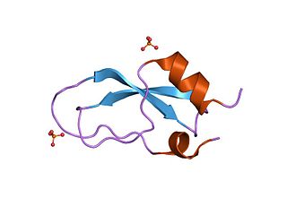
Kunitz domains are the active domains of proteins that inhibit the function of protein degrading enzymes or, more specifically, domains of Kunitz-type are protease inhibitors. They are relatively small with a length of about 50 to 60 amino acids and a molecular weight of 6 kDa. Examples of Kunitz-type protease inhibitors are aprotinin, Alzheimer's amyloid precursor protein (APP), and tissue factor pathway inhibitor (TFPI). Kunitz STI protease inhibitor, the trypsin inhibitor initially studied by Moses Kunitz, was extracted from soybeans.

Ribonuclease E is a bacterial ribonuclease that participates in the processing of ribosomal RNA and the chemical degradation of bulk cellular RNA.

References

  1. Raines RT (1998). "Ribonuclease A". Chem. Rev. 98 (3): 1045–1066. doi:10.1021/cr960427h. PMID   11848924.
  2. "The Nobel Prize in Chemistry 1972". Nobelprize.org. Retrieved 10 February 2015.
  3. "The Nobel Prize in Chemistry 1984". Nobelprize.org. Retrieved 10 February 2015.
  4. 1 2 Richards FM (1972). "The 1972 nobel prize for chemistry". Science. 178 (4060): 492–3. Bibcode:1972Sci...178..492R. doi:10.1126/science.178.4060.492. PMID   17754377.
  5. Marshall, G. R.; Feng, J. A.; Kuster, D. J. (2008). "Back to the future: Ribonuclease A". Biopolymers. 90 (3): 259–77. doi: 10.1002/bip.20845 . PMID   17868092.
  6. Cuchillo CM, Nogués MV, Raines RT (2011). "Bovine pancreatic ribonuclease: fifty years of the first enzymatic reaction mechanism". Biochemistry. 50 (37): 7835–7841. doi:10.1021/bi201075b. PMC   3172371 . PMID   21838247.
  7. Schmid, FX; Baldwin, RL (October 1978). "Acid catalysis of the formation of the slow-folding species of RNase A: evidence that the reaction is proline isomerization". Proceedings of the National Academy of Sciences of the United States of America. 75 (10): 4764–8. Bibcode:1978PNAS...75.4764S. doi: 10.1073/pnas.75.10.4764 . PMC   336200 . PMID   283390.
  8. Wyckoff HW, Hardman KD, Allewell NM, Inagami T, Johnson LN, Richards FM (1967). "The structure of ribonuclease-S at 3.5 A resolution". J. Biol. Chem. 242 (17): 3984–8. doi: 10.1016/S0021-9258(18)95844-8 . PMID   6037556.
  9. Volkin E, Cohn WE (1953). "On the structure of ribonucleic acids. II. The products of ribonuclease action". J. Biol. Chem. 205 (2): 767–82. doi: 10.1016/S0021-9258(18)49221-6 . PMID   13129256.
  10. 1 2 Krystal Worthington. "Ribonuclease - Worthington Enzyme Manual" . Retrieved 2011-09-26.
  11. Selwood T, Jaffe EK (2012). "Dynamic dissociating homo-oligomers and the control of protein function". Arch. Biochem. Biophys. 519 (2): 131–43. doi:10.1016/j.abb.2011.11.020. PMC   3298769 . PMID   22182754.

Further reading