Dog anatomy comprises the anatomical study of the visible parts of the body of a domestic dog. Details of structures vary tremendously from breed to breed, more than in any other animal species, wild or domesticated,[1] as dogs are highly variable in height and weight. The smallest known adult dog was a Yorkshire Terrier that stood only 6.3cm (2.5in) at the shoulder, 9.5cm (3.7in) in length along the head and body, and weighed only 113 grams (4.0oz). The heaviest dog was an English Mastiff named Zorba, which weighed 314 pounds (142kg).[2] The tallest known adult dog is a Great Dane that stands 106.7cm (42.0in) at the shoulder.[3]
The vertebrae have muscles attached to the pedicles, the laminae, the spinous, transverse, and articular processes, the vertebral and intervertebral foramina, the atlas (C1), axis (C2), dens, and ventral lamina (C6).[citation needed]
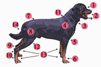
Dog skeletal features
Lateral view of a dog skeleton
Lateral view of a dog skull, jaw opened
Lateral view of a dog skull, jaw closed
Frontal view of a dog skull
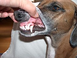
A dog's teeth
Skull
In 1986, a study of skull morphology found that the domestic dog is morphologically distinct from all other canids except the wolf-like canids. The difference in size and proportion between some breeds are as great as those between any wild genera, but all dogs are clearly members of the same species.[5] In 2010, a study of dog skull shape compared to extant carnivorans proposed that "The greatest shape distances between dog breeds clearly surpass the maximum divergence between species in the Carnivora. Moreover, domestic dogs occupy a range of novel shapes outside the domain of wild carnivorans."[6]
The domestic dog compared to the wolf shows the greatest variation in the size and shape of the skull (Evans 1979) that ranges from 7 to 28cm in length (McGreevy 2004). Wolves are dolichocephalic (long-skulled) but not as extreme as some breeds of dogs, such as greyhounds and Russian wolfhounds (McGreevy 2004). Canine brachycephaly (short-skulledness) is found only in domestic dogs and is related to paedomorphosis (Goodwin 1997). Puppies are born with short snouts, with the longer skull of dolichocephalic dogs emerging in later development (Coppinger 1995). Other differences in head shape between brachycephalic and dolichocephalic dogs include changes in the craniofacial angle (angle between the basilar axis and hard palate) (Regodón 1993), morphology of the temporomandibular joint (Dickie 2001), and radiographic anatomy of the cribriform plate (Schwarz 2000).[7]
One study found that the relative reduction in dog skull length compared to its width (the cephalic index) was significantly correlated to both the position and the angle of the brain within the skull, regardless of the brain size or the body weight of the dog.[8]
A wolf mandible diagram showing the names and positions of the teeth
The respiratory system is the set of organs responsible for the intake of oxygen and the expelling of carbon dioxide. As dogs have few sweat glands in their skin, the respiratory system also plays an important role in body thermoregulation.[10]
Dogs are mammals with two large lungs that are further divided into lobes. They have a spongy appearance due to the presence of a system of delicate branches of the bronchioles in each lung, ending in closed, thin-walled chambers (the points of gas exchange) called alveoli. The presence of a muscular structure, the diaphragm, exclusive to mammals, divides the peritoneal cavity from the pleural cavity, besides assisting the lungs during inhalation.
Sixty percent of the dog's body mass falls on the front legs.[14]
The dog has a cardiovascular system. The dog's muscles provide the dog with the ability to jump and leap. Their legs can propel them to leap forward rapidly to chase and overcome prey. They have small, tight feet and walk on their toes (thus having a digitigrade stance and locomotion). Their rear legs are fairly rigid and sturdy. The front legs are loose and flexible, with only muscle attaching them to the torso.
The dog's muzzle size will vary with the breed. Dogs with medium muzzles, such as the German Shepherd Dog, are called mesocephalic and dogs with a pushed in muzzle, such as the Pug, are called brachycephalic. Today's toy breeds have skeletons that mature in only a few months, while giant breeds, such as the Mastiffs, take 16 to 18months for the skeleton to mature. Dwarfism has affected the proportions of some breeds' skeletons, as in the Basset Hound.
All living Canidae have a ligament connecting the spinous process of their first thoracic (or chest) vertebra to the back of the axis bone (second cervical or neck bone), which supports the weight of the head without active muscle exertion, thus saving energy.[15] This ligament is analogous in function (but different in exact structural detail) to the nuchal ligament found in ungulates.[15] This ligament allows dogs to carry their heads while running long distances, such as while following scent trails with their nose to the ground, without expending much energy.[15]
Dogs have disconnected shoulder bones (lacking the collar bone of the human skeleton) that allow a greater stride length for running and leaping. They walk on four toes, front and back, and have vestigial dewclaws on their front legs and on their rear legs. When a dog has extra dewclaws in addition to the usual one in the rear, the dog is said to be "double dewclawed."
Size
The difference in body size between a Cane Corso (Italian mastiff) and a Yorkshire Terrier is over 30-fold; both are members of the same species.
Dogs are highly variable in height and weight. The smallest known adult dog was a Yorkshire Terrier that stood only 6.3cm (2.5in) at the shoulder, 9.5cm (3.7in) in length along the head and body, and weighed only 113 grams (4.0oz). The largest known adult dog was an English Mastiff, which weighed 155.6kg (343lb).[2] The tallest known adult dog is a Great Dane that stands 106.7cm (42.0in) at the shoulder.[3]
In 2007, a study identified a gene that was proposed to be responsible for dog size. The study found a regulatory sequence next to the gene Insulin-like growth factor 1 (IGF1), which, together with the gene and regulatory sequence, "is a major contributor to body size in all small dogs." Two variants of this gene were found in large dogs, making a more complex reason for the large breed size. The researchers concluded that this gene's instructions to make dogs small must be at least 12,000years old and it is not found in wolves.[16] Another study has proposed that lap dogs (small dogs) are among the oldest existing dog types.[17]
Domestic dogs often display the remnants of countershading, a common natural camouflage pattern. The general theory of countershading is that an animal that is lit from above will appear lighter on its upper half and darker on its lower half, where it will usually be in its own shade.[18][19] This is a pattern that predators can learn to watch for. A counter-shaded animal will have dark coloring on its upper surfaces and light coloring below.[18] This reduces the general visibility of the animal. In this pattern, many breeds will have the occasional "blaze", stripe, or "star" of white fur on their chests or undersides.[19]
A study found that the genetic basis that explains coat colors in horse coats and cat coats did not apply to dog coats.[20] The project took samples from 38 different breeds to find the gene (a beta defensin gene) responsible for dog coat color. One version produces yellow dogs and a mutation produces black dogs. All dog coat colors are modifications of black or yellow.[21] For example, the white in white miniature schnauzers is a cream color, not albinism (a genotype of E/E' at MC1R).
Modern dog breeds exhibit a diverse array of fur coats, including dogs without fur, such as the Mexican Hairless Dog. Dog coats vary in texture, color, and markings, and a specialized vocabulary has evolved to describe each characteristic.[22]
Tail
There are many different shapes of dog tails: straight, straight up, sickle, curled and cork-screw. In some breeds, the tail is traditionally docked to avoid injuries (especially for hunting dogs).[23] It can happen that some puppies are born with a short tail or no tail in some breeds. The T-box gene mutation (C189G) is responsible for bobtail breeds having no tail to short tail.[24][25] Dogs have a violet gland or supracaudal gland on the dorsal (upper) surface of their tails.
Footpad
The dog's footpad is a fatty tissue locomotive-supporting organ, present at the bottom of the four legs, consisting of digital pads, a metacarpal pad, and a carpal pad, with dewclaw near the footpad.[26] When a dog's footpad is exposed to the cold, heat loss is prevented by an adaptation of the blood system that recirculates heat back into the body. It brings blood from the skin surface and retains warm blood on the pad surface.[27]
Like most mammals, dogs have only two types of cone photoreceptors, making them dichromats.[28][29][30][31] These cone cells are maximally sensitive between 429nm and 555nm. Behavioural studies have shown that the dog's visual world consists of yellows, blues and grays,[31] but they have difficulty differentiating between red and green, making their color vision equivalent to red–green color blindness in humans (deuteranopia). When a human perceives an object as "red," this object appears as "yellow" to the dog, and the human perception of "green" appears as "white," a shade of gray. This white region (the neutral point) occurs around 480nm, the part of the spectrum that appears blue-green to humans. For dogs, wavelengths longer than the neutral point cannot be distinguished from each other, and all appear yellow.[31]
Dogs use color instead of brightness to differentiate between light or dark blue/yellow.[32][33][34] They are less sensitive to differences in gray shades than humans and can also detect brightness with about half the accuracy of humans.[35]:140 The dog's visual system has evolved to aid in hunting.[28] Dogs have been shown to be able to discriminate between humans (e.g., identifying their human guardian) at a range of between 800 and 900 metres (2,600 and 3,000ft); however, this range decreases to 500–600 metres (1,600–2,000ft) if the object is stationary.[28] Dogs can detect a change in movement that exists in a single diopter of space within their eye. Humans, by comparison, require a change of between 10 and 20 diopters to detect movement.[36] A test has estimated poodles' visual acuity to have a Snellen rating of 20/75, a relatively low score compared to humans' vision.[28]
As crepuscular hunters, dogs often rely on their vision in low light situations: They have very large pupils, a high density of rods in the fovea, an increased flicker rate, and a tapetum lucidum.[28] The tapetum is a reflective surface behind the retina that reflects light to give the photoreceptors a second chance to catch the photons. There is also a relationship between body size and the overall diameter of the eye. A range of 9.5 and 11.6mm can be found between various breeds of dogs. This 20% variance is associated with an adaptation toward superior night vision.[35]:139
The eyes of different breeds of dogs have different shapes, dimensions, and retina configurations.[37] Many long-nosed breeds have a "visual streak"—a wide foveal region that runs across the width of the retina and gives them a very wide field of excellent vision. Some long-muzzled breeds, in particular, the sighthounds, have a field of vision up to 270° (compared to 180° for humans). Short-nosed breeds, on the other hand, have an "area centralis", a central patch with up to three times the density of nerve endings as the visual streak, giving them detailed sight much more like a human's. Some broad-headed breeds with short noses have a field of vision similar to that of humans.[29][30]
Most breeds have good vision, but some show a genetic predisposition for myopia—such as Rottweilers, with which one out of every two has been found to be myopic.[28] Dogs also have a greater divergence of the eye axis than humans, enabling them to rotate their pupils farther in any direction. The divergence of the eye axis of dogs ranges from 12–25°, depending on the breed.[36] Experimentation has found that dogs can distinguish between complex visual images such as those of a cube or a prism. Dogs also show attraction to static visual images such as the silhouette of a dog on a screen, their own reflections, or videos of dogs; however, their interest declines sharply once they are unable to make social contact with the image.[35]:142
Hearing
"Dog hearing" redirects here. For dogs that assist people with hearing difficulties, see Hearing dog.
Schematic anatomy of the ear. In dogs, the ear canal has a "L" shape, with the vertical canal (first half) and the horizontal canal (deeper half, ending with the eardrum)
The frequency range of dog hearing is between 16–40Hz (compared to 20–70Hz for humans) and up to 45–60kHz (compared to 13–20kHz for humans), which means that dogs can detect sounds beyond the upper limit of the human auditory spectrum.[30][38][39][40]
Dogs have ear mobility that allows them to rapidly pinpoint the exact location of a sound. Eighteen or more muscles can tilt, rotate, raise, or lower a dog's ear. A dog can identify a sound's location much faster than a human can, as well as hear sounds at four times the distance.[41] Dogs can lose their hearing from age or an ear infection.[42]
Dogs have around 1,700 taste buds compared to humans, with around 9,000. The sweet taste buds in dogs respond to furaneol. It appears that dogs do like this flavor, and it probably evolved because, in a natural environment, dogs frequently supplement their diet of small animals with whatever fruits are available. Because of dogs' dislike of bitter tastes, various sprays, and gels have been designed to keep dogs from chewing on furniture or other objects. Dogs also have taste buds that are tuned for water, which is something they share with other carnivores but is not found in humans. This taste sense is found at the tip of the dog's tongue, which is the part of the tongue that they curl to lap water. This area responds to water at all times, but when the dog has eaten salty or sugary foods, the sensitivity to the taste of water increases. It is proposed that this ability to taste water evolved as a way for the body to keep internal fluids in balance after the animal has eaten things that will either result in more urine being passed or will require more water to adequately process. It appears that when these special water taste buds are active, dogs seem to get an extra pleasure out of drinking water, and will drink copious amounts of it.[43]
Touch
A dog's whiskers
Dogs have specialized whiskers known as vibrissae, sensing organs present above the dog's eyes, below their jaw, and on their muzzle. Vibrissae are more rigid, embedded much more deeply in the skin than other hairs, and have a greater number of receptor cells at their base. They can detect air currents, subtle vibrations, and objects in the dark. They provide an early warning system for objects that might strike the face or eyes, and probably help direct food and objects towards the mouth.[44]
A study found that dogs may prefer, when they are off the leash and the Earth's magnetic field is calm, to urinate and defecate with their bodies aligned on a north-south axis. Dogs are sensitive to changes in the Earth's magnetic field polarity.[45] No significant differences between males and females in angular preferences were found. Some studies have detected cryptochrome 1 in some dogs' photoreceptors' blue-sensitive cones.[46][47]
Temperature regulation
The nose of a dog
Primarily, dogs regulate their body temperature through panting[48] and sweating via their paws. Panting moves cooling air over the moist surfaces of the tongue and lungs, transferring heat to the atmosphere.
Dogs and other canids also possess a set of nasal turbinates, an elaborate set of bones and associated soft-tissue structures (including arteries and veins) in the nasal cavities. These turbinates allow for heat exchange between small arteries and veins on their maxilloturbinate surfaces (the surfaces of turbinates positioned on maxilla bone) in a counter-current heat-exchange system. Compared to the ambush predation of cats, dogs are capable of prolonged chases due to these turbinates (cats possess a much smaller and less-developed set of nasal turbinates).[49]:88 This same turbinate structure helps conserve water in arid environments. The water conservation and thermoregulatory capabilities of these turbinates in dogs may have allowed dogs (including both domestic dogs and their wild prehistoric ancestors) to survive in the Arctic environment and other cold areas of northern Eurasia and North America, which are dry and cold.[49]:87
↑Wayne, Robert K. (1986). "Cranial Morphology of Domestic and Wild Canids: The Influence of Development on Morphological Change". Evolution. 40 (2): 243–261. doi:10.2307/2408805. JSTOR2408805. PMID28556057.
This page is based on this Wikipedia article Text is available under the CC BY-SA 4.0 license; additional terms may apply. Images, videos and audio are available under their respective licenses.