Hydrodynamic theory (dentistry)

Last updated
Hydrodynamic theory
Biological system Nervous system
HealthHarmful
StimuliThermal (hot and cold), mechanical, osmotic, chemical, electrical
MethodStimuli exposure causes increased fluid flow in dentinal tubules
OutcomeMay cause dentine hypersensitivity
DurationOccurs in short, transient bursts

In dentistry, the hydrodynamic or fluid movement theory is one of three main theories developed to explain dentine hypersensitivity, which is a sharp, transient pain arising from stimuli exposure. [1] It states that different types of stimuli act on exposed dentine, causing increased fluid flow through the dentinal tubules. In response to this movement, mechanoreceptors on the pulp nerves trigger the acute, temporary pain of dentine hypersensitivity. [2]

Contents

The fluid flow mechanism behind hydrodynamic theory was first introduced by Alfred Gysi in 1900, and subsequently developed by Martin Brännström in the 1960s through a series of experimental studies. [3] Further supporting evidence has since been collected from epidemiological surveys and experimental data comparing dentinal tubule numbers in hypersensitive and non-hypersensitive teeth. [4]

Alternate theories include the “dentine innervation” and “odontoblast transduction” theories, [5] both of which lack substantial supporting evidence. The hydrodynamic theory is currently the accepted explanation for dentine hypersensitivity, upon which several treatment and diagnostic strategies have been built by dental practitioners. [6]

Overview of theory

Dental erosion and loss of enamel increase the patency of dentinal tubules Tooth erosion 2.jpg
Dental erosion and loss of enamel increase the patency of dentinal tubules

The hydrodynamic theory proposes that when dentinal tubules are exposed at the pulp and dentine surface, external stimuli cause changes in fluid flow. [7] Dentinal tubules may become exposed due to various reasons: e.g. dental erosion, enamel loss and periodontal diseases. [8]

When exposed dentinal tubules are then subject to stimuli, the direction and rate of fluid flow movement within the tubules change. This causes shifts in pressure within the dentine, which stimulate the myelinated nerves located in the pulp, causing the sensation of pain. [9] The main nerve fibres responsible for responding to the stimuli are mostly type A-beta (Aβ), [10] which carry tactile information, [11] and some A-delta (Aδ) nerves, [12] which relay pain and temperature information to the brain. [13]

Impact of various stimuli on fluid flow in dentine [14] [15]
Types of stimuliExample(s)Effect
Mechanical Over-brushing, abrasions from sharp, dental instrumentsOutward fluid flow, arising from compressed surface tissue
Cold thermal Cold foods and liquidsOutward fluid flow
Heat thermalHot food and liquidsInward fluid flow, caused by fluid contraction in tubules
Evaporative Air blasts, windy weatherOutward fluid flow caused by fluid evaporation at surface of tubule
Osmotic Sugar, acidic solutionsOutward fluid flow
Chemical [16] Cosmetic dental procedures e.g. teeth whitening Outward fluid flow

Origin and development

Alfred Gysi - 1900

Portrait of Dr. Alfred Gysi taken in 1914, University of Zurich (UAZ) AB.1.0361.08 Gysi.jpg
Portrait of Dr. Alfred Gysi taken in 1914, University of Zurich

Dr. Gysi (1865-1957) was a Swiss dentist who specialised in jaw movement and structure, as well as prosthodontics. He studied dentistry in Geneva, Switzerland and the Philadelphia Dental College, eventually becoming a dentistry professor at the University of Zurich. [17] At the time, it was still believed that dentine hypersensitivity was caused by either nerve fibre stimulation within dentine or odontoblast processes acting as pain receptors. [18]

In 1900, Gysi first introduced the concept of fluid-flow movement as an explanation for dentine hypersensitivity in his publication for the British Journal of Dental Science: An attempt to explain the sensitiveness of dentine. [19] In his clinical practice, Gysi observed that the removal of fluid from the cavity floor of his patients' teeth produced the sharp, short pain of dentine hypersensitivity. He subsequently concluded that drying the cavity fluid caused an interruption in the fluid flow movement within the dentinal tubules. [20] This led to Gysi's hypothesis that within the dentinal tubules, there was a natural outward flow of fluid. Physical and thermal forms of stimuli would cause an increase change in the direction of fluid flow, which activated the pulpal nerve endings. [21]

Martin Brännström - 1960s

Dr.Brännström (1922-2001) was a Swedish dentist, specialising in Oral Pathology and the mechanism of tooth sensitivity. [22] In the 1960s, Brännström provided evidence to support Gysi's hydrodynamic theory through a series of experimental studies in vitro to show that various stimuli caused shifts in fluid movement across dentine, producing pain. [23] Brännström's 1965 experimental study indicated that hypersensitive dentine exhibited a higher number of exposed, patent tubules than non-sensitive dentine. This hypersensitivity was noticeably lessened when the tubules were then deliberately occluded to restrict fluid flow. [24]

Brännström conducted trials using different stimuli. It was shown that various types of cold thermal, evaporative cooling, osmotic stimuli and hypertonic chemical substances could cause an increase in outward fluid flow along the dentinal tubules. Contrastingly, thermal hot stimuli caused an inward fluid flow. [25] In vitro studies indicated that stimuli causing an outward flow of fluid from the dentine-pulp complex led to a greater intensity of dentine hypersensitivity than stimuli triggering inward flow. [26] Brännström's multiple publications of both experimental and observational data provided significance evidence to support the theory of hydrodynamic mechanism causing dentine hypersensitivity. [27]

Supporting experiments

SEM image of dentine with exposed dentinal tubules (Daood et al. 2018; Electron Microscope Unit, Queen Mary Hospital, The University of Hong Kong) Figure 1 Scanning electron micrograph of dentin with open dentinal tubules.jpg
SEM image of dentine with exposed dentinal tubules (Daood et al. 2018; Electron Microscope Unit, Queen Mary Hospital, The University of Hong Kong)

Epidemiological surveys have shown that dentine hypersensitivity arises when the dentinal tubules are both exposed and patent. It was proposed that if the hydrodynamic fluid flow was responsible for hypersensitivity, then there must be higher numbers of dentinal tubules exposed at the surface of the root and patent to the dental pulp. A strong positive correlation has been identified between the levels of hypersensitivity in exposed dentine and the number and density of dentinal tubules. [28]

The patency of dentinal tubules 1987

In 1987, A study of the patency of dentinal tubules in sensitive and non-sensitive cervical dentine, [29] was published in the Journal of Clinical Periodontology. This experiment was conducted by E.G. Absi, M. Addy and D.Adams from the Dental School of Cardiff University. It compared the patency of sensitive and non-sensitive cervical dentine and provided significant evidence to support the hydrodynamic theory. [30]

The aim of the experiment was to investigate whether patients with dentine hypersensitivity would have higher numbers of dentinal tubules exposed at the surface of the root. There had been little evidence to support the theory prior to this experiment, with the exception of Brännström's in vitro studies. [31] With the use of a scanning electron microscope and dye penetration technology, the findings concluded that the dentinal tubules were eight times greater in number and 2 times wider in diameter in dentine of “hypersensitive” classified patients than in non-hypersensitive dentin. [32] The results supported the theory in that hypersensitive dentin had wider, more exposed dentinal tubules and provided evidence of stimuli transmission via a hydrodynamic movement along dentinal tubules. [33]

Intradental nerves and the rate of fluid flow 2007

In 2007, The relationship between the discharge of intradental nerves and the rate of fluid flow through dentine in the cat [34] was published in the Archives of Oral Biology. The experiment was conducted by Noppakun Vongsavan and Bruce Matthews from the Department of Physiology at the University of Bristol.

The aim of the experiment was to investigate the impact of fluid flow movement on intradental nerves, to provide evidence for the hydrodynamic theory. [35] Hydrostatic pressure stimuli were applied to exposed dentine in cats under anaesthetic. Recordings of the resulting nerve impulses were taken from the dentine and filaments located in the inferior alveolar nerve. [36] The results showed that outward fluid flow produced a greater discharge of intradental nerves than inward fluid flow and were more likely to stimulate the pain receptors. This supports Brännström's findings that stimuli causing an outwards fluid flow (e.g. cold thermal), generate more hypersensitivity than stimuli for inward flow (e.g. heat thermal). [37]

Criticisms and alternative theories

Criticisms

Although there is sufficient evidence for a fluid flow mechanism within dentinal tubules, hydrodynamic theory is criticised for being unable to adequately explain the neurosensory mechanisms behind dentine hypersensitivity. It is uncertain how the physical changes in direction and rate of fluid flow within dentinal tubules actually causes the stimulation of pain receptors in pulp nerves. [38]

Evidence against the theory was presented in an experiment in which dentinal tubules that had been purposely obstructed to prevent fluid flow still exhibited hypersensitivity upon the application of thermal stimuli. [39] Additionally, it has been noted in clinical cases that hypersensitivity worsened even after dental cavities had significantly destroyed dentinal tubules and made fluid flow impossible. [40]

It has since been suggested that the hydrodynamic theory may account for some, but not all types of stimuli. [41] As a result, an addition to the theory has been proposed that the pain may be stimulated by the generation of an electric potential arising from the fluid flow movement. However, it is unclear whether the fluid movement is capable of generating sufficient hydrostatic pressure to electrically stimulate pain receptors. [42]

Dentine innervation theory

The dentine innervation theory is based on the premise that dentine is innervated and that the stimulation of the nerve endings within dentinal tubules causes hypersensitivity. [43] It states that damage to local dentinal tubules stimulates an action potential, which travels along the nerve to inner dentine. Initially, histological stain experimentation was carried out using silver to demonstrate the existence of nerve fibres in dentinal tubules. [44] However, further experimentation indicated that although there were rare instances of nerve fibres in inner dentine, there was no indication of innervation within the actual tubules. [45] Transmission electron microscopy and immune-histochemical analyses later indicated that areas of inner dentine up to the dentin-enamel junction may be innervated. [46] However, there has been no evidence supporting the innervation of outer dentine, which is considered the main area of hypersensitivity. [47]

Odontoblast transduction theory

The odontoblast transduction theory proposes that odontoblast processes act as pain receptors that conduct these stimuli impulses to the pulpal nerve endings of inner dentine. The theory arose from research that showed the odontoblast sending processes from the outer pulp layer to the dentin-enamel junction. [48] Rapp & Avery published evidence supporting the existence of acetylcholinesterase on the dental odontoblast processes. [49] As acetylcholinesterase is responsible for terminating neurotransmission and synapse signalling, they hypothesised that it helped conduct sensory information to the pulpal nerves from the odontoblast processes. However, there has been a lack of evidence that there are synapses between odontoblasts and the pulpal nerve endings, or that there exist neurotransmitter vesicles in odontoblast processes. [50] Further criticism arose from clinical observations indicating that hypersensitivity continued even after the odontoblast layer was destructed by dental cavities and nerve injuries. [51]

Applications

Dental practitioners have applied the hydrodynamic theory to prevent and treat dentine hypersensitivity in patients. [52] Brännström's studies on the impact of various stimuli on hypersensitivity have led to new techniques for diagnosis and management.

As mechanical stimuli have proven to be a major cause of hypersensitivity, preventative strategies include proper hygiene and brushing technique instruction. [53] Patients have been advised to use non-abrasive toothpastes, select soft-bristle toothbrushes and avoid overstimulating the surface of dentine. To avoid increasing the patency of tubules, patients have been encouraged to schedule regular appointments to check for dental erosion, tooth decay and periodontal disease. [54]

Consumption of acidic drinks e.g. vinegar, causes dental erosion, thus chemical stimulation 216 pH Scale-01.jpg
Consumption of acidic drinks e.g. vinegar, causes dental erosion, thus chemical stimulation

To avoid chemical stimulation and dental erosion, dental practitioners have prescribed reduced consumption of erosive agents and acids from soft drinks, fruit and vinegar. Certain chemicals have now been advised against for cosmetic procedures, such as teeth whitening. [55]

Brännström's initial studies on hydrodynamic theory indicate that occlusion of the dentinal tubules reduces the severity of dentine hypersensitivity. This has led to several treatment methods to block or reduce the diameter of the tubules, either mechanically or chemically. [56]

One notable non-invasive method is the depolarisation of the pupal nerve using a dentifrice with chemical treatments e.g. strontium chloride and potassium nitrate. Desensitising treatments are separated into two groups: at-home procedures in the form of mouthwash and toothpaste, or in-office products such as resin sealers, glass ionomers and dentine solutions. These desensitising agents are prescribed to block nerve impulses by physically reducing the width of dentinal tubules or chemically preventing the transmission of the stimuli across dentin. [57]

Recently, laser treatment has been proposed as a potential method of treating dentine hypersensitivity. Low-output lasers may be used to block the electric transmission of stimuli to pulpal nerves whereas high-output lasers are being considered as an alternate method of occluding the dentinal tubules. [58] Occlusion using lasers may occur in two forms: through partial melting, or through clotting the fluid proteins within the tubules. [59]

See also

Related Research Articles

<span class="mw-page-title-main">Cementum</span> Specialized calcified substance covering the root of a tooth

Cementum is a specialized calcified substance covering the root of a tooth. The cementum is the part of the periodontium that attaches the teeth to the alveolar bone by anchoring the periodontal ligament.

<span class="mw-page-title-main">Tooth decay</span> Deformation of teeth due to acids produced by bacteria

Tooth decay, also known as cavities or caries, is the breakdown of teeth due to acids produced by bacteria. The cavities may be a number of different colors from yellow to black. Symptoms may include pain and difficulty with eating. Complications may include inflammation of the tissue around the tooth, tooth loss and infection or abscess formation.

<span class="mw-page-title-main">Dentin</span> Calcified tissue of the body; one of the four major components of teeth

Dentin or dentine is a calcified tissue of the body and, along with enamel, cementum, and pulp, is one of the four major components of teeth. It is usually covered by enamel on the crown and cementum on the root and surrounds the entire pulp. By volume, 45% of dentin consists of the mineral hydroxyapatite, 33% is organic material, and 22% is water. Yellow in appearance, it greatly affects the color of a tooth due to the translucency of enamel. Dentin, which is less mineralized and less brittle than enamel, is necessary for the support of enamel. Dentin rates approximately 3 on the Mohs scale of mineral hardness. There are two main characteristics which distinguish dentin from enamel: firstly, dentin forms throughout life; secondly, dentin is sensitive and can become hypersensitive to changes in temperature due to the sensory function of odontoblasts, especially when enamel recedes and dentin channels become exposed.

<span class="mw-page-title-main">Toothache</span> Medical condition of the teeth

Toothache, also known as dental pain or tooth pain, is pain in the teeth or their supporting structures, caused by dental diseases or pain referred to the teeth by non-dental diseases. When severe it may impact sleep, eating, and other daily activities.

<span class="mw-page-title-main">Pulp (tooth)</span> Part in the center of a tooth made up of living connective tissue and cells called odontoblasts

The pulp is the connective tissue, nerves, blood vessels, and odontoblasts that comprise the innermost layer of a tooth. The pulp's activity and signalling processes regulate its behaviour.

<span class="mw-page-title-main">Bone canaliculus</span> Canal system in bones

Bone canaliculi are microscopic canals between the lacunae of ossified bone. The radiating processes of the osteocytes project into these canals. These cytoplasmic processes are joined together by gap junctions. Osteocytes do not entirely fill up the canaliculi. The remaining space is known as the periosteocytic space, which is filled with periosteocytic fluid. This fluid contains substances too large to be transported through the gap junctions that connect the osteocytes.

<span class="mw-page-title-main">Dental erosion</span> Medical condition

Acid erosion is a type of tooth wear. It is defined as the irreversible loss of tooth structure due to chemical dissolution by acids not of bacterial origin. Dental erosion is the most common chronic condition of children ages 5–17, although it is only relatively recently that it has been recognised as a dental health problem. There is generally widespread ignorance of the damaging effects of acid erosion; this is particularly the case with erosion due to consumption of fruit juices because they tend to be considered as healthy. Acid erosion begins initially in the enamel, causing it to become thin, and can progress into dentin, giving the tooth a dull yellow appearance and leading to dentin hypersensitivity.

<span class="mw-page-title-main">Odontoblast</span> Type of cell that produces dentin in teeth

In vertebrates, an odontoblast is a cell of neural crest origin that is part of the outer surface of the dental pulp, and whose biological function is dentinogenesis, which is the formation of dentin, the substance beneath the tooth enamel on the crown and the cementum on the root.

Pulpitis is inflammation of dental pulp tissue. The pulp contains the blood vessels, the nerves, and connective tissue inside a tooth and provides the tooth's blood and nutrients. Pulpitis is mainly caused by bacterial infection which itself is a secondary development of caries. It manifests itself in the form of a toothache.

<span class="mw-page-title-main">Dentine bonding agents</span>

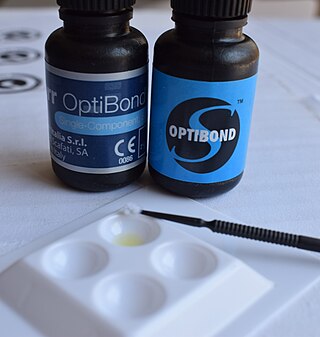
Also known as a "bonderizer" bonding agents are resin materials used to make a dental composite filling material adhere to both dentin and enamel.

<span class="mw-page-title-main">Dentinogenesis imperfecta</span> Medical condition

Dentinogenesis imperfecta (DI) is a genetic disorder of tooth development. It is inherited in an autosomal dominant pattern, as a result of mutations on chromosome 4q21, in the dentine sialophosphoprotein gene (DSPP). It is one of the most frequently occurring autosomal dominant features in humans. Dentinogenesis imperfecta affects an estimated 1 in 6,000-8,000 people.

Dentin hypersensitivity is dental pain which is sharp in character and of short duration, arising from exposed dentin surfaces in response to stimuli, typically thermal, evaporative, tactile, osmotic, chemical or electrical; and which cannot be ascribed to any other dental disease.

<span class="mw-page-title-main">Dentin dysplasia</span> Medical condition

Dentin dysplasia (DD) is a rare genetic developmental disorder affecting dentine production of the teeth, commonly exhibiting an autosomal dominant inheritance that causes malformation of the root. It affects both primary and permanent dentitions in approximately 1 in every 100,000 patients. It is characterized by the presence of normal enamel but atypical dentin with abnormal pulpal morphology. Witkop in 1972 classified DD into two types which are Type I (DD-1) is the radicular type, and type II (DD-2) is the coronal type. DD-1 has been further divided into 4 different subtypes (DD-1a,1b,1c,1d) based on the radiographic features.

<span class="mw-page-title-main">Sensodyne</span> Dental hygiene product for sensitive teeth

Sensodyne is a brand name of toothpaste and mouthwash targeted at people with sensitive teeth. Sensodyne is owned by Haleon and is marketed under the name Shumitect in Japan.

<span class="mw-page-title-main">Fluoride varnish</span> Highly concentrated form of fluoride

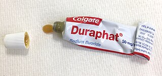
Fluoride varnish is a highly concentrated form of fluoride which is applied to the tooth's surface, by a dentist, dental hygienist or other health care professional, as a type of topical fluoride therapy. It is not a permanent varnish but due to its adherent nature it is able to stay in contact with the tooth surface for several hours. It may be applied to the enamel, dentine or cementum of the tooth and can be used to help prevent decay, remineralise the tooth surface and to treat dentine hypersensitivity. There are more than 30 fluoride-containing varnish products on the market today, and they have varying compositions and delivery systems. These compositional differences lead to widely variable pharmacokinetics, the effects of which remain largely untested clinically.

<span class="mw-page-title-main">Cracked tooth syndrome</span> Medical condition

Cracked tooth syndrome (CTS) is where a tooth has incompletely cracked but no part of the tooth has yet broken off. Sometimes it is described as a greenstick fracture. The symptoms are very variable, making it a notoriously difficult condition to diagnose.

Dental pulpal testing is a clinical and diagnostic aid used in dentistry to help establish the health of the dental pulp within the pulp chamber and root canals of a tooth. Such investigations are important in aiding dentists in devising a treatment plan for the tooth being tested.

In dentistry, the smear layer is a layer found on root canal walls after root canal instrumentation. It consists of microcrystalline and organic particle debris. It was first described in 1975 and research has been performed since then to evaluate its importance in bacteria penetration into the dentinal tubules and its effects on endodontic treatment. More broadly, it is the organic layer found over all hard tooth surfaces.

<span class="mw-page-title-main">Pulp capping</span>

Pulp capping is a technique used in dental restorations to prevent the dental pulp from necrosis, after being exposed, or nearly exposed during a cavity preparation, from a traumatic injury, or by a deep cavity that reaches the center of the tooth causing the pulp to die. When dental caries is removed from a tooth, all or most of the infected and softened enamel and dentin are removed. This can lead to the pulp of the tooth either being exposed or nearly exposed which causes pulpitis (inflammation). Pulpitis, in turn, can become irreversible, leading to pain and pulp necrosis, and necessitating either root canal treatment or extraction. The ultimate goal of pulp capping or stepwise caries removal is to protect a healthy dental pulp and avoid the need for root canal therapy.

Silver diammine fluoride (SDF), also known as silver diamine fluoride in most of the dental literature, is a topical medication used to treat and prevent dental caries and relieve dentinal hypersensitivity. It is a colorless or blue-tinted, odourless liquid composed of silver, ammonium and fluoride ions at a pH of 10.4 or 13. Ammonia compounds reduce the oxidative potential of SDF, increase its stability and helps to maintain a constant concentration over a period of time, rendering it safe for use in the mouth. Silver and fluoride ions possess antimicrobial properties and are used in the remineralization of enamel and dentin on teeth for preventing and arresting dental caries.

References

  1. Absi, E.; Addy, M.; Adams, D. (1987). "Dentine hypersensitivity. A study of the patency of dentinal tubules in sensitive and non-sensitive cervical dentine". Journal of Clinical Periodontology. 14 (5): 280–284. doi:10.1111/j.1600-051x.1987.tb01533.x. PMID   3475295.
  2. West, N.; Lussi, A.; Seong, J.; Hellwig, E. (2012). "Dentin hypersensitivity: pain mechanisms and aetiology of exposed cervical dentin". Clinical Oral Investigation. 17 (1): 9–19. doi: 10.1007/s00784-012-0887-x . PMID   23224116.
  3. West, N.; Lussi, A.; Seong, J.; Hellwig, E. (2012). "Dentin hypersensitivity: pain mechanisms and aetiology of exposed cervical dentin". Clinical Oral Investigation. 17 (1): 9–19. doi: 10.1007/s00784-012-0887-x . PMID   23224116.
  4. Migliani, S.; Aggarwal, V.; Ahuja, B. (2010). "Dentin hypersensitivity: Recent trends in management". Journal of Conservative Dentistry. 13 (4): 218–24. doi: 10.4103/0972-0707.73385 . PMC   3010026 . PMID   21217949.
  5. Charles, F Cox; Suzuki, Keizo; Yamaguchi, Hiroyasu; Ruby, John; Suzuki, Shiro; Akimoto, Naotake; Maeda, Nobuko; Momoi, Yasuko (2017). "Sensory mechanisms in dentine: A literature review of light microscopy (LM), transmission microscopy (TEM), scanning microscopy (SEM) & electro physiological (EP) tooth sensitivity: Is the ciliary organelle on the odontoblast the elusive primary nociceptor?". Dental, Oral and Craniofacial Research. 4 (3): 2–5.
  6. West, N.; Lussi, A.; Seong, J.; Hellwig, E. (2012). "Dentin hypersensitivity: pain mechanisms and aetiology of exposed cervical dentin". Clinical Oral Investigation. 17 (1): 9–19. doi: 10.1007/s00784-012-0887-x . PMID   23224116.
  7. Sasaki, Keiichi; Suzuki, Osamu; Takahashi, Nobuhiro (December 2014). Interface Oral Health Science 2014: Innovative Research on Biosis-Abiosis Intelligent Interface. Tokyo, Japan: Springer. pp. 325–333. ISBN   978-4431551256.
  8. Loveren, Cor V. (December 2012). "Exposed cervical dentin and dentin hypersensitivity summary of the discussion and recommendations". Clinical Oral Investigations. 17 (Suppl 1): 73–76. doi:10.1007/s00784-012-0902-2. PMC   3585836 . PMID   23224117.
  9. Sasaki, Keiichi; Suzuki, Osamu; Takahashi, Nobuhiro (December 2014). Interface Oral Health Science 2014: Innovative Research on Biosis-Abiosis Intelligent Interface. Tokyo, Japan: Springer. pp. 325–333. ISBN   978-4431551256.
  10. Bekes, Katrin; Robinson, Peter G. (2015). Dentine Hypersensitivity. Academic Press. pp. 21–32. ISBN   9780128016312.
  11. Woessner, James. "A Conceptual Model of Pain: Measurement and Diagnosis". Practical Pain Management. Remedy Health Media. Retrieved 20 November 2020.
  12. Bekes, Katrin; Robinson, Peter G. (2015). Dentine Hypersensitivity. Academic Press. pp. 21–32. ISBN   9780128016312.
  13. Woessner, James. "A Conceptual Model of Pain: Measurement and Diagnosis". Practical Pain Management. Remedy Health Media. Retrieved 20 November 2020.
  14. Sasaki, Keiichi; Suzuki, Osamu; Takahashi, Nobuhiro (December 2014). Interface Oral Health Science 2014: Innovative Research on Biosis-Abiosis Intelligent Interface. Tokyo, Japan: Springer. pp. 325–333. ISBN   978-4431551256.
  15. Bekes, Katrin; Robinson, Peter G. (2015). Dentine Hypersensitivity. Academic Press. pp. 21–32. ISBN   9780128016312.
  16. Pashley, David H. (1986). "Sensitivity of dentin to chemical stimuli". Dental Traumatology. 2 (4): 130–137. doi:10.1111/j.1600-9657.1986.tb00599.x. PMID   3464416.
  17. Phoenix, Rodney D.; Engelmeier, Robert L. (November 2016). "The Contributions of Dr. Alfred Gysi". Journal of Prosthodontics. 27 (3): 276–277. doi:10.1111/jopr.12536. PMID   27883359.
  18. Charles, F Cox; Suzuki, Keizo; Yamaguchi, Hiroyasu; Ruby, John; Suzuki, Shiro; Akimoto, Naotake; Maeda, Nobuko; Momoi, Yasuko (2017). "Sensory mechanisms in dentine: A literature review of light microscopy (LM), transmission microscopy (TEM), scanning microscopy (SEM) & electro physiological (EP) tooth sensitivity: Is the ciliary organelle on the odontoblast the elusive primary nociceptor?". Dental, Oral and Craniofacial Research. 4 (3): 4.
  19. Gysi, Alfred (1900). "An attempt to explain the sensitiveness of dentin". British Journal of Dental Science. 43: 865–868.
  20. Charles, F Cox; Suzuki, Keizo; Yamaguchi, Hiroyasu; Ruby, John; Suzuki, Shiro; Akimoto, Naotake; Maeda, Nobuko; Momoi, Yasuko (2017). "Sensory mechanisms in dentine: A literature review of light microscopy (LM), transmission microscopy (TEM), scanning microscopy (SEM) & electro physiological (EP) tooth sensitivity: Is the ciliary organelle on the odontoblast the elusive primary nociceptor?". Dental, Oral and Craniofacial Research. 4 (3): 4.
  21. Miglani, S.; Aggarwal, V.; Ahuja, B. (2010). "Dentin hypersensitivity: Recent trends in management". Journal of Conservative Dentistry. 13 (4): 218–24. doi: 10.4103/0972-0707.73385 . PMC   3010026 . PMID   21217949.
  22. Charles, F Cox; Suzuki, Keizo; Yamaguchi, Hiroyasu; Ruby, John; Suzuki, Shiro; Akimoto, Naotake; Maeda, Nobuko; Momoi, Yasuko (2017). "Sensory mechanisms in dentine: A literature review of light microscopy (LM), transmission microscopy (TEM), scanning microscopy (SEM) & electro physiological (EP) tooth sensitivity: Is the ciliary organelle on the odontoblast the elusive primary nociceptor?". Dental, Oral and Craniofacial Research. 4 (3): 4.
  23. Absi, E.; Addy, M.; Adams, D. (1987). "Dentine hypersensitivity. A study of the patency of dentinal tubules in sensitive and non-sensitive cervical dentine". Journal of Clinical Periodontology. 14 (5): 280–284. doi:10.1111/j.1600-051x.1987.tb01533.x. PMID   3475295.
  24. Miglani, S.; Aggarwal, V.; Ahuja, B. (2010). "Dentin hypersensitivity: Recent trends in management". Journal of Conservative Dentistry. 13 (4): 218–24. doi: 10.4103/0972-0707.73385 . PMC   3010026 . PMID   21217949.
  25. West, N.; Lussi, A.; Seong, J.; Hellwig, E. (2012). "Dentin hypersensitivity: pain mechanisms and aetiology of exposed cervical dentin". Clinical Oral Investigation. 17 (1): 9–19. doi: 10.1007/s00784-012-0887-x . PMID   23224116.
  26. Liu, X.; Tenenbaum, H.; Wilder, R.; Quock, R.; Hewlett, E.; Ren, Y. (2020). "Pathogenesis, diagnosis and management of dentin hypersensitivity: an evidence-based overview for dental practitioners". BMC Oral Health. 20 (1): 220–230. doi: 10.1186/s12903-020-01199-z . PMC   7409672 . PMID   32762733.
  27. Charles, F Cox; Suzuki, Keizo; Yamaguchi, Hiroyasu; Ruby, John; Suzuki, Shiro; Akimoto, Naotake; Maeda, Nobuko; Momoi, Yasuko (2017). "Sensory mechanisms in dentine: A literature review of light microscopy (LM), transmission microscopy (TEM), scanning microscopy (SEM) & electro physiological (EP) tooth sensitivity: Is the ciliary organelle on the odontoblast the elusive primary nociceptor?". Dental, Oral and Craniofacial Research. 4 (3): 4.
  28. West, N.; Lussi, A.; Seong, J.; Hellwig, E. (2012). "Dentin hypersensitivity: pain mechanisms and aetiology of exposed cervical dentin". Clinical Oral Investigation. 17 (1): 9–19. doi: 10.1007/s00784-012-0887-x . PMID   23224116.
  29. Absi, E.; Addy, M.; Adams, D. (1987). "Dentine hypersensitivity. A study of the patency of dentinal tubules in sensitive and non-sensitive cervical dentine". Journal of Clinical Periodontology. 14 (5): 280–284. doi:10.1111/j.1600-051x.1987.tb01533.x. PMID   3475295.
  30. Absi, E.; Addy, M.; Adams, D. (1987). "Dentine hypersensitivity. A study of the patency of dentinal tubules in sensitive and non-sensitive cervical dentine". Journal of Clinical Periodontology. 14 (5): 280–284. doi:10.1111/j.1600-051x.1987.tb01533.x. PMID   3475295.
  31. Liu, X.; Tenenbaum, H.; Wilder, R.; Quock, R.; Hewlett, E.; Ren, Y. (2020). "Pathogenesis, diagnosis and management of dentin hypersensitivity: an evidence-based overview for dental practitioners". BMC Oral Health. 20 (1): 220–230. doi: 10.1186/s12903-020-01199-z . PMC   7409672 . PMID   32762733.
  32. Absi, E.; Addy, M.; Adams, D. (1987). "Dentine hypersensitivity. A study of the patency of dentinal tubules in sensitive and non-sensitive cervical dentine". Journal of Clinical Periodontology. 14 (5): 280–4. doi:10.1111/j.1600-051x.1987.tb01533.x. PMID   3475295.
  33. Absi, E.; Addy, M.; Adams, D. (1987). "Dentine hypersensitivity. A study of the patency of dentinal tubules in sensitive and non-sensitive cervical dentine". Journal of Clinical Periodontology. 14 (5): 284. doi:10.1111/j.1600-051x.1987.tb01533.x. PMID   3475295.
  34. Vongsavan, Noppakun; Matthews, Bruce (2007). "The relationship between the discharge of intradental nerves and the rate of fluid flow through dentine in the cat". Archives of Oral Biology. 52 (7): 640–647. doi:10.1016/j.archoralbio.2006.12.019. PMID   17303068.
  35. Vongsavan, Noppakun; Matthews, Bruce (2007). "The relationship between the discharge of intradental nerves and the rate of fluid flow through dentine in the cat". Archives of Oral Biology. 52 (7): 640–7. doi:10.1016/j.archoralbio.2006.12.019. PMID   17303068.
  36. Vongsavan, Noppakun; Matthews, Bruce (2007). "The relationship between the discharge of intradental nerves and the rate of fluid flow through dentine in the cat". Archives of Oral Biology. 52 (7): 640–642. doi:10.1016/j.archoralbio.2006.12.019. PMID   17303068.
  37. Miglani, S.; Aggarwal, V.; Ahuja, B. (2010). "Dentin hypersensitivity: Recent trends in management". Journal of Conservative Dentistry. 13 (4): 218–24. doi: 10.4103/0972-0707.73385 . PMC   3010026 . PMID   21217949.
  38. Liu, X.; Tenenbaum, H.; Wilder, R.; Quock, R.; Hewlett, E.; Ren, Y. (2020). "Pathogenesis, diagnosis and management of dentin hypersensitivity: an evidence-based overview for dental practitioners". BMC Oral Health. 20 (1): 222–223. doi: 10.1186/s12903-020-01199-z . PMC   7409672 . PMID   32762733.
  39. Linsuwanont, P.; Versluis, A.; Palamara, J.; Messer, H. (2008). "Thermal stimulation causes tooth deformation: A possible alternative to the hydrodynamic theory?". Archives of Oral Biology. 53 (3): 261–272. doi:10.1016/j.archoralbio.2007.10.006. PMID   18037388.
  40. Liu, X.; Tenenbaum, H.; Wilder, R.; Quock, R.; Hewlett, E.; Ren, Y. (2020). "Pathogenesis, diagnosis and management of dentin hypersensitivity: an evidence-based overview for dental practitioners". BMC Oral Health. 20 (1): 223. doi: 10.1186/s12903-020-01199-z . PMC   7409672 . PMID   32762733.
  41. West, N.; Lussi, A.; Seong, J.; Hellwig, E. (2012). "Dentin hypersensitivity: pain mechanisms and aetiology of exposed cervical dentin". Clinical Oral Investigation. 17 (1): 9–19. doi: 10.1007/s00784-012-0887-x . PMID   23224116.
  42. West, N.; Lussi, A.; Seong, J.; Hellwig, E. (2012). "Dentin hypersensitivity: pain mechanisms and aetiology of exposed cervical dentin". Clinical Oral Investigation. 17 (1): 9–19. doi: 10.1007/s00784-012-0887-x . PMID   23224116.
  43. Charles, F Cox; Suzuki, Keizo; Yamaguchi, Hiroyasu; Ruby, John; Suzuki, Shiro; Akimoto, Naotake; Maeda, Nobuko; Momoi, Yasuko (2017). "Sensory mechanisms in dentine: A literature review of light microscopy (LM), transmission microscopy (TEM), scanning microscopy (SEM) & electro physiological (EP) tooth sensitivity: Is the ciliary organelle on the odontoblast the elusive primary nociceptor?". Dental, Oral and Craniofacial Research. 4 (3): 2–5.
  44. West, N.; Lussi, A.; Seong, J.; Hellwig, E. (2012). "Dentin hypersensitivity: pain mechanisms and aetiology of exposed cervical dentin". Clinical Oral Investigation. 17 (1): 9–19. doi: 10.1007/s00784-012-0887-x . PMID   23224116.
  45. Liu, X.; Tenenbaum, H.; Wilder, R.; Quock, R.; Hewlett, E.; Ren, Y. (2020). "Pathogenesis, diagnosis and management of dentin hypersensitivity: an evidence-based overview for dental practitioners". BMC Oral Health. 20 (1): 220–230. doi: 10.1186/s12903-020-01199-z . PMC   7409672 . PMID   32762733.
  46. Addy, M. (2002). "Dentine hypersensitivity: New perspectives on an old problem". International Dental Journal. 52 (S5P2): 367–375. doi: 10.1002/j.1875-595x.2002.tb00936.x .
  47. Yoshiba, K.; Yoshiba, N.; Ejiri, S.; Iwaku, M.; Ozawa, H. (2002). "Odontoblast processes in human dentin revealed by fluorescence labeling and transmission electron microscopy". Histochemistry and Cell Biology. 118 (3): 205–212. doi:10.1007/s00418-002-0442-y. PMID   12271356. S2CID   3069920.
  48. Liu, X.; Tenenbaum, H.; Wilder, R.; Quock, R.; Hewlett, E.; Ren, Y. (2020). "Pathogenesis, diagnosis and management of dentin hypersensitivity: an evidence-based overview for dental practitioners". BMC Oral Health. 20 (1): 220–230. doi: 10.1186/s12903-020-01199-z . PMC   7409672 . PMID   32762733.
  49. Rapp, Robert; Avery, James K.; Strachan, Donald S. (1967). "The Distribution of Nerves in Human Primary Teeth". The Anatomical Record. 159 (1): 89–103. doi:10.1002/ar.1091590113. hdl: 2027.42/49808 . PMID   6062789. S2CID   28547170.
  50. Yoshiba, K.; Yoshiba, N.; Ejiri, S.; Iwaku, M.; Ozawa, H. (2002). "Odontoblast processes in human dentin revealed by fluorescence labeling and transmission electron microscopy". Histochemistry and Cell Biology. 118 (3): 205–212. doi:10.1007/s00418-002-0442-y. PMID   12271356. S2CID   3069920.
  51. Liu, X.; Tenenbaum, H.; Wilder, R.; Quock, R.; Hewlett, E.; Ren, Y. (2020). "Pathogenesis, diagnosis and management of dentin hypersensitivity: an evidence-based overview for dental practitioners". BMC Oral Health. 20 (1): 220–230. doi: 10.1186/s12903-020-01199-z . PMC   7409672 . PMID   32762733.
  52. Liu, X.; Tenenbaum, H.; Wilder, R.; Quock, R.; Hewlett, E.; Ren, Y. (2020). "Pathogenesis, diagnosis and management of dentin hypersensitivity: an evidence-based overview for dental practitioners". BMC Oral Health. 20 (1): 220–230. doi: 10.1186/s12903-020-01199-z . PMC   7409672 . PMID   32762733.
  53. Loveren, Cor V. (2012). "Exposed cervical dentin and dentin hypersensitivity summary of the discussion and recommendations". Clinical Oral Investigations. 17 (Suppl 1): 73–76. doi:10.1007/s00784-012-0902-2. PMC   3585836 . PMID   23224117.
  54. Davari, A.R.; Ataei, E.; Assarzadeh, H. (2013). "Dentin Hypersensitivity: Etiology, Diagnosis and Treatment; A Literature Review". J Dent (Shiraz). 14 (3): 136–145. PMC   3927677 . PMID   24724135.
  55. Liu, X.; Tenenbaum, H.; Wilder, R.; Quock, R.; Hewlett, E.; Ren, Y. (2020). "Pathogenesis, diagnosis and management of dentin hypersensitivity: an evidence-based overview for dental practitioners". BMC Oral Health. 20 (1): 220–230. doi: 10.1186/s12903-020-01199-z . PMC   7409672 . PMID   32762733.
  56. Addy, M. (2002). "Dentine hypersensitivity: New perspectives on an old problem". International Dental Journal. 52 (S5P2): 367–375. doi: 10.1002/j.1875-595x.2002.tb00936.x .
  57. Miglani, S.; Aggarwal, V.; Ahuja, B. (2010). "Dentin hypersensitivity: Recent trends in management". Journal of Conservative Dentistry. 13 (4): 218–24. doi: 10.4103/0972-0707.73385 . PMC   3010026 . PMID   21217949.
  58. Liu, X.; Tenenbaum, H.; Wilder, R.; Quock, R.; Hewlett, E.; Ren, Y. (2020). "Pathogenesis, diagnosis and management of dentin hypersensitivity: an evidence-based overview for dental practitioners". BMC Oral Health. 20 (1): 220–230. doi: 10.1186/s12903-020-01199-z . PMC   7409672 . PMID   32762733.
  59. Davari, A.R.; Ataei, E.; Assarzadeh, H. (2013). "Dentin Hypersensitivity: Etiology, Diagnosis and Treatment; A Literature Review". J Dent (Shiraz). 14 (3): 136–145. PMC   3927677 . PMID   24724135.