Heterochromia iridum

Last updated
Heterochromia
Cuando el color llama la atencion.JPG
Complete heterochromia iridum
Specialty Ophthalmology   OOjs UI icon edit-ltr-progressive.svg
Symptoms Different or partially different eye color
DurationLifelong
TreatmentIris implant surgery (controversial for cosmetic purposes)

Heterochromia is a variation in coloration most often used to describe color differences of the iris, but can also be applied to color variation of hair [1] or skin. Heterochromia is determined by the production, delivery, and concentration of melanin (a pigment). It may be inherited, or caused by genetic mosaicism, chimerism, disease, or injury. [2] It occurs in humans and certain breeds of domesticated animals.

Contents

Heterochromia of the eye is called heterochromia iridum or heterochromia iridis. It can be complete, sectoral, or central. In complete heterochromia, one iris is a different color from the other. In sectoral heterochromia, part of one iris is a different color from its remainder. In central heterochromia, there is a ring around the pupil or possibly spikes of different colors radiating from the pupil.

Though multiple causes have been posited, the scientific consensus is that a lack of genetic diversity is the primary reason behind heterochromia, at least in domestic animals. This is due to a mutation of the genes that determine melanin distribution at the 8-HTP pathway, which usually only become corrupted due to chromosomal homogeneity. [3] Though common in some breeds of cats, dogs, cattle and horses due to inbreeding, heterochromia is uncommon in humans, affecting fewer than 200,000 people in the United States, and is not associated with lack of genetic diversity. [4] [5]

The affected eye may be hyperpigmented (hyperchromic) or hypopigmented (hypochromic). [3] In humans, an increase of melanin production in the eyes indicates hyperplasia of the iris tissues, whereas a lack of melanin indicates hypoplasia.

The term is derived from Ancient Greek: ἕτερος, héteros "different" and χρῶμα, chrôma "color". [6]

Background

Domestic cat with complete heterochromia June odd-eyed-cat cropped.jpg
Domestic cat with complete heterochromia

Eye color, specifically the color of the irises, is determined primarily by the concentration and distribution of melanin. Although the processes determining eye color are not fully understood, it is known that inherited eye color is determined by multiple genes. Environmental or acquired factors can alter these inherited traits. [7]

The color of the mammalian, including human, iris is very variable. However, there are only two pigments present, eumelanin and pheomelanin. The overall concentration of these pigments, the ratio between them, variation in the distribution of pigment in the layers of the stroma of the iris and the effects of light scattering all play a part in determining eye color. [8] In the United States, July 12 is observed by some as National Different Colored Eyes Day. [9]

Classification

Congenital heterochromia: inherited in autosomal dominant fashion (from men or women) Autosomal Dominant Pedigree Chart.svg
Congenital heterochromia: inherited in autosomal dominant fashion (from men or women)

Heterochromia is classified primarily by onset: as either genetic or acquired. Although a distinction is frequently made between heterochromia that affects an eye completely or only partially (sectoral heterochromia), it is often classified as either genetic (due to mosaicism or congenital) or acquired, with mention as to whether the affected iris or portion of the iris is darker or lighter. [10] Most cases of heterochromia are hereditary, or caused by genetic factors such as chimerism, and are entirely benign and unconnected to any pathology, however, some are associated with certain diseases and syndromes. Sometimes one eye may change color following disease or injury. [11] [12] [13]

Genetic

Abnormal iris darker

Abnormal iris lighter

Individual with Waardenburg Syndrome Type II exhibiting complete heterochromia iridum Heterochromia plos.png
Individual with Waardenburg Syndrome Type II exhibiting complete heterochromia iridum
  • Simple heterochromia – a rare condition characterized by the absence of other ocular or systemic problems. The lighter eye is typically regarded as the affected eye as it usually shows iris hypoplasia. It may affect an iris completely or only partially.
  • Congenital Horner's syndrome [16] – sometimes inherited, although usually acquired.
  • Waardenburg syndrome [16] – a syndrome in which heterochromia is expressed as a bilateral iris hypochromia in some cases. A Japanese review of 11 children with albinism found that the condition was present. All had sectoral/partial heterochromia. [17]
  • Piebaldism – similar to Waardenburg's syndrome, a rare disorder of melanocyte development characterized by a white forelock and multiple symmetrical hypopigmented or depigmented macules.
  • Hirschsprung's disease – a bowel disorder associated with heterochromia in the form of a sector hypochromia. The affected sectors have been shown to have reduced numbers of melanocytes and decreased stromal pigmentation. [18]
  • Incontinentia pigmenti [3]
  • Parry–Romberg syndrome [3]

Acquired

An Alaskan husky sled dog with heterochromia. Huskies are a breed known to have a high incidence of heterochromia. Sled dog on Svalbard with heterochromia.jpg
An Alaskan husky sled dog with heterochromia. Huskies are a breed known to have a high incidence of heterochromia.

Acquired heterochromia is usually due to injury, inflammation, the use of certain eyedrops that damage the iris, [19] or tumors.

Abnormal iris darker

  • Deposition of material
    • Siderosis – iron deposition within ocular tissues due to a penetrating injury and a retained iron-containing, intraocular foreign body.
    • Hemosiderosis – long standing hyphema (blood in the anterior chamber) following blunt trauma to the eye may lead to iron deposition from blood products.
  • Certain eyedrops – prostaglandin analogues (latanoprost, isopropyl unoprostone, travoprost, and bimatoprost) are used topically to lower intraocular pressure in glaucoma patients. A concentric heterochromia has developed in some patients applying these drugs. The stroma around the iris sphincter muscle becomes darker than the peripheral stroma.[ medical citation needed ] A stimulation of melanin synthesis within iris melanocytes has been postulated. [20]
  • Neoplasm – Nevi and melanomatous tumors.
  • Iridocorneal endothelium syndrome [3]
  • Iris ectropion syndrome [3] [21]

Abnormal iris lighter

  • Fuchs heterochromic iridocyclitis – a condition characterized by a low grade, asymptomatic uveitis in which the iris in the affected eye becomes hypochromic and has a washed-out, somewhat moth eaten appearance. The heterochromia can be very subtle, especially in patients with lighter colored irides. It is often most easily seen in daylight. The prevalence of heterochromia associated with Fuchs has been estimated in various studies [22] [23] [24] with results suggesting that there is more difficulty recognizing iris color changes in dark-eyed individuals. [24] [25]
  • Acquired Horner's syndrome – usually acquired, as in neuroblastoma, [26] although sometimes inherited.
  • NeoplasmMelanomas can also be very lightly pigmented, and a lighter colored iris may be a rare manifestation of metastatic disease to the eye.
  • Parry–Romberg syndrome – due to tissue loss. [27]

Heterochromia has also been observed in those with Duane syndrome. [28] [29]

Partial heterochromia – different colors in the same iris

Partial heterochromia is most often a benign trait of genetic origins, but, like complete heterochromia, can be acquired or be related to clinical syndromes.

Sectoral heterochromia Sectoral heterochromia in left eye.png
Sectoral heterochromia

Sectoral

In sectoral heterochromia, areas of the same iris contain two different colors, the contrasting colors being demarcated in a radial, or sectoral, manner. Sectoral heterochromia may affect one or both eyes. [30] It is unknown how rare sectoral heterochromia is in humans, but it is considered to be less common than complete heterochromia.
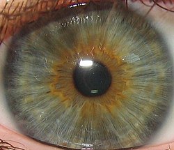
Central

Central heterochromia, green with brown Green eyes with brown central heterochromia.jpg
Central heterochromia, green with brown

Central heterochromia is also an eye condition where there are two colors in the same iris; but the arrangement is concentric, rather than sectoral. The central (pupillary) zone of the iris is a different color than the mid-peripheral (ciliary) zone. Central heterochromia is more noticeable in irises containing low amounts of melanin. [31]

In history and culture

Heterochromia of the eye was first described as a human condition by Aristotle, who termed it heteroglaucos. [12]

Notable historical figures thought to have heterochromia include the Byzantine emperor Anastasius the First, dubbed dikoros (Greek for 'having two pupils'). "His right eye was light blue, while the left was black, nevertheless his eyes were most attractive", is the description of the historian John Malalas. [32] [33] [34] A more recent example is the German poet, playwright, novelist, scientist, statesman, theatre director, and critic, Johann Wolfgang Goethe. [35]

The Alexander Romance , an early literary treatment of the life of Alexander the Great, attributes heterochromia to him. In it he is described as having one eye light and one eye dark. However, no ancient historical source mentions this. It is used to emphasise the otherworldly and heroic qualities of Alexander. [36] [37]

In the Ars Amatoria, the Roman poet Ovid describes the witch Dipsas as having 'double pupils'. Kirby Flower Smith suggested that this could be understood as heterochromia, though other scholars have disagreed. The Roman jurist and writer Cicero also mentions the same feature of 'double pupils' as being found in some Italic women. Pliny the Elder related this feature to the concept of 'the evil eye'. [38]

The twelfth-century scholar Eustathius, in his commentary on the Iliad, reports a tradition in which the Thracian Thamyris (son of the nymph Argiope), who was famed for his musical abilities, had one eye that was grey, whilst the other was black. W. B. McDaniel suggests that this should be interpreted as heterochromia. [39]

In other animals

A Turkish Angora cat with complete heterochromia. Angoraturco.jpg
A Turkish Angora cat with complete heterochromia.

Although infrequently seen in humans, complete heterochromia is more frequently observed in species of domesticated mammals. The blue eye occurs within a white spot, where melanin is absent from the skin and hair (see Leucism). These species include the cat, particularly breeds such as Turkish Van, Turkish Angora, Khao Manee and (rarely) Japanese Bobtail. These so-called odd-eyed cats are white, or mostly white, with one normal eye (copper, orange, yellow, green), and one blue eye. Among dogs, complete heterochromia is seen often in the Siberian Husky and few other breeds, usually Australian Shepherd and Catahoula Leopard Dog and rarely in Shih Tzu. Horses with complete heterochromia have one brown and one white, gray, or blue eye—complete heterochromia is more common in horses with pinto coloring. Complete heterochromia occurs also in cattle and even water buffalo. [40] It can also be seen in ferrets with Waardenburg syndrome, although it can be very hard to tell at times as the eye color is often a midnight blue.

Sectoral heterochromia, usually sectoral hypochromia, is often seen in dogs, specifically in breeds with merle coats. These breeds include the Australian Shepherd, Border Collie, Collie, Shetland Sheepdog, Welsh Corgi, Pyrenean Shepherd, Mudi, Beauceron, Catahoula Cur, Dunker, Great Dane, Dachshund and Chihuahua. It also occurs in certain breeds that do not carry the merle trait, such as the Siberian Husky, Dalmatian, and rarely, Shih Tzu. There are examples of cat breeds that have the condition such as Van cat. [41]

See also

Related Research Articles

<span class="mw-page-title-main">Albinism in humans</span> Condition characterized by partial or complete absence of pigment in the skin, hair and eyes

Albinism is a congenital condition characterized in humans by the partial or complete absence of pigment in the skin, hair and eyes. Albinism is associated with a number of vision defects, such as photophobia, nystagmus, and amblyopia. Lack of skin pigmentation makes for more susceptibility to sunburn and skin cancers. In rare cases such as Chédiak–Higashi syndrome, albinism may be associated with deficiencies in the transportation of melanin granules. This also affects essential granules present in immune cells, leading to increased susceptibility to infection.

<span class="mw-page-title-main">Melanin</span> Group of natural pigments found in most organisms

Melanin consist of oligomers or polymers arranged in a manner which among other functions provide the pigments of many organisms. Melanin pigments are produced in a specialized group of cells known as melanocytes. They have been described as "among the last remaining biological frontiers with the unknown".

<span class="mw-page-title-main">Iris (anatomy)</span> Colored part of an eye

The iris is a thin, annular structure in the eye in most mammals and birds, responsible for controlling the diameter and size of the pupil, and thus the amount of light reaching the retina. In optical terms, the pupil is the eye's aperture, while the iris is the diaphragm. Eye color is defined by the iris.

<span class="mw-page-title-main">Tiger eye</span> Color of horses eyes

Tiger eye or goat eye is a gene causing diluted eye color in horses. There are two variants, Tiger-eye 1 (TE1) and Tiger-eye 2 (TE2), which are both recessive. Horses displaying tiger eye typically have a yellow, orange, or amber iris. Tiger eye has only been found in Puerto Rican Paso Fino horses. Horses of related breeds were tested, and none were found to have either tiger eye allele. No obvious link between eye shade and coat color was seen, making this the first studied gene in horses to affect eye color but not coat color. Tiger eye does not appear to affect vision, and there were no signs of reduced pigment on the retina or retinal pigment epithelium.

<span class="mw-page-title-main">Waardenburg syndrome</span> Genetic condition involving hearing loss and depigmentation

Waardenburg syndrome is a group of rare genetic conditions characterised by at least some degree of congenital hearing loss and pigmentation deficiencies, which can include bright blue eyes, a white forelock or patches of light skin. These basic features constitute type 2 of the condition; in type 1, there is also a wider gap between the inner corners of the eyes called telecanthus, or dystopia canthorum. In type 3, which is rare, the arms and hands are also malformed, with permanent finger contractures or fused fingers, while in type 4, the person also has Hirschsprung's disease. There also exist at least two types that can result in central nervous system (CNS) symptoms such as developmental delay and muscle tone abnormalities.

<span class="mw-page-title-main">Eye color</span> Polygenic phenotypic characteristic

Eye color is a polygenic phenotypic trait determined by two factors: the pigmentation of the eye's iris and the frequency-dependence of the scattering of light by the turbid medium in the stroma of the iris.

<span class="mw-page-title-main">Uveitis</span> Inflammation of the uvea of the eye

Uveitis is inflammation of the uvea, the pigmented layer of the eye between the inner retina and the outer fibrous layer composed of the sclera and cornea. The uvea consists of the middle layer of pigmented vascular structures of the eye and includes the iris, ciliary body, and choroid. Uveitis is described anatomically, by the part of the eye affected, as anterior, intermediate or posterior, or panuveitic if all parts are involved. Anterior uveitis (iridocyclitis) is the most common, with the incidence of uveitis overall affecting approximately 1:4500, most commonly those between the ages of 20-60. Symptoms include eye pain, eye redness, floaters and blurred vision, and ophthalmic examination may show dilated ciliary blood vessels and the presence of cells in the anterior chamber. Uveitis may arise spontaneously, have a genetic component, or be associated with an autoimmune disease or infection. While the eye is a relatively protected environment, its immune mechanisms may be overcome resulting in inflammation and tissue destruction associated with T-cell activation.

<span class="mw-page-title-main">Leucism</span> Partial loss of pigmentation in an animal

Leucism is a wide variety of conditions that result in partial loss of pigmentation in an animal—causing white, pale, or patchy coloration of the skin, hair, feathers, scales, or cuticles, but not the eyes. It is occasionally spelled leukism. Some genetic conditions that result in a "leucistic" appearance include piebaldism, Waardenburg syndrome, vitiligo, Chédiak–Higashi syndrome, flavism, isabellinism, xanthochromism, axanthism, amelanism, and melanophilin mutations. Pale patches of skin, feathers, or fur can also result from injury.

<span class="mw-page-title-main">Hyphema</span> Hemorrhage in the front chamber of the eye

Hyphema is the medical condition of bleeding in the anterior chamber of the eye between the iris and the cornea. People usually first notice a loss or decrease in vision. The eye may also appear to have a reddish tinge, or it may appear as a small pool of blood at the bottom of the iris in the cornea. A traumatic hyphema is caused by a blow to the eye. A hyphema can also occur spontaneously.

Ocular melanosis (OM) is a blue-gray and/or brown lesion of the conjunctiva that can be separated into benign conjunctival epithelial melanosis (BCEM) and primary acquired melanosis (PAM), of which the latter is considered a risk factor for uveal melanoma. The disease is caused by an increase of melanocytes in the iris, choroid, and surrounding structures. Overproduction of pigment by these cells can block the trabecular meshwork through which fluid drains from the eye. The increased fluid in the eye leads to increased pressure, which can lead to glaucoma. In humans, this is sometimes known as pigment dispersion syndrome.

Albinism-black lock-cell migration disorder is the initialism for the following terms and concepts that describe a condition affecting a person's physical appearance and physiology: (1) A – albinism, (2) B – black lock of hair, (3) C – cell migration disorder of the neurocytes of the gut, and (4) D – sensorineural deafness. The syndrome is caused by mutation in the endothelin B receptor gene (EDNRB).

<span class="mw-page-title-main">Piebaldism</span> Medical condition

Piebaldism refers to the absence of mature melanin-forming cells (melanocytes) in certain areas of the skin and hair. It is a rare autosomal dominant disorder of melanocyte development. Common characteristics include a congenital white forelock, scattered normal pigmented and hypopigmented macules and a triangular shaped depigmented patch on the forehead. There is nevertheless great variation in the degree and pattern of presentation, even within affected families. In some cases, piebaldism occurs together with severe developmental problems, as in Waardenburg syndrome and Hirschsprung's disease.

<span class="mw-page-title-main">White horse</span> Horse coat color

A white horse is born predominantly white and stays white throughout its life. A white horse has mostly pink skin under its hair coat, and may have brown, blue, or hazel eyes. "True white" horses, especially those that carry one of the dominant white (W) genes, are rare. Most horses that are commonly referred to as "white" are actually "gray" horses whose hair coats are completely white. Gray horses may be born of any color and their hairs gradually turn white as time goes by and take on a white appearance. Nearly all gray horses have dark skin, except under any white markings present at birth. Skin color is the most common method for an observer to distinguish between mature white and gray horses.

Polycoria is a pathological condition of the eye characterized by more than one pupillary opening in the iris. It may be congenital or result from a disease affecting the iris. It results in decreased function of the iris and pupil, affecting the physical eye and visualization.

Oculocutaneous albinism type I or type 1A is an autosomal recessive skin disease. This subtype of oculocutaneous albinism is caused when the gene for tyrosinase does not function properly.

<span class="mw-page-title-main">Ocular albinism type 1</span> Most common type of ocular albinism

Ocular albinism type 1(OA1) is the most common type of ocular albinism, with a prevalence rate of 1:50,000. It is an inheritable classical Mendelian type X-linked recessive disorder wherein the retinal pigment epithelium lacks pigment while hair and skin appear normal. Since it is usually an X-linked disorder, it occurs mostly in males, while females are carriers unless they are homozygous. About 60 missense and nonsense mutations, insertions, and deletions have been identified in Oa1. Mutations in OA1 have been linked to defective glycosylation and thus improper intracellular transportation.

Petrus Johannes Waardenburg was a Dutch ophthalmologist, geneticist, and pioneer in the application of genetics to ophthalmology. Waardenburg syndrome is named after him.

<span class="mw-page-title-main">Amelanism</span> Pigmentation abnormality

Amelanism is a pigmentation abnormality characterized by the lack of pigments called melanins, commonly associated with a genetic loss of tyrosinase function. Amelanism can affect fish, amphibians, reptiles, birds, and mammals including humans. The appearance of an amelanistic animal depends on the remaining non-melanin pigments. The opposite of amelanism is melanism, a higher percentage of melanin.

Waardenburg syndrome type 4A is an extremely rare congenital disorder caused by a mutation in an endothelin receptor gene. It results in common Waardenburg syndrome symptoms such as abnormal hair and skin pigmentation and heterochromia, but also present with symptoms of Hirschsprung's disease. Symptoms include abdominal pain and bowel obstruction. Waardenburg syndrome type 4A is the rarest among the types, appearing only once in about every 1,000,000 individuals. There have only been a total of 50 cases reported in total as of 2016.

Ocular albinism late onset sensorineural deafness (OASD) is a rare, X-linked recessive disease characterized by intense visual impairments, reduced retinal pigments, translucent pale-blue irises and moderately severe hearing loss from adolescence to middle-age. It is a subtype of Ocular Albinism (OA) that is linked to Ocular albinism type I (OA1). OA1 is the most common form of ocular albinism, affecting at least 1/60,000 males.

References

  1. Kumar P (2017). "Focal Scalp Hair Heterochromia in an Infant". Sultan Qaboos University Medical Journal. 17 (1): e116–118. doi:10.18295/squmj.2016.17.01.022. PMC   5380409 . PMID   28417041.
  2. Imesch PD, Wallow IH, Albert DM (February 1997). "The color of the human eye: a review of morphologic correlates and of some conditions that affect iridial pigmentation throughout life". Survey of Ophthalmology. 41 (Suppl 2): S117–23. doi:10.1016/S0039-6257(97)80018-5. PMID   9154287.
  3. 1 2 3 4 5 6 7 8 9 10 Loewenstein, John; Scott Lee (2004). Ophthalmology: Just the Facts. New York: McGraw-Hill. ISBN   0-07-140332-9.
  4. Konovalova EN, Gladyr EA, Kostiunina OV, Zinovieva LK (2017). "Congenital Defects of Beef Cattle and General Principles of their Prevention". Journal of Agriculture and Environment. 2 (3). doi:10.23649/jae.2017.2.3.1.{{cite journal}}: CS1 maint: multiple names: authors list (link)
  5. Ur Rehman H (2008). "Heterochromia". CMAJ. 179 (5): 447–448. doi: 10.1503/cmaj.070497 . PMC   2518194 . PMID   18725617.
  6. "heterochromia iridis - definition of heterochromia iridis in the Medical dictionary - by the Free Online Medical Dictionary, Thesaurus and Encyclopedia". Medical-dictionary.thefreedictionary.com. Retrieved 2014-04-27.
  7. Wielgus AR, Sarna T (December 2005). "Melanin in human irides of different color and age of donors". Pigment Cell Research. 18 (6): 454–64. doi:10.1111/j.1600-0749.2005.00268.x. PMID   16280011.
  8. Prota G, Hu DN, Vincensi MR, McCormick SA, Napolitano A (September 1998). "Characterization of melanins in human irides and cultured uveal melanocytes from eyes of different colors". Experimental Eye Research. 67 (3): 293–9. doi:10.1006/exer.1998.0518. PMID   9778410.
  9. National Day Calendar "National Different Colored Eyes Day - July 12" Archived 2023-03-31 at the Wayback Machine National Day Calendar. November 8, 2022. Retrieved July 12, 2023.
  10. Swann P. "Heterochromia." Archived 2006-01-08 at the Wayback Machine Optometry Today. January 29, 1999. Retrieved November 1, 2006.
  11. "Heterochromia: MedlinePlus Medical Encyclopedia". Nlm.nih.gov. Retrieved 2014-04-27.
  12. 1 2 Gladstone RM (1969). "Development and Significance of Heterochromia of the Iris". Arch Neurol. 21 (2): 184–192. doi:10.1001/archneur.1969.00480140084008. PMID   5797351.
  13. Guha M, Maity D (2014). "Heterochromia iridis - a case study". Explor Anim Med Res. 4 (2): 240–245.
  14. van Emelen C, Goethals M, Dralands L, Casteels I (Jan–Feb 2000). "Treatment of glaucoma in children with Sturge-Weber syndrome". Journal of Pediatric Ophthalmology and Strabismus. 37 (1): 29–34. doi:10.3928/0191-3913-20000101-08. PMID   10714693.
  15. "Sturge-Weber syndrome: Definition and Much More from Answers.com". Answers.com<!. Retrieved 2009-11-19.
  16. 1 2 Wallis DH, Granet DB, Levi L (June 2003). "When the darker eye has the smaller pupil". Journal of American Association for Pediatric Ophthalmology and Strabismus. 7 (3): 215–6. doi:10.1016/S1091-8531(02)42020-4. PMID   12825064.
  17. Ohno N, Kiyosawa M, Mori H, Wang WF, Takase H, Mochizuki M (Jan–Feb 2003). "Clinical findings in Japanese patients with Waardenburg syndrome type 2". Japanese Journal of Ophthalmology. 47 (1): 77–84. doi:10.1016/S0021-5155(02)00629-9. PMID   12586183.
  18. Brazel SM, Sullivan TJ, Thorner PS, Clarke MP, Hunter WS, Morin JD (February 1992). "Iris sector heterochromia as a marker for neural crest disease". Archives of Ophthalmology. 110 (2): 233–5. doi:10.1001/archopht.1992.01080140089033. PMID   1736874.
  19. Liu CSC (August 1999). "A case of acquired iris depigmentation as a possible complication of levobunolol eye drops". British Journal of Ophthalmology. 83 (12): 1405–6. doi:10.1136/bjo.83.12.1403c. PMC   1722884 . PMID   10660314.
  20. Stjernschantz, Johan W.; Albert, Daniel M.; Hu, Dan-Ning; Drago, Filippo; Wistrand, Per J. (August 2002). "Mechanism and Clinical Significance of Prostaglandin-Induced Iris Pigmentation". Survey of Ophthalmology . 47: S162–S175. doi:10.1016/S0039-6257(02)00292-8. PMID   12204714.
  21. Sridhar, Uma; Tripathy, Koushik (January 2023). "Iris Ectropion Syndrome". StatPearls. StatPearls Publishing, Treasure Island, FL. PMID   35593818.
  22. Yang P, Fang W, Jin H, Li B, Chen X, Kijlstra A (March 2006). "Clinical features of Chinese patients with Fuchs' syndrome". Ophthalmology. 113 (3): 473–80. doi:10.1016/j.ophtha.2005.10.028. PMID   16458965.
  23. Arellanes-Garcia L, del Carmen Preciado-Delgadillo M, Recillas-Gispert C (June 2002). "Fuchs' heterochromic iridocyclitis: clinical manifestations in dark-eyed Mexican patients". Ocular Immunology and Inflammation. 10 (2): 125–31. doi:10.1076/ocii.10.2.125.13976. PMID   12778348. S2CID   21171244.
  24. 1 2 Tabbut BR, Tessler HH, Williams D (December 1988). "Fuchs' heterochromic iridocyclitis in blacks". Archives of Ophthalmology. 106 (12): 1688–90. doi:10.1001/archopht.1988.01060140860027. PMID   3196209.
  25. Bloch-Michel E (1983). "Fuchs heterochromic cyclitis: current concepts". Journal Français d'Ophtalmologie (in French). 6 (10): 853–8. PMID   6368659.
  26. Mehta K, Haller JO, Legasto AC (2003). "Imaging neuroblastoma in children". Critical Reviews in Computed Tomography. 44 (1): 47–61. doi:10.1080/10408370390808469. PMID   12627783.
  27. Genetic and Rare Diseases Information Center (GARD). "Heterochromia iridis".
  28. Khan AO, Aldamesh M (June 2006). "Bilateral Duane syndrome and bilateral aniridia". Journal of American Association for Pediatric Ophthalmology and Strabismus. 10 (3): 273–4. doi:10.1016/j.jaapos.2006.02.002. PMID   16814183.
  29. Shauly Y, Weissman A, Meyer E (May–Jun 1993). "Ocular and systemic characteristics of Duane syndrome". Journal of Pediatric Ophthalmology and Strabismus. 30 (3): 178–83. doi:10.3928/0191-3913-19930501-12. PMID   8350229.
  30. "Heterochromia iridis". Genetic and Rare Diseases Information Center (GARD) – an NCATS Program. NIH. 8 April 2015. Retrieved 9 February 2019.
  31. Edwards, Melissa; Cha, David; Krithika, S.; Johnson, Monique; Cook, Gillian; Parra, Esteban J. (2015-11-07). "Iris pigmentation as a quantitative trait: variation in populations of European, East Asian and South Asian ancestry and association with candidate gene polymorphisms". Pigment Cell & Melanoma Research . 29 (2): 156. doi: 10.1111/pcmr.12435 . PMID   26547379.
  32. Baldwin, Barry (1981). "Physical Descriptions of Byzantine Emperors". Byzantion. 51 (1): 8–21. ISSN   0378-2506. JSTOR   44170668.
  33. Fronimopoulos, John; Lascaratos, John (1992-03-01). "Some Byzantine chroniclers and historians on ophthalmological topics". Documenta Ophthalmologica. 81 (1): 121–132. doi:10.1007/BF00155022. ISSN   1573-2622. PMID   1473460. S2CID   26240821.
  34. Head, C. (1980)"PHYSICAL DESCRIPTIONS OF THE EMPERORS IN BYZANTINE HISTORICAL WRITING", Byzantion, Vol. 50, No. 1 (1980), pp. 226-24, Peeters Publishers, p. 229
  35. Friedenthal, Richard (1963). Goethe: His Life and Times. Transaction Publishers. p. 472. ISBN   978-1-412-84321-8.
  36. Nawotka, Krzysztof (2010) Alexander the Great, Cambridge Scholarship Publishing, ISBN   9781443818117, p. 44
  37. Boardman, J. (2019) Alexander the Great: From His Death to the Present Day, Princeton University Press, Princeton N.J., p. 40
  38. Alvar Nuño, A. (2012) "Ocular Pathologies and the Evil Eye in the Early Roman Principate", Numen, Vol. 59, No. 4 (2012), pp. 295-321, pp. 299-301
  39. McDaniel, W,B. (1918) "The Pupula Duplex and Other Tokens of an "Evil Eye" in the Light of Ophthalmology", Classical Philology, Vol. 13, No. 4 (Oct., 1918), pp. 335-346, The University of Chicago Press, p.338
  40. Misk NA, Semieka MA, Fathy A (1998). "Heterochromia iridis in water buffaloes (Bubalus bubalis)". Veterinary Ophthalmology. 1 (4): 195–201. doi:10.1046/j.1463-5224.1998.00036.x. PMID   11397231.
  41. "Turkish Van Cats" . Retrieved 8 April 2014. This tertiary source reuses information from other sources but does not name them.