
In human anatomy, the inferior epigastric vessels [1] refers to the inferior epigastric artery and the inferior epigastric vein.
In human anatomy, the inferior epigastric vessels [1] refers to the inferior epigastric artery and the inferior epigastric vein.
| | This cardiovascular system article is a stub. You can help Wikipedia by expanding it. |

The external iliac arteries are two major arteries which bifurcate off the common iliac arteries anterior to the sacroiliac joint of the pelvis.

In human anatomy, the inguinal triangle is a region of the abdominal wall. It is also known by the eponym Hesselbach's triangle, after Franz Kaspar Hesselbach.

In human anatomy, the inferior epigastric artery is an artery that arises from the external iliac artery. It is accompanied by the inferior epigastric vein; inferiorly, these two inferior epigastric vessels together travel within the lateral umbilical fold The inferior epigastric artery then traverses the arcuate line of rectus sheath to enter the rectus sheath, then anastomoses with the superior epigastric artery within the rectus sheath.

In human anatomy, inferior epigastric vein are 1-2 veins accompanying the inferior epigastric artery. They drain into the external iliac vein just proximal to the inguinal ligament.

In human anatomy, the superior epigastric artery is a terminal branch of the internal thoracic artery that provides arterial supply to the abdominal wall, and upper rectus abdominis muscle. It enters the rectus sheath to descend upon the inner surface of the rectus abdominis muscle. It ends by anastomosing with the inferior epigastric artery.
In human anatomy, the superior epigastric veins are two or more venae comitantes which accompany either superior epigastric artery before emptying into the internal thoracic vein. They participate in the drainage of the superior surface of the diaphragm.

Lateral to the conjoint tendon, previously known as the inguinal aponeurotic falx, there is a ligamentous band originating from the lower margin of the transversalis fascia and extending down in front of the inferior epigastric artery to the superior ramus of the pubis; it is termed the interfoveolar ligament of Hesselbach and sometimes contains a few muscular fibers.
The conjoint tendon is a sheath of connective tissue formed from the lower part of the common aponeurosis of the abdominal internal oblique muscle and the transversus abdominis muscle, joining the muscle to the pelvis. It forms the medial part of the posterior wall of the inguinal canal.
The obturator artery is a branch of the internal iliac artery that passes antero-inferiorly on the lateral wall of the pelvis, to the upper part of the obturator foramen, and, escaping from the pelvic cavity through the obturator canal, it divides into an anterior branch and a posterior branch.

The lumbar arteries are arteries located in the lower back or lumbar region. The lumbar arteries are in parallel with the intercostals.
In human anatomy, the inguinal region refers to either the groin or the lower lateral regions of the abdomen. It may also refer to:

The sternocostal triangle are small zones lying between the costal and sternal attachments of the thoracic diaphragm. No vascular elements are present within this space. The borders of this space are:

The lateral umbilical fold is an elevation of the peritoneum lining the inner/posterior surface of the lower anterior abdominal wall formed by the underlying inferior epigastric artery and inferior epigastric vein which the peritoneum covers. Superiorly, the lateral umbilical fold ends where the vessels reach and enter the rectus sheath at the arcuate line of rectus sheath; in spite of the name, the lateral umbilical folds do not extend as far superiorly as the umbilicus. Inferiorly, it extends to just medial to the deep inguinal ring.

The superficial epigastric artery arises from the front of the femoral artery about 1 cm below the inguinal ligament, and, passing through the femoral sheath and the fascia cribrosa, turns upward in front of the inguinal ligament, and ascends between the two layers of the superficial fascia of the abdominal wall nearly as far as the umbilicus.
In anatomy, arterial tree is used to refer to all arteries and/or the branching pattern of the arteries. This article regards the human arterial tree. Starting from the aorta:

The cremasteric artery is a branch of the inferior epigastric artery which accompanies the spermatic cord to supply the cremaster muscle as well as other coverings of the spermatic cord in the male.

The medial umbilical ligament is a paired structure found in human anatomy. It is on the deep surface of the anterior abdominal wall, and is covered by the medial umbilical folds. It is different from the median umbilical ligament, a structure that represents the remnant of the embryonic urachus.

The deep circumflex iliac artery is an artery in the pelvis that travels along the iliac crest of the pelvic bone.
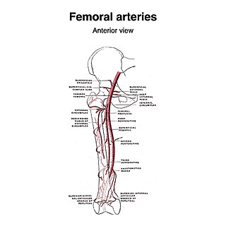
The superficial iliac circumflex artery, the smallest of the cutaneous branches of the femoral artery, arises close to the superficial epigastric artery, and, piercing the fascia lata, runs lateralward, parallel with the inguinal ligament, as far as the crest of the ilium.

The following outline is provided as an overview of and topical guide to human anatomy: