Phage ecology

Last updated

Bacteriophages (phages), potentially the most numerous "organisms" on Earth, are the viruses of bacteria (more generally, of prokaryotes [1] ). Phage ecology is the study of the interaction of bacteriophages with their environments. [2]

Contents

Introduction to phage ecology

Vastness of phage ecology

Phages are obligate intracellular parasites meaning that they are able to reproduce only while infecting bacteria. Phages therefore are found only within environments that contain bacteria. Most environments contain bacteria, including our own bodies (called normal flora). Often these bacteria are found in large numbers. As a consequence, phages are found almost everywhere.[ citation needed ]

As a rule of thumb, many phage biologists expect that phage population densities will exceed bacterial densities by a ratio of 10-to-1 or more (VBR or virus-to-bacterium ratio; see [3] for a summary of actual data). As there exist estimates of bacterial numbers on Earth of approximately 1030, [4] there consequently is an expectation that 1031 or more individual virus (mostly phage [5] ) particles exist , making phages the most numerous category of "organisms" on our planet.

Bacteria (along with archaea) appear to be highly diverse and there possibly are millions of species. [6] Phage-ecological interactions therefore are quantitatively vast: huge numbers of interactions. Phage-ecological interactions are also qualitatively diverse: There are huge numbers of environment types, bacterial-host types, [7] and also individual phage types [8]

Studying phage ecology

The study of phage ecology reflects established scientific disciplines in ecological studies in scope, the most obvious being general ecology. Accordingly, phage ecology is treated under the following heads "organismal" ecology, population ecology, community ecology, and ecosystem ecology. Phage ecology also may be considered (though mostly less well formally explored) from perspectives of phage behavioral ecology, evolutionary ecology, functional ecology, landscape ecology, mathematical ecology, molecular ecology, physiological ecology (or ecophysiology), and spatial ecology. Phage ecology additionally draws (extensively) from microbiology, particularly in terms of environmental microbiology, but also from an enormous catalog (90 years) of study of phage and phage-bacterial interactions in terms of their physiology and, especially, their molecular biology.[ citation needed ]

Phage "organismal" ecology

Phage "organismal" ecology is primarily the study of the evolutionary ecological impact of phage growth parameters:

Another way of envisioning phage "organismal" ecology is that it is the study of phage adaptations that contribute to phage survival and transmission to new hosts or environments. Phage "organismal" ecology is the most closely aligned of phage ecology disciplines with the classical molecular and molecular genetic analyses of bacteriophage.

From the perspective of ecological subdisciplines, we can also consider phage behavioral ecology, functional ecology, and physiological ecology under the heading of phage "organismal" ecology. However, as noted, these subdisciplines are not as well developed as more general considerations of phage "organismal" ecology. Phage growth parameters often evolve over the course of phage experimental adaptation studies.

Historical overview

In the mid 1910s, when phage were first discovered, the concept of phage was very much a whole-culture phenomenon (like much of microbiology [11] ), where various types of bacterial cultures (on solid media, in broth) were visibly cleared by phage action. Though from the start there was some sense, especially by Fėlix d'Hėrelle, that phage consisted of individual "organisms", in fact it wasn't until the late 1930s through the 1940s that phages were studied, with rigor, as individuals, e.g., by electron microscopy and single-step growth experiments. [12] Note, though, that for practical reasons much of "organismal" phage study is of their properties in bulk culture (many phage) rather than the properties of individual phage virions or individual infections.[ citation needed ]

This somewhat whole-organismal view of phage biology saw its heyday during the 1940s and 1950s, before giving way to much more biochemical, molecular genetic, and molecular biological analyses of phages, as seen during the 1960s and onward. This shift, paralleled in much of the rest of microbiology , represented a retreat from a much more ecological view of phages (first as bacterial killers, and then as organisms unto themselves). However, the organismal view of phage biology lives on as a foundation of phage ecological understanding. Indeed, it represents a key thread that ties together the ecological thinking on phage ecology with the more "modern" considerations of phage as molecular model systems.[ citation needed ]

Methods

The basic experimental toolkit of phage "organismal" ecology consists of the single-step growth (or one-step growth; [12] ) experiment and the phage adsorption curve. [13] Single-step growth is a means of determining the phage latent period (example), which is approximately equivalent (depending on how it is defined) to the phage period of infection. Single-step growth experiments also are employed to determine a phage's burst size, which is the number of phage (on average) that are produced per phage-infected bacterium.[ citation needed ]

The adsorption curve is obtained by measuring the rate at which phage virion particles (see Virion#Structure) attach to bacteria. This is usually done by separating free phage from phage-infected bacteria in some manner so that either the loss of not currently infecting (free) phage or the gain of infected bacteria may be measured over time.[ citation needed ]

Phage population ecology

A population is a group of individuals which either do or can interbreed or, if incapable of interbreeding, then are recently derived from a single individual (a clonal population). Population ecology considers characteristics that are apparent in populations of individuals but either are not apparent or are much less apparent among individuals. These characteristics include so-called intraspecific interactions, that is between individuals making up the same population, and can include competition as well as cooperation. Competition can be either in terms of rates of population growth (as seen especially at lower population densities in resource-rich environments) or in terms of retention of population sizes (seen especially at higher population densities where individuals are directly competing over limited resources). Respectively, these are population-density independent and dependent effects.[ citation needed ]

Phage population ecology considers issues of rates of phage population growth, but also phage-phage interactions as can occur when two or more phage adsorb an individual bacterium.

Phage community ecology

A community consists of all of the biological individuals found within a given environment (more formally, within an ecosystem), particularly when more than one species is present. Community ecology studies those characteristics of communities that either are not apparent or which are much less apparent if a community consists of only a single population. Community ecology thus deals with interspecific interactions. Interspecific interactions, like intraspecific interactions, can range from cooperative to competitive but also to quite antagonistic (as are seen, for example, with predator-prey interactions). An important consequence of these interactions is coevolution.

Relationship with bacteria

The interaction of phage with bacteria is the primary concern of phage community ecologists. Bacteria have developed mechanisms that prevent phages from having an effect on them, which has led to this evolutionary arms race between the phages and their host bacteria. [14] Bacterial resistance to phages puts pressure on the phages to develop stronger effects on the bacteria. The Red Queen hypothesis describes this relationship, as the organisms must constantly adapt and evolve in order to survive. [15] This relationship is important to understand as phages are now being used for more practical and medicinal purposes.

Bacteria have developed multiple defense mechanisms to fight off the effects of bacteriophages. [16] In experimentation, amount of resistance can be determined by how much of a plate (generally agar with bacteria, infected with phages) ends up being clear. The clearer, the less resistant as more bacteria have been lysed. [17] The most common of these defense mechanisms is called the restriction-modification system (RM system). In this system, foreign DNA trying to enter the bacterial host is restricted by endonucleases that recognize specific base pairs within the DNA, while the DNA of the cell is protected from restriction due to methylase. [16] RM systems have evolved to keep up with the ever-changing bacteria and phage. In general, these RM types differ in the nucleotide sequences that they recognize. [18] However, there is an occasional slip where the endonuclease misses the DNA sequence of the phage and the phage DNA is able to enter the cell anyway, becoming methylated and protected against the endonuclease. This accident is what can spur the evolution of the RM system. Phages can acquire or use the enzyme from the host cell to protect their own DNA, or sometimes they will have proteins that dismantle the enzyme that is meant to restrict the phage DNA. [16] Another option is for the phage to insert different base pairs into its DNA, thereby confusing the enzyme.

Another mechanism employed by bacteria is referred to as CRISPR. This stands for “clustered regularly interspersed palindromic repeats” which means that the immunity to phages by bacteria has been acquired via adding spacers of DNA that are identical to that of the DNA from the phage. Some phages have been found to be immune to this mechanism as well. In some way or another, the phages have managed to get rid of the sequence that would be replicated.

A third way that bacteria have managed to escape the effects of bacteriophages is by abortive infection. This is a last resort option- when the host cell has already been infected by the phage. This method is not ideal for the host cell, as it still leads to its death. The redeeming feature of this mechanism is the fact that it interferes with the phage processes and prevents it from then moving on to infect other cells. [16]

On top of the above mentioned strategies, a growing arsenal of anti-phage immune systems has been described and quantified in bacteria. [19]

Phages are also capable of interacting with species other than bacteria, e.g., such as phage-encoded exotoxin interaction with animals. [20] Phage therapy is an example of applied phage community ecology.[ citation needed ]

Phage ecosystem ecology

An ecosystem consists of both the biotic and abiotic components of an environment. Abiotic entities are not alive and so an ecosystem essentially is a community combined with the non-living environment within which that ecosystem exists. Ecosystem ecology naturally differs from community ecology in terms of the impact of the community on these abiotic entities, and vice versa. In practice, the portion of the abiotic environment of most concern to ecosystem ecologists is inorganic nutrients and energy.

Phages impact the movement of nutrients and energy within ecosystems primarily by lysing bacteria. Phages can also impact abiotic factors via the encoding of exotoxins (a subset of which are capable of solubilizing the biological tissues of living animals ). Phage ecosystem ecologists are primarily concerned with the phage impact on the global carbon cycle, especially within the context of a phenomenon known as the microbial loop.

Notes

  1. The term "prokaryotes" is useful to mean the sum of the bacteria and archaea but otherwise can be controversial, as discussed by Woese CR (June 2004). "A new biology for a new century". Microbiol. Mol. Biol. Rev. 68 (2): 173–86. doi:10.1128/MMBR.68.2.173-186.2004. PMC   419918 . PMID   15187180. The Dismantling of Bacteriology and a Deconstruction of the Procaryote {{cite journal}}: External link in |quote= (help); see also pp. 103–4 of Sapp, Jan (2004). "Evolving biological organization". Microbial phylogeny and evolution: concepts and controversies . Oxford [Oxfordshire]: Oxford University Press. pp.  99–118. ISBN   978-0-19-516877-8.
    Sapp J (September 2006). "Two faces of the prokaryote concept" (PDF). Int. Microbiol. 9 (3): 163–72. PMID   17061206. provides a history.
  2. This article on phage ecology was expanded from a stub during the writing of the first chapter of the edited monograph, Bacteriophage Ecology (forecasted publication date: March, 2008, Cambridge University Press), in order to be cited by that chapter especially as a repository of phage ecology review chapters and articles.
  3. Weinbauer MG (May 2004). "Ecology of prokaryotic viruses". FEMS Microbiol. Rev. 28 (2): 127–81. doi: 10.1016/j.femsre.2003.08.001 . PMID   15109783.
  4. Whitman WB, Coleman DC, Wiebe WJ (June 1998). "Prokaryotes: the unseen majority". Proc. Natl. Acad. Sci. U.S.A. 95 (12): 6578–83. Bibcode:1998PNAS...95.6578W. doi: 10.1073/pnas.95.12.6578 . PMC   33863 . PMID   9618454.
  5. Wommack KE, Colwell RR (March 2000). "Virioplankton: viruses in aquatic ecosystems". Microbiol. Mol. Biol. Rev. 64 (1): 69–114. doi:10.1128/MMBR.64.1.69-114.2000. PMC   98987 . PMID   10704475.
  6. Curtis TP, Sloan WT, Scannell JW (August 2002). "Estimating prokaryotic diversity and its limits". Proc. Natl. Acad. Sci. U.S.A. 99 (16): 10494–9. Bibcode:2002PNAS...9910494C. doi: 10.1073/pnas.142680199 . PMC   124953 . PMID   12097644.
  7. Sogin ML, Morrison HG, Huber JA, et al. (August 2006). "Microbial diversity in the deep sea and the underexplored "rare biosphere"". Proc. Natl. Acad. Sci. U.S.A. 103 (32): 12115–20. Bibcode:2006PNAS..10312115S. doi: 10.1073/pnas.0605127103 . PMC   1524930 . PMID   16880384.
  8. Breitbart M, Salamon P, Andresen B, et al. (October 2002). "Genomic analysis of uncultured marine viral communities". Proc. Natl. Acad. Sci. U.S.A. 99 (22): 14250–5. Bibcode:2002PNAS...9914250B. doi: 10.1073/pnas.202488399 . PMC   137870 . PMID   12384570.
  9. Barksdale L, Arden SB (1974). "Persisting bacteriophage infections, lysogeny, and phage conversions". Annu. Rev. Microbiol. 28: 265–99. doi:10.1146/annurev.mi.28.100174.001405. PMID   4215366.
  10. Miller, R. V. & S. A. Ripp (2002). "Pseudolysogeny: A bacteriophage strategy for increasing longevity in situ". In Kado, Clarence I. & Syvanen, Michael (eds.). Horizontal gene transfer (2nd ed.). Boston: Academic Press. pp.  81–91. ISBN   978-0-12-680126-2.
  11. Summers WC (1991). "From culture as organism to organism as cell: historical origins of bacterial genetics". J Hist Biol. 24 (2): 171–90. doi:10.1007/bf00209428. PMID   11612551. S2CID   36544748.
  12. 1 2 You L, Suthers PF, Yin J (April 2002). "Effects of Escherichia coli physiology on growth of phage T7 in vivo and in silico". J. Bacteriol. 184 (7): 1888–94. doi:10.1128/JB.184.7.1888-1894.2002. PMC   134924 . PMID   11889095.
  13. Abedon ST, Hyman P, Thomas C (December 2003). "Experimental examination of bacteriophage latent-period evolution as a response to bacterial availability". Appl. Environ. Microbiol. 69 (12): 7499–506. Bibcode:2003ApEnM..69.7499A. doi:10.1128/AEM.69.12.7499-7506.2003. PMC   310036 . PMID   14660403.
  14. Stern, Adi; Sorek, Rotem (2011). "The phage-host arms race: Shaping the evolution of microbes". BioEssays. 33 (1): 43–51. doi:10.1002/bies.201000071. PMC   3274958 . PMID   20979102.
  15. Lenski, Richard E.; Levin, Bruce R. (1 January 1985). "Constraints on the Coevolution of Bacteria and Virulent Phage: A Model, Some Experiments, and Predictions for Natural Communities". The American Naturalist. 125 (4): 585–602. doi:10.1086/284364. JSTOR   2461275. S2CID   82562085.
  16. 1 2 3 4 ISSN   0265-9247 issue v33i0001 article 43
  17. Buckling, Angus; Rainey, Paul B. (1 January 2002). "Antagonistic Coevolution between a Bacterium and a Bacteriophage". Proceedings: Biological Sciences. 269 (1494): 931–936. doi:10.1098/rspb.2001.1945. JSTOR   3067783. PMC   1690980 . PMID   12028776.
  18. ISSN   0092-8240 issue v62i0004 article 759
  19. Beavogui, Angelina; Lacroix, Auriane; Wiart, Nicolas; Poulain, Julie; Delmont, Tom O.; Paoli, Lucas; Wincker, Patrick; Oliveira, Pedro H. (2024-03-08). "The defensome of complex bacterial communities". Nature Communications. 15 (1). doi:10.1038/s41467-024-46489-0. ISSN   2041-1723.
  20. "Evolutionary Bioinformatics Online 2005". Libertas Academica. Archived from the original on 2006-05-26.

Related Research Articles

<span class="mw-page-title-main">Bacteriophage</span> Virus that infects and replicates within bacteria

A bacteriophage, also known informally as a phage, is a virus that infects and replicates within bacteria and archaea. The term was derived from "bacteria" and the Greek φαγεῖν, meaning "to devour". Bacteriophages are composed of proteins that encapsulate a DNA or RNA genome, and may have structures that are either simple or elaborate. Their genomes may encode as few as four genes and as many as hundreds of genes. Phages replicate within the bacterium following the injection of their genome into its cytoplasm.

<span class="mw-page-title-main">DNA virus</span> Virus that has DNA as its genetic material

A DNA virus is a virus that has a genome made of deoxyribonucleic acid (DNA) that is replicated by a DNA polymerase. They can be divided between those that have two strands of DNA in their genome, called double-stranded DNA (dsDNA) viruses, and those that have one strand of DNA in their genome, called single-stranded DNA (ssDNA) viruses. dsDNA viruses primarily belong to two realms: Duplodnaviria and Varidnaviria, and ssDNA viruses are almost exclusively assigned to the realm Monodnaviria, which also includes some dsDNA viruses. Additionally, many DNA viruses are unassigned to higher taxa. Reverse transcribing viruses, which have a DNA genome that is replicated through an RNA intermediate by a reverse transcriptase, are classified into the kingdom Pararnavirae in the realm Riboviria.

<span class="mw-page-title-main">Hershey–Chase experiment</span> DNA experiment

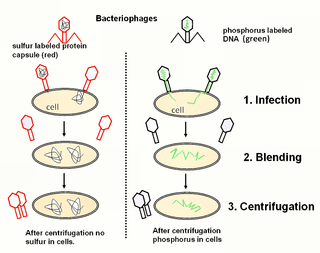
The Hershey–Chase experiments were a series of experiments conducted in 1952 by Alfred Hershey and Martha Chase that helped to confirm that DNA is genetic material.

<span class="mw-page-title-main">Prophage</span> Bacteriophage genome that is integrated into a bacterial cell

A prophage is a bacteriophage genome that is integrated into the circular bacterial chromosome or exists as an extrachromosomal plasmid within the bacterial cell. Integration of prophages into the bacterial host is the characteristic step of the lysogenic cycle of temperate phages. Prophages remain latent in the genome through multiple cell divisions until activation by an external factor, such as UV light, leading to production of new phage particles that will lyse the cell and spread. As ubiquitous mobile genetic elements, prophages play important roles in bacterial genetics and evolution, such as in the acquisition of virulence factors.

<span class="mw-page-title-main">Transduction (genetics)</span> Transfer process in genetics

Transduction is the process by which foreign DNA is introduced into a cell by a virus or viral vector. An example is the viral transfer of DNA from one bacterium to another and hence an example of horizontal gene transfer. Transduction does not require physical contact between the cell donating the DNA and the cell receiving the DNA, and it is DNase resistant. Transduction is a common tool used by molecular biologists to stably introduce a foreign gene into a host cell's genome.

<i>Escherichia virus T4</i> Species of bacteriophage

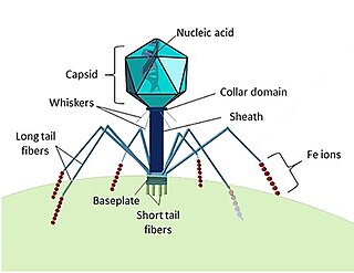
Escherichia virus T4 is a species of bacteriophages that infect Escherichia coli bacteria. It is a double-stranded DNA virus in the subfamily Tevenvirinae from the family Myoviridae. T4 is capable of undergoing only a lytic life cycle and not the lysogenic life cycle. The species was formerly named T-even bacteriophage, a name which also encompasses, among other strains, Enterobacteria phage T2, Enterobacteria phage T4 and Enterobacteria phage T6.

<span class="mw-page-title-main">Filamentous bacteriophage</span> Family of viruses

Filamentous bacteriophages are a family of viruses (Inoviridae) that infect bacteria, or bacteriophages. They are named for their filamentous shape, a worm-like chain, about 6 nm in diameter and about 1000-2000 nm long. This distinctive shape reflects their method of replication: the coat of the virion comprises five types of viral protein, which are located in the inner membrane of the host bacterium during phage assembly, and these proteins are added to the nascent virion's DNA as it is extruded through the membrane. The simplicity of filamentous phages makes them an appealing model organism for research in molecular biology, and they have also shown promise as tools in nanotechnology and immunology.

<span class="mw-page-title-main">Lysogenic cycle</span> Process of virus reproduction

Lysogeny, or the lysogenic cycle, is one of two cycles of viral reproduction. Lysogeny is characterized by integration of the bacteriophage nucleic acid into the host bacterium's genome or formation of a circular replicon in the bacterial cytoplasm. In this condition the bacterium continues to live and reproduce normally, while the bacteriophage lies in a dormant state in the host cell. The genetic material of the bacteriophage, called a prophage, can be transmitted to daughter cells at each subsequent cell division, and later events can release it, causing proliferation of new phages via the lytic cycle.

<span class="mw-page-title-main">T7 phage</span> Species of virus

Bacteriophage T7 is a bacteriophage, a virus that infects bacteria. It infects most strains of Escherichia coli and relies on these hosts to propagate. Bacteriophage T7 has a lytic life cycle, meaning that it destroys the cell it infects. It also possesses several properties that make it an ideal phage for experimentation: its purification and concentration have produced consistent values in chemical analyses; it can be rendered noninfectious by exposure to UV light; and it can be used in phage display to clone RNA binding proteins.

Salmonella virus P22 is a bacteriophage in the Podoviridae family that infects Salmonella typhimurium. Like many phages, it has been used in molecular biology to induce mutations in cultured bacteria and to introduce foreign genetic material. P22 has been used in generalized transduction and is an important tool for investigating Salmonella genetics.

<span class="mw-page-title-main">Cyanophage</span> Virus that infects cyanobacteria

Cyanophages are viruses that infect cyanobacteria, also known as Cyanophyta or blue-green algae. Cyanobacteria are a phylum of bacteria that obtain their energy through the process of photosynthesis. Although cyanobacteria metabolize photoautotrophically like eukaryotic plants, they have prokaryotic cell structure. Cyanophages can be found in both freshwater and marine environments. Marine and freshwater cyanophages have icosahedral heads, which contain double-stranded DNA, attached to a tail by connector proteins. The size of the head and tail vary among species of cyanophages. Cyanophages infect a wide range of cyanobacteria and are key regulators of the cyanobacterial populations in aquatic environments, and may aid in the prevention of cyanobacterial blooms in freshwater and marine ecosystems. These blooms can pose a danger to humans and other animals, particularly in eutrophic freshwater lakes. Infection by these viruses is highly prevalent in cells belonging to Synechococcus spp. in marine environments, where up to 5% of cells belonging to marine cyanobacterial cells have been reported to contain mature phage particles.

<span class="mw-page-title-main">Bacteriophage MS2</span> Species of virus

Bacteriophage MS2, commonly called MS2, is an icosahedral, positive-sense single-stranded RNA virus that infects the bacterium Escherichia coli and other members of the Enterobacteriaceae. MS2 is a member of a family of closely related bacterial viruses that includes bacteriophage f2, bacteriophage Qβ, R17, and GA.

The phage group was an informal network of biologists centered on Max Delbrück that contributed heavily to bacterial genetics and the origins of molecular biology in the mid-20th century. The phage group takes its name from bacteriophages, the bacteria-infecting viruses that the group used as experimental model organisms. In addition to Delbrück, important scientists associated with the phage group include: Salvador Luria, Alfred Hershey, Seymour Benzer, Charles Steinberg, Gunther Stent, James D. Watson, Frank Stahl, and Renato Dulbecco.

<span class="mw-page-title-main">Virus</span> Infectious agent that replicates in cells

A virus is a submicroscopic infectious agent that replicates only inside the living cells of an organism. Viruses infect all life forms, from animals and plants to microorganisms, including bacteria and archaea. Viruses are found in almost every ecosystem on Earth and are the most numerous type of biological entity. Since Dmitri Ivanovsky's 1892 article describing a non-bacterial pathogen infecting tobacco plants and the discovery of the tobacco mosaic virus by Martinus Beijerinck in 1898, more than 11,000 of the millions of virus species have been described in detail. The study of viruses is known as virology, a subspeciality of microbiology.

<span class="mw-page-title-main">Archaea</span> Domain of single-celled organisms

Archaea is a domain of single-celled organisms. These microorganisms lack cell nuclei and are therefore prokaryotes. Archaea were initially classified as bacteria, receiving the name archaebacteria, but this term has fallen out of use.

Experimental evolution studies are a means of testing evolutionary theory under carefully designed, reproducible experiments. Given enough time, space, and money, any organism could be used for experimental evolution studies. However, those with rapid generation times, high mutation rates, large population sizes, and small sizes increase the feasibility of experimental studies in a laboratory context. For these reasons, bacteriophages are especially favored by experimental evolutionary biologists. Bacteriophages, and microbial organisms, can be frozen in stasis, facilitating comparison of evolved strains to ancestors. Additionally, microbes are especially labile from a molecular biologic perspective. Many molecular tools have been developed to manipulate the genetic material of microbial organisms, and because of their small genome sizes, sequencing the full genomes of evolved strains is trivial. Therefore, comparisons can be made for the exact molecular changes in evolved strains during adaptation to novel conditions.

<span class="mw-page-title-main">Corynebacteriophage</span> Virus of bacteria

A corynebacteriophage is a DNA-containing bacteriophage specific for bacteria of genus Corynebacterium as its host. Corynebacterium diphtheriae virus strain Corynebacterium diphtheriae phage introduces toxigenicity into strains of Corynebacterium diphtheriae as it encodes diphtheria toxin, it has subtypes beta c and beta vir. According to proposed taxonomic classification, corynephages β and ω are unclassified members of the genus Lambdavirus, family Siphoviridae.

<span class="mw-page-title-main">Ff phages</span> Group of viruses

Ff phages is a group of almost identical filamentous phage including phages f1, fd, M13 and ZJ/2, which infect bacteria bearing the F fertility factor. The virion is a flexible filament measuring about 6 by 900 nm, comprising a cylindrical protein tube protecting a single-stranded circular DNA molecule at its core. The phage codes for only 11 gene products, and is one of the simplest viruses known. It has been widely used to study fundamental aspects of molecular biology. George Smith and Greg Winter used f1 and fd for their work on phage display for which they were awarded a share of the 2018 Nobel Prize in Chemistry. Early experiments on Ff phages used M13 to identify gene functions, and M13 was also developed as a cloning vehicle, so the name M13 is sometimes used as an informal synonym for the whole group of Ff phages.

<span class="mw-page-title-main">Kill the Winner hypothesis</span> Microbiological population model hypothesis

The "Kill the Winner" hypothesis (KtW) is an ecological model of population growth involving prokaryotes, viruses and protozoans that links trophic interactions to biogeochemistry. The model is related to the Lotka–Volterra equations. It assumes that prokaryotes adopt one of two strategies when competing for limited resources: priority is either given to population growth ("winners") or survival ("defenders"). As "winners" become more abundant and active in their environment, their contact with host-specific viruses increases, making them more susceptible to viral infection and lysis. Thus, viruses moderate the population size of "winners" and allow multiple species to coexist. Current understanding of KtW primarily stems from studies of lytic viruses and their host populations.

<span class="mw-page-title-main">Marine viruses</span> Viruses found in marine environments

Marine viruses are defined by their habitat as viruses that are found in marine environments, that is, in the saltwater of seas or oceans or the brackish water of coastal estuaries. Viruses are small infectious agents that can only replicate inside the living cells of a host organism, because they need the replication machinery of the host to do so. They can infect all types of life forms, from animals and plants to microorganisms, including bacteria and archaea.