Ion channel (eukaryotic) | |||||||||||
---|---|---|---|---|---|---|---|---|---|---|---|
Identifiers | |||||||||||
Symbol | Ion_trans | ||||||||||
Pfam | PF00520 | ||||||||||
InterPro | IPR005821 | ||||||||||
SCOP2 | 1bl8 / SCOPe / SUPFAM | ||||||||||
TCDB | 1.A.1 | ||||||||||
OPM superfamily | 8 | ||||||||||
OPM protein | 2a79 | ||||||||||
|
Ion channel (bacterial) | |||||||||||
---|---|---|---|---|---|---|---|---|---|---|---|
Identifiers | |||||||||||
Symbol | Ion_trans_2 | ||||||||||
Pfam | PF07885 | ||||||||||
InterPro | IPR013099 | ||||||||||
SCOP2 | 1bl8 / SCOPe / SUPFAM | ||||||||||
OPM protein | 1r3j | ||||||||||
|
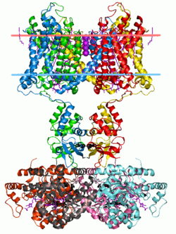
The transmembrane cation channel superfamily was defined in InterPro and Pfam as the family of tetrameric ion channels. These include the sodium, potassium, [1] calcium, ryanodine receptor, HCN, CNG, CatSper, and TRP channels. This large group of ion channels apparently includes families 1.A.1 , 1.A.2 , 1.A.3 , and 1.A.4 of the TCDB transporter classification.
They are described as minimally having two transmembrane helices flanking a loop which determines the ion selectivity of the channel pore. Many eukaryotic channels have four additional transmembrane helices (TM) (Pfam PF00520), related to or vestigial of voltage gating. The proteins with only two transmembrane helices (Pfam PF07885) are most commonly found in bacteria. This also includes the 2-TM inward-rectifier potassium channels (Pfam PF01007) found primarily in eukaryotes. There are commonly additional regulatory domains which serve to regulate ion conduction and channel gating. The pores may also be homotetramers or heterotetramers; where heterotetramers may be encoded as distinct genes or as multiple pore domains within a single polypeptide. The HVCN1 and Putative tyrosine-protein phosphatase proteins do not contain an expected ion conduction pore domain, but rather have homology only to the voltage sensor domain of voltage gated ion channels.
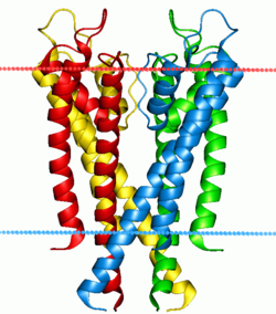
Ion channels are pore-forming membrane proteins that allow ions to pass through the channel pore. Their functions include establishing a resting membrane potential, shaping action potentials and other electrical signals by gating the flow of ions across the cell membrane, controlling the flow of ions across secretory and epithelial cells, and regulating cell volume. Ion channels are present in the membranes of all cells. Ion channels are one of the two classes of ionophoric proteins, the other being ion transporters.
Potassium channels are the most widely distributed type of ion channel found in virtually all organisms. They form potassium-selective pores that span cell membranes. Potassium channels are found in most cell types and control a wide variety of cell functions.
In neuroscience, repolarization refers to the change in membrane potential that returns it to a negative value just after the depolarization phase of an action potential which has changed the membrane potential to a positive value. The repolarization phase usually returns the membrane potential back to the resting membrane potential. The efflux of potassium (K+) ions results in the falling phase of an action potential. The ions pass through the selectivity filter of the K+ channel pore.
Voltage-gated ion channels are a class of transmembrane proteins that form ion channels that are activated by changes in the electrical membrane potential near the channel. The membrane potential alters the conformation of the channel proteins, regulating their opening and closing. Cell membranes are generally impermeable to ions, thus they must diffuse through the membrane through transmembrane protein channels. They have a crucial role in excitable cells such as neuronal and muscle tissues, allowing a rapid and co-ordinated depolarization in response to triggering voltage change. Found along the axon and at the synapse, voltage-gated ion channels directionally propagate electrical signals. Voltage-gated ion-channels are usually ion-specific, and channels specific to sodium (Na+), potassium (K+), calcium (Ca2+), and chloride (Cl−) ions have been identified. The opening and closing of the channels are triggered by changing ion concentration, and hence charge gradient, between the sides of the cell membrane.
The shaker (Sh) gene, when mutated, causes a variety of atypical behaviors in the fruit fly, Drosophila melanogaster. Under ether anesthesia, the fly’s legs will shake ; even when the fly is unanaesthetized, it will exhibit aberrant movements. Sh-mutant flies have a shorter lifespan than regular flies; in their larvae, the repetitive firing of action potentials as well as prolonged exposure to neurotransmitters at neuromuscular junctions occurs.
Potassium voltage-gated channel subfamily A member 1 also known as Kv1.1 is a shaker related voltage-gated potassium channel that in humans is encoded by the KCNA1 gene. Isaacs syndrome is a result of an autoimmune reaction against the Kv1.1 ion channel.
Calcium-activated potassium channels are potassium channels gated by calcium, or that are structurally or phylogenetically related to calcium gated channels. They were first discovered in 1958 by Gardos who saw that calcium levels inside of a cell could affect the permeability of potassium through that cell membrane. Then in 1970, Meech was the first to observe that intracellular calcium could trigger potassium currents. In humans they are divided into three subtypes: large conductance or BK channels, which have very high conductance which range from 100 to 300 pS, intermediate conductance or IK channels, with intermediate conductance ranging from 25 to 100 pS, and small conductance or SK channels with small conductances from 2-25 pS.
Voltage-gated potassium channels (VGKCs) are transmembrane channels specific for potassium and sensitive to voltage changes in the cell's membrane potential. During action potentials, they play a crucial role in returning the depolarized cell to a resting state.
Potassium voltage-gated channel subfamily E member 1 is a protein that in humans is encoded by the KCNE1 gene.
K+ channel tetramerisation domain is the N-terminal, cytoplasmic tetramerisation domain (T1) of voltage-gated K+ channels. It defines molecular determinants for subfamily-specific assembly of alpha-subunits into functional tetrameric channels. It is distantly related to the BTB/POZ domain Pfam PF00651.
Calcium-activated potassium channel subunit alpha-1 also known as large conductance calcium-activated potassium channel, subfamily M, alpha member 1 (KCa1.1), or BK channel alpha subunit, is a voltage gated potassium channel encoded by the KCNMA1 gene and characterized by their large conductance of potassium ions (K+) through cell membranes.
Potassium voltage-gated channel subfamily D member 2 is a protein that in humans is encoded by the KCND2 gene. It contributes to the cardiac transient outward potassium current (Ito1), the main contributing current to the repolarizing phase 1 of the cardiac action potential.
Potassium voltage-gated channel subfamily A member 4 also known as Kv1.4 is a protein that in humans is encoded by the KCNA4 gene. It contributes to the cardiac transient outward potassium current (Ito1), the main contributing current to the repolarizing phase 1 of the cardiac action potential.
Potassium voltage-gated channel, shaker-related subfamily, member 3, also known as KCNA3 or Kv1.3, is a protein that in humans is encoded by the KCNA3 gene.
Potassium voltage-gated channel subfamily E member 4, originally named MinK-related peptide 3 or MiRP3 when it was discovered, is a protein that in humans is encoded by the KCNE4 gene.
Potassium voltage-gated channel, Shaw-related subfamily, member 4 (KCNC4), also known as Kv3.4, is a human gene.
Potassium voltage-gated channel subfamily C member 1 is a protein that in humans is encoded by the KCNC1 gene.
Potassium voltage-gated channel subfamily G member 1 is a protein that in humans is encoded by the KCNG1 gene.
Potassium voltage-gated channel, Shaw-related subfamily, member 3 also known as KCNC3 or Kv3.3 is a protein that in humans is encoded by the KCNC3.
Lipid-gated ion channels are a class of ion channels whose conductance of ions through the membrane depends directly on lipids. Classically the lipids are membrane resident anionic signaling lipids that bind to the transmembrane domain on the inner leaflet of the plasma membrane with properties of a classic ligand. Other classes of lipid-gated channels include the mechanosensitive ion channels that respond to lipid tension, thickness, and hydrophobic mismatch. A lipid ligand differs from a lipid cofactor in that a ligand derives its function by dissociating from the channel while a cofactor typically derives its function by remaining bound.